. 2024 Jan;300(1):105545.
doi: 10.1016/j.jbc.2023.105545. Epub 2023 Dec 9.

Tau seeding without tauopathy

Affiliations

Tau seeding without tauopathy

Michael S LaCroix et al. J Biol Chem. 2024 Jan.

Abstract

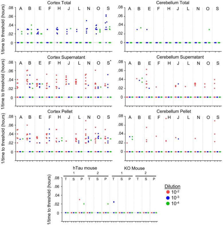
Neurodegenerative tauopathies such as Alzheimer's disease (AD) are caused by brain accumulation of tau assemblies. Evidence suggests tau functions as a prion, and cells and animals can efficiently propagate unique, transmissible tau pathologies. This suggests a dedicated cellular replication machinery, potentially reflecting a normal physiologic function for tau seeds. Consequently, we hypothesized that healthy control brains would contain seeding activity. We have recently developed a novel monoclonal antibody (MD3.1) specific for tau seeds. We used this antibody to immunopurify tau from the parietal and cerebellar cortices of 19 healthy subjects without any neuropathology, ranging 19 to 65 years. We detected seeding in lysates from the parietal cortex, but not in the cerebellum. We also detected no seeding in brain homogenates from wildtype or human tau knockin mice, suggesting that cellular/genetic context dictates development of seed-competent tau. Seeding did not correlate with subject age or brain tau levels. We confirmed our essential findings using an orthogonal assay, real-time quaking-induced conversion, which amplifies tau seeds in vitro. Dot blot analyses revealed no AT8 immunoreactivity above background levels in parietal and cerebellar extracts and ∼1/100 of that present in AD. Based on binding to a panel of antibodies, the conformational characteristics of control seeds differed from AD, suggesting a unique underlying assembly, or structural ensemble. Tau's ability to adopt self-replicating conformations under nonpathogenic conditions may reflect a normal function that goes awry in disease states.

Keywords: Alzheimer’s disease; FRET biosensor; healthy brain; prion; tau seeding activity; tauopathy.


Conflict of interest statement

Conflict of interest The authors declare that they have no conflicts of interest with the contents of this article.

Figures

Figure 1
Figure 1
Tau seeding is present in the parietal lobe of 19 control subjects. Parietal cortex fresh-frozen samples from 19 individuals (AS) without any known neurological diagnoses were used to create total (T) clarified lysate (10% [wt/vol]) followed by immunoprecipitation with the MD3.1 antibody to generate a tau-depleted supernatant (S) and tau-enriched pellet (P). Tau seeding was reliably detected in 16 of 19 cortical immunoprecipitation pellets. Columns represent the mean fluorescence resonance energy transfer (FRET) positivity from three technical replicates (dots). Statistical significance was determined by performing one-way ANOVA followed by Dunnett’s multiple comparisons testing of all samples compared against Lipofectamine-treated negative controls, ∗p < 0.05, ∗∗p < 0.01, ∗∗∗p < 0.001, ∗∗∗∗p < 0.0001. Error bars = SD.
Figure 2
Figure 2
No reliable tau seeding activity is detected in the cerebellum of 19 control subjects. Fresh-frozen cerebellum samples from 19 subjects without any known neurological diagnoses (referenced AS) were used to create total (T) clarified lysate (10% [wt/vol]) followed by immunoprecipitation with the MD3.1 antibody to generate a tau-depleted supernatant (S) and tau-enriched pellet (P). No reliable tau seeding was detected across all cerebellum samples. Columns indicate the mean fluorescence resonance energy transfer (FRET) positivity from three technical replicates (dots). Statistical significance was determined by performing one-way ANOVA followed by Dunnett’s multiple comparisons testing of all samples compared against Lipofectamine-treated negative controls, ∗∗∗∗p < 0.0001. Error bars = SD. Note alteration of seeding scale to encompass the positive control.
Figure 3
Figure 3
Human and mouse tau expressed in mouse cortex does not form detectable seeds. Of the groups tested, only human tau enriched via immunoprecipitation from human cortex, and not human cerebellum, formed seeds that were detectable at a statistically significant level. Tau immunoprecipitated from the cortex of mice expressing human tau (hTau, n = 10, F = 5, M = 5) and wildtype mouse tau (WT, n = 9, M = 4, F = 5) did not show significant seeding activity. Statistical significance was determined by performing one-way ANOVA followed by Dunnett’s multiple comparisons testing of all samples compared against Lipofectamine-treated negative controls, ∗∗∗∗p < 0.0001. Error bars = SD. CB, cerebellum; Ctx, cortex; FRET, fluorescence resonance energy transfer.
Figure 4
Figure 4
RT-QuIC detects tau seeds in control brain. Total homogenates (Total), supernatants (Supernatant), and immunoprecipitated pellets (Pellet) were diluted in KO/N2/Hepes buffer as indicated: 10-2, 10-3, 10-4 and used to seed the K12 RT-QuIC assay. The reciprocal of time to threshold thioflavin T fluorescence is plotted for eight replicate reactions except for sample marked S∗, which had only four replicates. Reactions for which 1/time to threshold values are marked as zero refer to those that failed to reach the threshold within 48 h. Nonzero data points are distinguishable as separate data points, whereas remaining replicates (out of a total of 8) are overlapped on the baseline. RT-QuIC, real-time quaking-induced conversion.
Figure 5
Figure 5
Comparative AT8 staining of brain tissues. Fresh-frozen brain samples were prepared from an Alzheimer’s disease (AD) control, an aged PS19 tauopathy mouse, and 10 control brain samples that had the highest levels of tau seeding and were imaged at 40× magnification (A, B, D, E, G, IK, O, S). We easily detected tau pathology in AD and PS19 brain. We detected no AT8 signal in any control brain. The scale bar represents 50 μm.
Figure 6
Figure 6
Control brains contain markedly less AT8 positivity relative to Alzheimer’s disease (AD). Two micrograms of soluble protein lysate from AD was used for 1:1, followed by serial dilutions. Two micrograms of total soluble protein was used for control samples, loaded in left to right order starting with sample A. Dashes represent no loading.
Figure 7
Figure 7
ELISA quantification of tau in brain samples.A, quantification of soluble tau in total clarified lysates from the parietal cortex and cerebellum. B, quantification of tau in the pellet following immunoprecipitation using the MD3.1 antibody. Statistical significance was determined by performing Student’s t test, ∗∗p < 0.01, ∗∗∗∗p < 0.0001.
Figure 8
Figure 8
Lack of correlation of tau seeding with age or tau concentration.A, initial tau concentration in total soluble protein fractions did not correlate with seeding in immunoprecipitation pellets. B, the final tau concentration from immunoprecipitation pellets did not correlate with seeding in immunoprecipitation pellets. C, age did not correlate with seeding in immunoprecipitation pellets. Data were analyzed using Pearson correlation, ns, not significant. FRET, fluorescence resonance energy transfer.
Figure 9
Figure 9
Differential seed capture efficiency from control and Alzheimer’s disease (AD) brain. A custom antibody panel reveals unique epitope exposure of tau seeds in control brain versus AD. A, epitopes of antibodies used. B, MD3.1 immunoprecipitation pellets from AD brain have roughly 1000× seeding activity versus MD3.1 pellets from control brain. MD3.1 pellets from AD were diluted 1000-fold prior to seeding on v2H biosensors while control pellets were used undiluted. No significant difference between undiluted control pellets and 1000-fold diluted AD pellets was found (p < 0.3692), Student’s t test, error bars = SD. (C) MD3.1 was most efficient at isolating tau seeds from control brain. D, multiple antibodies efficiently isolated seeds from AD brain. FRET, fluorescence resonance energy transfer.

References

    1. Lee V.M., Trojanowski J.Q. Neurodegenerative tauopathies: human disease and transgenic mouse models. Neuron. 1999;24:507–510. - PubMed
    1. Frost B., Ollesch J., Wille H., Diamond M.I. Conformational diversity of wild-type Tau fibrils specified by templated conformation change. J. Biol. Chem. 2009;284:3546–3551. - PMC - PubMed
    1. Frost B., Jacks R.L., Diamond M.I. Propagation of tau misfolding from the outside to the inside of a cell. J. Biol. Chem. 2009;284:12845–12852. - PMC - PubMed
    1. Clavaguera F., Bolmont T., Crowther R.A., Abramowski D., Frank S., Probst A., et al. Transmission and spreading of tauopathy in transgenic mouse brain. Nat. Cell Biol. 2009;11:909–913. - PMC - PubMed
    1. Clavaguera F., Akatsu H., Fraser G., Crowther R.A., Frank S., Hench J., et al. Brain homogenates from human tauopathies induce tau inclusions in mouse brain. Proc. Natl. Acad. Sci. U. S. A. 2013;110:9535–9540. - PMC - PubMed

Publication types