. 2023 Dec;12(12):e12392.
doi: 10.1002/jev2.12392.

Metabolic labelling of a subpopulation of small extracellular vesicles using a fluorescent palmitic acid analogue

Affiliations

Metabolic labelling of a subpopulation of small extracellular vesicles using a fluorescent palmitic acid analogue

Valeria Barreca et al. J Extracell Vesicles. 2023 Dec.

Abstract

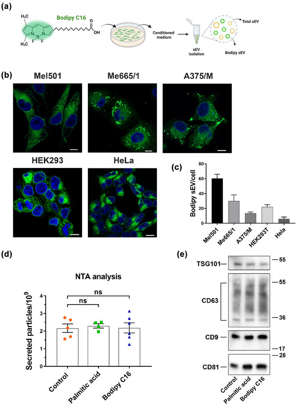
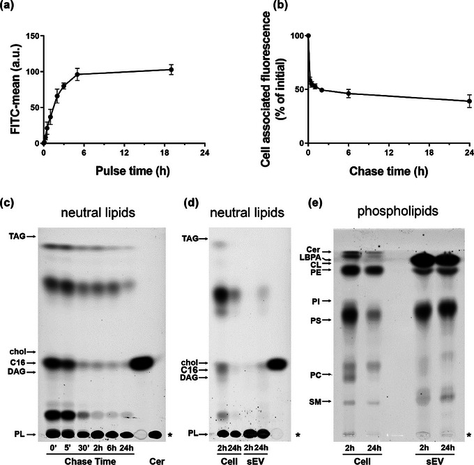
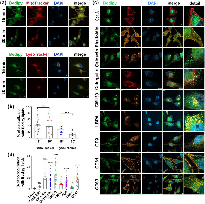
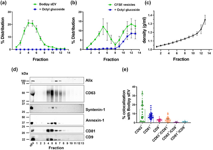
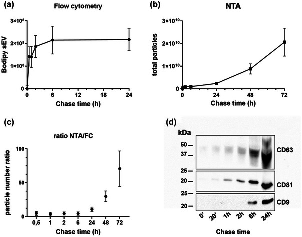
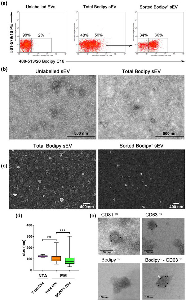
Exosomes are among the most puzzling vehicles of intercellular communication, but several crucial aspects of their biogenesis remain elusive, primarily due to the difficulty in purifying vesicles with similar sizes and densities. Here we report an effective methodology for labelling small extracellular vesicles (sEV) using Bodipy FL C16, a fluorescent palmitic acid analogue. In this study, we present compelling evidence that the fluorescent sEV population derived from Bodipy C16-labelled cells represents a discrete subpopulation of small exosomes following an intracellular pathway. Rapid cellular uptake and metabolism of Bodipy C16 resulted in the incorporation of fluorescent phospholipids into intracellular organelles specifically excluding the plasma membrane and ultimately becoming part of the exosomal membrane. Importantly, our fluorescence labelling method facilitated accurate quantification and characterization of exosomes, overcoming the limitations of nonspecific dye incorporation into heterogeneous vesicle populations. The characterization of Bodipy-labelled exosomes reveals their enrichment in tetraspanin markers, particularly CD63 and CD81, and in minor proportion CD9. Moreover, we employed nanoFACS sorting and electron microscopy to confirm the exosomal nature of Bodipy-labelled vesicles. This innovative metabolic labelling approach, based on the fate of a fatty acid, offers new avenues for investigating exosome biogenesis and functional properties in various physiological and pathological contexts.

Keywords: MVB; exosome biogenesis; exosomes; extracellular vesicles; lipid metabolism; sEV.


Conflict of interest statement

The authors declare no conflicts of interest.

Figures

FIGURE 1
FIGURE 1
(a) Scheme of the structure of Bodipy C16 and experimental strategy. (b) Confocal microscopy images of cells labelled for 2 h with Bodipy C16 at steady state. (c) Quantification by Flow Cytometry (FC) of sEV secreted in the conditioned medium of different cell lines. (d) Measurement by Nanoparticle Tracking Analysis (NTA) of total sEV released by Mel501 cells untreated or treated with Bodipy C16 or with unlabelled palmitic acid for 5 h. SEV were purified from conditioned media after 24 h of chase. Bars represent the mean ± SEM (n > 3). (e) Western blot analysis of total sEV released by untreated or treated with Bodipy C16 or with unlabelled palmitic acid.
FIGURE 2
FIGURE 2
Dynamics of Bodipy C16 uptake and transformation into phospholipids. (a) Mel501 cells were treated with 7 µM Bodipy C16 for the indicated times or (b) treated for 15 min, washed with H‐BSA and chased in complete medium. Cell associated fluorescence was measured by FC. Data are expressed as mean  ±  SEM (n = 3). (c) Total lipids from cells pulsed with Bodipy C16 for 15 min and chased for different times were extracted and analyzed by HPTLC. The amount of lipid spotted per lane was equivalent to 5 × 104 cells. (d) Lipids from 2 × 104 cells pulsed with Bodipy C16 for 5 h and chased for 2 h and 24 h and Bodipy sEV isolated from the conditioned medium were extracted and analyzed by HPTLC for neutral lipids and (e) phospholipids. The amount of lipid spotted per lane was equivalent to 4 × 104 cells and 8,6 × 108 sEV. Asterisk indicates the sample origin line. Lipids were identified based on comparison to the respective lipid standards. Neutral lipids: TAG, triacylglycerols; DAG, diacylglycerol; chol, cholesterol; C16, Bodipy C16; PL, Polar Lipids. Phospholipids: Cer, ceramide; LBPA, Lysobisphosphatidic acid; CL, cardiolipin; PE, phosphatidylethanolamine; PI, phosphatidylinositol; PS, phosphatidylserine; PC phosphatidylcholine; SM, sphingomyelin.
FIGURE 3
FIGURE 3
Co‐localization analysis of Bodipy lipids with intracellular markers. (a) Colocalization with mitochondria and lysosomes at different pulse time. Cells were labelled with 1 µM Bodipy C16 and Mitotracker Deep Red or Lysotracker Deep Red for 15 or 30 min, fixed and mounted with DAPI. (b) Quantification of colocalization with Bodipy lipids. Quantification of 3 independent experiments showing the mean ± SEM (≥20 fields). (c) Colocalization of Bodipy lipids with intracellular organelles markers and tetraspanins after a 2 h pulse with Bodipy C16 at steady‐state. Fixed cells were permeabilized with 0,1% saponin prior to incubation with antibodies or non permeabilized when incubated with Con A. Percentage of colocalization is calculated by summing the pixels in the colocalized region and then dividing by the sum of Bodipy C16 pixels. All data are expressed as means ± SEM (n ≥ 3) (n ≥ 20 fields). Individual unpaired t‐test was performed to compare two groups. Significance of colocalization in (d) was calculated by comparing percent of individual markers colocalization with Con A colocalization. *p < 0.05, **p < 0.01, ***p < 0.001, ****p < 0.0001, ns, no statistical significance. Scale bar = 10 µm.
FIGURE 4
FIGURE 4
Bodipy sEV separate as a single peak enriched in tetraspanins. (a) Bodipy sEV 100K pellets were loaded at the bottom of an iodixanol density gradient and ultracentrifuged for 16 h. Fractions were collected from the top of the gradient and were analyzed for Bodipy sEV number by FC before and after the addition of Octyl‐beta‐glucoside (OG). Data are expressed as mean ± SEM (n = 14). (b) 100K pellets from conditioned media of untreated Mel501 cells were incubated with CFSE to non specifically label the total population of sEV and loaded at the bottom of an iodixanol density gradient. Data are expressed as mean ± SEM (n = 4). (c) Density of the fractions was measured by refractometry. Data are expressed as mean ± SEM (n = 17). (d) The total volume of each fraction was analyzed by Western blot. Data shown are representative of three independent experiments. (d) Analysis of colocalization of tetraspanins fluorescent antibodies with Bodipy sEV.
FIGURE 5
FIGURE 5
Secretion kinetics of Bodipy sEV. Cells were pulsed with Bodipy C16 for 2 h and chased for different times. The number of purified sEV purified by differential ultracentrifugation was measured by (a) FC or by (b) Nanoparticle Tracking Analysis (NTA). Results are expressed as means  ±  SEM of at least 6 independent experiments. (c) Ratio between the total number of sEV counted by NTA and Bodipy sEV counted by FC. A constant ratio is showed up to 6 h, while there is an increase of total vesicles over the time. Results are expressed as means  ±  SEM of at least six independent experiments. (d) Representative Western blot analysis of tetraspanins expression in Bodipy sEV secreted at different chase time. The total sEV released at each time point has been analyzed.
FIGURE 6
FIGURE 6
Bodipy sEV can be sorted in a homogenous subpopulation of small exosomes. (a) Fluorescent sEV pellets have been sorted with a MoFlow AstriosCell Sorter. Dot plot on the left shows unlabelled EVs. The dot plot in the middle shows total Bodipy+ sEV and the arrow indicates the region used for sort decision. The post sorting analysis shows purified Bodipy+ sEV (dot plot on the right). Dot plots are representative of three independent experiments. (b) Unlabelled sEV and total Bodipy+ sEV were analyzed by TEM showing the presence of a discrete population of membrane vesicles in both samples. (c) FACS sorted Bodipy+ sEV analyzed by SEM revealed a homogeneous population of small vesicles in a very low background (right), particularly appreciable when compared directly to the total Bodipy‐labelled sEV population. (left). (d) Total sEV size was determined by NTA and electron microscopy (EM) The semi‐quantitative EM analysis (N > 100) showed that the sorted Bodipy+ sEV had a median size of 80 nm, significantly smaller than total Bodipy‐labelled sEV population (*** p < 0.001). (e) Total sEV were immunolabeled with anti CD81, CD63 and Bodipy antibodies, revealed by 10 nm colloidal gold conjugated secondary antibodies (upper left, upper right and lower left TEM micrographs). Colocalization of CD63 (10 nm) and Bodipy (5 nm, white arrow) antigens on the same vesicle is shown in the lower right image.

References

    1. Akoumi, A. , Haffar, T. , Mousterji, M. , Kiss, R. S. , & Bousette, N. (2017). Palmitate mediated diacylglycerol accumulation causes endoplasmic reticulum stress, Plin2 degradation, and cell death in H9C2 cardiomyoblasts. Experimental Cell Research, 354(2), 85–94. 10.1016/j.yexcr.2017.03.032 - DOI - PubMed
    1. Clancy, J. W. , Schmidtmann, M. , & D'Souza‐Schorey, C. (2021). The ins and outs of microvesicles. FASEB BioAdvances, 3(6), 399–406. 10.1096/fba.2020-00127 - DOI - PMC - PubMed
    1. Colombo, M. , Raposo, G. , & Thery, C. (2014). Biogenesis, secretion, and intercellular interactions of exosomes and other extracellular vesicles. Annual Review of Cell and Developmental Biology, 30, 255–289. 10.1146/annurev-cellbio-101512-122326 - DOI - PubMed
    1. Coscia, C. , Parolini, I. , Sanchez, M. , Biffoni, M. , Boussadia, Z. , Zanetti, C. , Fiani, M. L. , & Sargiacomo, M. (2016). Generation, quantification, and tracing of metabolically labeled fluorescent exosomes. In Federico M., (ed.) Lentiviral vectors and exosomes as gene and protein delivery tools. pp. 217–235. Springer New York. - PubMed
    1. Escola, J. M. , Kleijmeer, M. J. , Stoorvogel, W. , Griffith, J. M. , Yoshie, O. , & Geuze, H. J. (1998). Selective enrichment of tetraspan proteins on the internal vesicles of multivesicular endosomes and on exosomes secreted by human B‐lymphocytes. Journal of Biological Chemistry, 273(32), 20121–20127. 10.1074/jbc.273.32.20121 - DOI - PubMed

Publication types

MeSH terms

Substances

LinkOut - more resources