Clinical Trial
. 2023 Dec 11;10(1):63.
doi: 10.1186/s40779-023-00493-5.

Adipsin inhibits Irak2 mitochondrial translocation and improves fatty acid β-oxidation to alleviate diabetic cardiomyopathy

Affiliations
Clinical Trial

Adipsin inhibits Irak2 mitochondrial translocation and improves fatty acid β-oxidation to alleviate diabetic cardiomyopathy

Meng-Yuan Jiang et al. Mil Med Res. .

Abstract

Background: Diabetic cardiomyopathy (DCM) causes the myocardium to rely on fatty acid β-oxidation for energy. The accumulation of intracellular lipids and fatty acids in the myocardium usually results in lipotoxicity, which impairs myocardial function. Adipsin may play an important protective role in the pathogenesis of DCM. The aim of this study is to investigate the regulatory effect of Adipsin on DCM lipotoxicity and its molecular mechanism.

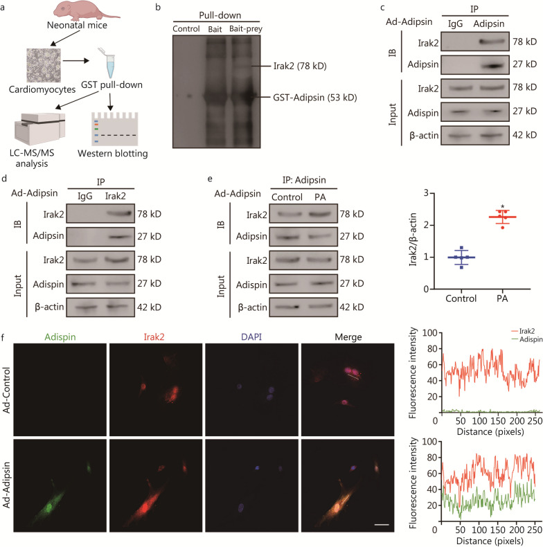
Methods: A high-fat diet (HFD)-induced type 2 diabetes mellitus model was constructed in mice with adipose tissue-specific overexpression of Adipsin (Adipsin-Tg). Liquid chromatography-tandem mass spectrometry (LC-MS/MS), glutathione-S-transferase (GST) pull-down technique, Co-immunoprecipitation (Co-IP) and immunofluorescence colocalization analyses were used to investigate the molecules which can directly interact with Adipsin. The immunocolloidal gold method was also used to detect the interaction between Adipsin and its downstream modulator.

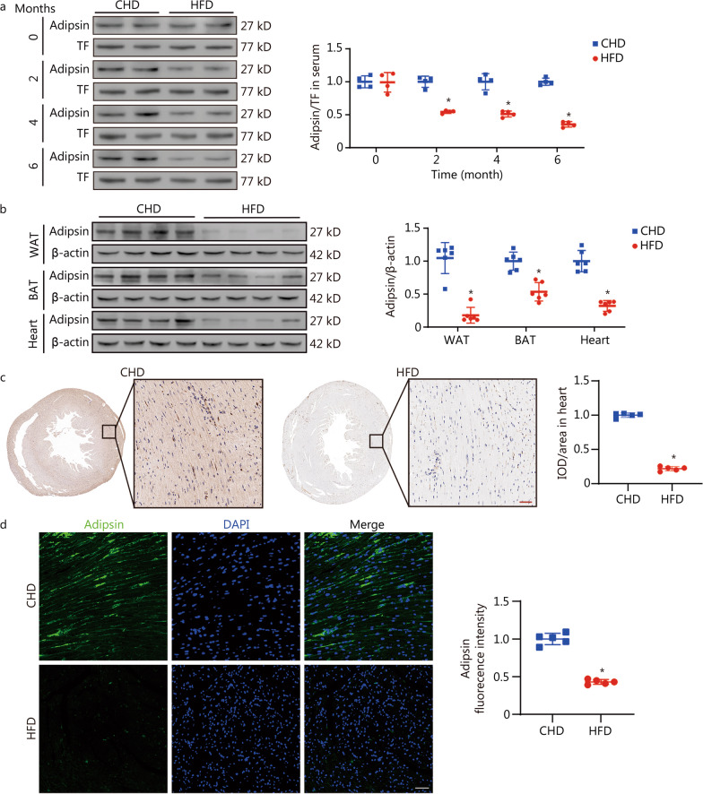
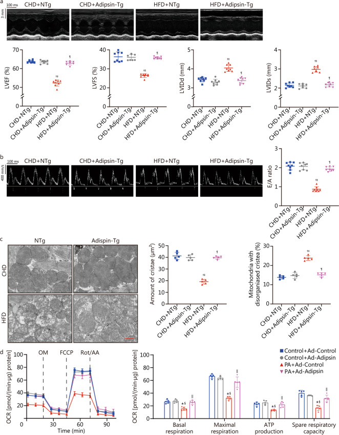
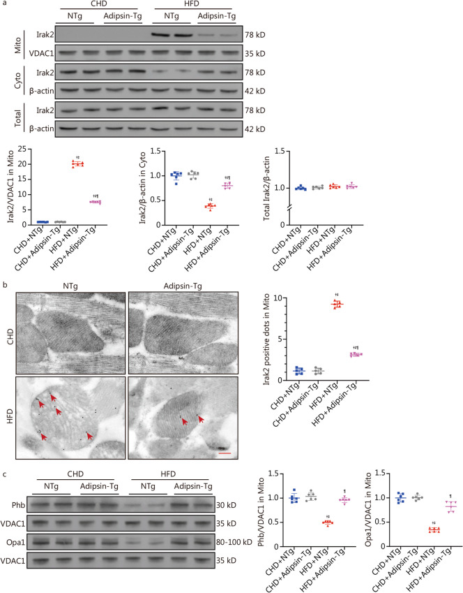
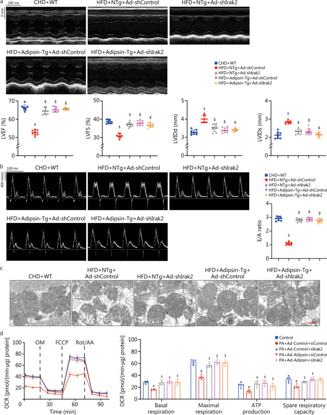
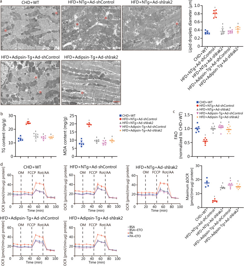
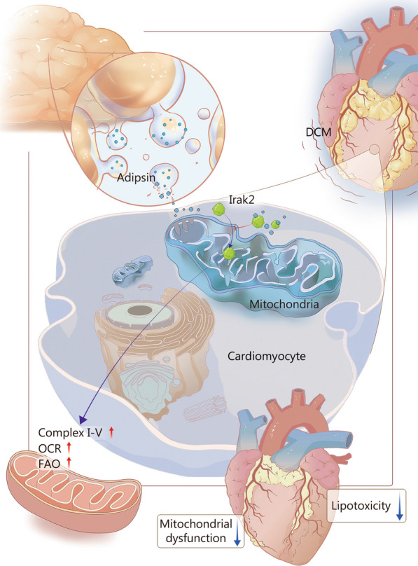
Results: The expression of Adipsin was significantly downregulated in the HFD-induced DCM model (P < 0.05). Adipose tissue-specific overexpression of Adipsin significantly improved cardiac function and alleviated cardiac remodeling in DCM (P < 0.05). Adipsin overexpression also alleviated mitochondrial oxidative phosphorylation function in diabetic stress (P < 0.05). LC-MS/MS analysis, GST pull-down technique and Co-IP studies revealed that interleukin-1 receptor-associated kinase-like 2 (Irak2) was a downstream regulator of Adipsin. Immunofluorescence analysis also revealed that Adipsin was co-localized with Irak2 in cardiomyocytes. Immunocolloidal gold electron microscopy and Western blotting analysis indicated that Adipsin inhibited the mitochondrial translocation of Irak2 in DCM, thus dampening the interaction between Irak2 and prohibitin (Phb)-optic atrophy protein 1 (Opa1) on mitochondria and improving the structural integrity and function of mitochondria (P < 0.05). Interestingly, in the presence of Irak2 knockdown, Adipsin overexpression did not further alleviate myocardial mitochondrial destruction and cardiac dysfunction, suggesting a downstream role of Irak2 in Adipsin-induced responses (P < 0.05). Consistent with these findings, overexpression of Adipsin after Irak2 knockdown did not further reduce the accumulation of lipids and their metabolites in the cardiac myocardium, nor did it enhance the oxidation capacity of cardiomyocytes expose to palmitate (PA) (P < 0.05). These results indicated that Irak2 may be a downstream regulator of Adipsin.

Conclusions: Adipsin improves fatty acid β-oxidation and alleviates mitochondrial injury in DCM. The mechanism is related to Irak2 interaction and inhibition of Irak2 mitochondrial translocation.

Keywords: Diabetic cardiomyopathy; Fatty acid β-oxidation; Mitochondrial function; Mitochondrial translocation.


Conflict of interest statement

The authors declare that they have no competing interests.

Figures

Fig. 1
Fig. 1
Adipsin is significantly downregulated in HFD-induced DCM. a Western blotting results and quantitative analysis of serum Adipsin levels in the treatment groups at 0, 2, 4, and 6 months (n = 4). b Western blotting results and quantitative analysis of Adipsin expression in WAT, interscapular BAT and heart tissue lysates from CHD- and HFD-fed mice (n = 6). c, d Immunohistochemical/immunofluorescence stainings and quantitative analysis of Adispin in mouse hearts from CHD- and HFD-fed groups (n = 5). Scale bar = 50 μm. Statistical significance was determined by two-tailed Student’s t test. All data are represented with mean ± SD. *P < 0.05 vs. CHD; CHD chow diet, HFD high-fat diet, WAT white adipose tissue, BAT brown adipose tissue, TF transferrin, IOD integrated option density, DAPI 4’,6-diamidino-2-phenylindole
Fig. 2
Fig. 2
Overexpression of Adipsin can improve cardiac function, reduce mitochondrial cristae damage, and improve mitochondrial function. a Echocardiographic results and quantitative analysis of echocardiographic data including LVEF, LVFS, LVIDd and LVIDs (n = 8). b Pulse-wave Doppler results and quantitative analysis of the E/A ratio (n = 8). c Transmission electron microscopic images, and quantitative analysis of cristae amount per μm2 and the proportion of mitochondria with disorganized cristae of myocardium from NTg and Adipsin-Tg groups with CHD- or HFD-feeding (n = 5). Scale bar = 500 nm. d OCR in cardiomyocytes from various treatment groups, and quantitative statistical analysis of OCR including basal respiration, maximal respiration, ATP production, and spare respiration capacity. Statistical significance was determined using one-way ANOVA analysis. All data are represented with mean ± SD. P < 0.05 vs. CHD + NTg; P < 0.05 vs. CHD + Adipsin-Tg; P < 0.05 vs. HFD + NTg; *P < 0.05 vs. Control + Ad-Control; §P < 0.05 vs. Control + Ad-Adipsin; ||P < 0.05 vs. PA + Ad-Control. CHD chow diet, HFD high-fat diet, Adipsin-Tg Adipsin tissue-specific transgenic mice, NTg nontransgenic mice, LVEF left ventricular ejection fraction, LVFS left ventricular fractional shortening, LVIDd end-diastolic left ventricular internal diameters, LVIDs end-systolic left ventricular internal diameters, E/A the ratio of mitral peak velocity of early filling (E) to mitral peak velocity of late filling (A), OCR oxygen consumption rate, OM oligomycin, FCCP fluoro-carbonyl cyanide phenylhydrazone, Rot/AA rotenone and antimycin A, PA palmitate
Fig. 3
Fig. 3
Adipsin and Irak2 bind directly in cardiomyocytes. a Schematic representation of the experimental protocol. The cardiomyocytes lysis prey protein was obtained by immobilizing GST-Adipsin with equilibrated glutathione agarose. Then, the eluted protein complexes were employed for mass spectrometry analysis and subsequent immunoblotting. b Proteins bound and pulled down by purified GST-Adipsin in cardiomyocytes. c, d Co-IP results indicating the interaction between Adipsin and Irak2 following the infection with Ad-Adipsin. e Co-IP results indicating the interaction between Adipsin and Irak2 with or without PA challenge following the infection with Ad-Adipsin (n = 5). f Colocalization images and analysis of Adipsin (green) and Irak2 (red) proteins in cardiomyocytes. Scale bar = 50 μm. Statistical significance was determined using a two-tailed Student’s t test. All data are represented with mean ± SD. *P < 0.05 vs. Control. LC–MS/MS liquid chromatography-tandem mass spectrometry, Co-IP Co-immunoprecipitation, DAPI 4′,6-diamidino-2-phenylindole, Irak2 interleukin-1 receptor-associated kinase-like 2, PA palmitate, GST glutathione-S-transferase, IB immunoblotting
Fig. 4
Fig. 4
Overexpression of Adipsin inhibits mitochondrial translocation of Irak2. a Western blotting results and quantitative analysis of Irak2 expression in cytoplasm and mitochondria of cardiomyocytes in the CHD + NTg, CHD + Adipsin-Tg, NTg + HFD, and Adipsin-Tg + HFD groups (n = 6). b Electron microscopy images of Irak2 stained with immunogold and quantitative analysis of Irak2-positive dots (red arrow) in the myocardial mitochondria of NTg or Adipsin-Tg mice fed with CHD or HFD (n = 5). Scale bar = 200 nm. c Western blotting results and quantitative analysis of Phb and Opa1 in myocardial mitochondria of NTg or Adipsin-Tg mice fed with CHD or HFD (n = 6). Statistical significance was determined using one-way ANOVA. All data are represented with mean ± SD. P < 0.05 vs. CHD + NTg; P < 0.05 vs. CHD + Adipsin-Tg; P < 0.05 vs. HFD + NTg. Mito mitochondria, Cyto cytosol, CHD chow diet, HFD high-fat diet, Adipsin-Tg Adipsin tissue-specific transgenic mice, NTg nontransgenic mice, Irak2 interleukin-1 receptor-associated kinase-like 2, VDAC1 voltage-dependent anion channel 1, Phb prohibitin, Opa1 optic atrophy protein 1
Fig. 5
Fig. 5
Adipsin improves cardiac function, reduces mitochondrial cristae damage, and improves mitochondrial function through Irak2 signaling pathway. a Echocardiographic results and quantitative analysis of echocardiographic data including LVEF, LVFS, LVIDd and LVIDs (n = 8). b Pulse-wave Doppler results and quantitative analysis of the E/A ratio (n = 8). c Transmission electron microscopic images of myocardium from different groups (n = 8). Scale bar = 500 nm. d OCR in cardiomyocytes from various treatment groups, and quantitative statistical analysis of OCR including basal respiration, ATP production, maximal respiration and spare respiration capacity. Statistical significance was determined using one-way ANOVA. All data are represented with mean ± SD. P < 0.05 vs. CHD + WT; P < 0.05 vs. HFD + NTg + Ad-shControl; *P < 0.05 vs. Con; §P < 0.05 vs. PA + Ad-Control + siControl. CHD chow diet, HFD high-fat diet, Adipsin-Tg Adipsin tissue-specific transgenic mice, NTg nontransgenic mice, LVEF left ventricular ejection fraction, LVFS left ventricular fractional shortening, LVIDd end-diastolic left ventricular internal diameters, LVIDs end-systolic left ventricular internal diameters, E/A the ratio of mitral peak velocity of early filling (E) to mitral peak velocity of late filling (A), OCR oxygen consumption rate, OM oligomycin, FCCP fluoro-carbonyl cyanide phenylhydrazone, Rot/AA rotenone and antimycin A, PA palmitate, Irak2 interleukin-1 receptor-associated kinase-like 2, siControl control small interfering RNA
Fig. 6
Fig. 6
Adipsin improves cardiomyocyte FAO through the Irak2 signaling pathway. a Transmission electron microscopic images and quantitative analyses of mouse hearts depicting larger lipid droplets accumulation in various treatment groups (n = 6). Lipid droplets were marked by asterisks. Scale bar = 1 μm. b TG and MDA content in myocardial tissues (n = 8). c FAO evaluating with [3H]-oleic acid uptake (n = 6). d Oxidation pressure test of palmitate in cardiomyocytes from various treatment groups, and evaluation of FAO using quantitative analysis of maximum ΔOCR. Statistical significance was determined using one-way ANOVA. All data are represented with mean ± SD. P < 0.05 vs. CHD + WT; P < 0.05 vs. HFD + NTg + Ad-shControl. CHD chow diet, HFD high-fat diet, Adipsin-Tg Adipsin tissue-specific transgenic mice, NTg nontransgenic mice, TG triglyceride, MDA malonaldehyde, FAO fatty acid β-oxidation, BSA bovine serum albumin, ETO etomoxir, PA palmitate, OCR oxygen consumption rate, OM oligomycin, FCCP fluoro-carbonyl cyanide phenylhydrazone, Rot/AA rotenone and antimycin A, △OCR oxygen consumption rate[PA-(PA+ETO)]—oxygen consumption rate[BSA-(BSA+ETO)], Irak2 interleukin-1 receptor-associated kinase-like 2
Fig. 7
Fig. 7
Schematic illustration of Adipsin-induced alleviation of diabetic cardiomyopathy (DCM) by inhibiting Irak2 mitochondrial translocation. Adipsin is secreted by adipose tissues and enters cardiomyocytes. Under HFD conditions, Adipsin enters cardiomyocytes to bind with Irak2 and inhibit its mitochondrial translocation. Reduction in Irak2 entry into mitochondria improves FAO, protects mitochondrial structure and function, reduces myocardial lipid accumulation, further improves cardiac function, and prevents DCM. HFD high-fat diet, OCR oxygen consumption rate, FAO fatty acid β-oxidation, Irak2 interleukin-1 receptor-associated kinase-like 2

Comment in

References

    1. Wild S, Roglic G, Green A, Sicree R, King H. Global prevalence of diabetes: estimates for the year 2000 and projections for 2030. Diabetes Care. 2004;27(5):1047–1053. - PubMed
    1. Razavi M, Wei YY, Rao XQ, Zhong JX. DPP-4 inhibitors and GLP-1RAs: cardiovascular safety and benefits. Mil Med Res. 2022;9(1):45. - PMC - PubMed
    1. Knapp M, Tu X, Wu R. Vascular endothelial dysfunction, a major mediator in diabetic cardiomyopathy. Acta Pharmacol Sin. 2019;40(1):1–8. - PMC - PubMed
    1. Chavali V, Tyagi SC, Mishra PK. Predictors and prevention of diabetic cardiomyopathy. Diabetes Metab Syndr Obes. 2013;6:151–160. - PMC - PubMed
    1. Nathan DM. Diabetes: advances in diagnosis and treatment. JAMA. 2015;314(10):1052–1062. - PubMed

Publication types

MeSH terms