. 2024 Mar 1;35(3):321-334.
doi: 10.1681/ASN.0000000000000277. Epub 2023 Dec 11.

Matrix Metalloproteinase-7 in Urinary Extracellular Vesicles Identifies Rapid Disease Progression in Autosomal Dominant Polycystic Kidney Disease

Collaborators, Affiliations

Matrix Metalloproteinase-7 in Urinary Extracellular Vesicles Identifies Rapid Disease Progression in Autosomal Dominant Polycystic Kidney Disease

Martijn H van Heugten et al. J Am Soc Nephrol. .

Abstract

Significance statement: There is an unmet need for biomarkers of disease progression in autosomal dominant polycystic kidney disease (ADPKD). This study investigated urinary extracellular vesicles (uEVs) as a source of such biomarkers. Proteomic analysis of uEVs identified matrix metalloproteinase 7 (MMP-7) as a biomarker predictive of rapid disease progression. In validation studies, MMP-7 was predictive in uEVs but not in whole urine, possibly because uEVs are primarily secreted by tubular epithelial cells. Indeed, single-nucleus RNA sequencing showed that MMP-7 was especially increased in proximal tubule and thick ascending limb cells, which were further characterized by a profibrotic phenotype. Together, these data suggest that MMP-7 is a biologically plausible and promising uEV biomarker for rapid disease progression in ADPKD.

Background: In ADPKD, there is an unmet need for early markers of rapid disease progression to facilitate counseling and selection for kidney-protective therapy. Our aim was to identify markers for rapid disease progression in uEVs.

Methods: Six paired case-control groups ( n =10-59/group) of cases with rapid disease progression and controls with stable disease were formed from two independent ADPKD cohorts, with matching by age, sex, total kidney volume, and genetic variant. Candidate uEV biomarkers were identified by mass spectrometry and further analyzed using immunoblotting and an ELISA. Single-nucleus RNA sequencing of healthy and ADPKD tissue was used to identify the cellular origin of the uEV biomarker.

Results: In the discovery proteomics experiments, the protein abundance of MMP-7 was significantly higher in uEVs of patients with rapid disease progression compared with stable disease. In the validation groups, a significant >2-fold increase in uEV-MMP-7 in patients with rapid disease progression was confirmed using immunoblotting. By contrast, no significant difference in MMP-7 was found in whole urine using ELISA. Compared with healthy kidney tissue, ADPKD tissue had significantly higher MMP-7 expression in proximal tubule and thick ascending limb cells with a profibrotic phenotype.

Conclusions: Among patients with ADPKD, rapid disease progressors have higher uEV-associated MMP-7. Our findings also suggest that MMP-7 is a biologically plausible biomarker for more rapid disease progression.

Trial registration: ClinicalTrials.gov NCT02497521.


Conflict of interest statement

R.T. Gansevoort reports Consultancy: AstraZeneca, Bayer, Galapagos, Mironid, and Sanofi-Genzyme; Research Funding: AstraZeneca, Abbvie, Bayer, Galapagos, Otsuka Pharmaceuticals, Roche, and Sanofi-Genzyme; Honoraria: Galapagos, Mironid, and Otsuka Pharmaceuticals; and Advisory or Leadership Role: Editor of American Journal of Kidney Diseases, CJASN, Journal of Nephrology, Kidney360 Member of the Council of the European Renal Association, Nephron Clinical Practice, and Nephrology Dialysis Transplantation. S. Hayat reports Consultancy: Sequantrix GmbH; Ownership Interest: Sequantrix GmbH; and Research Funding: Askbio GmbH and Novo Nordisk. E.J. Hoorn reports Research Funding: Aurinia; Honoraria: UpToDate; and Advisory or Leadership Role: Editorial Boards: American Journal of Physiology-Renal Physiology, JASN, and Journal of Nephrology; Other committees: Board Member, Dutch Federation of Nephrology and ERA Working Group Genes and Kidney. R. Kramann reports Consultancy: Bayer Healthcare, Gruenenthal, Novo Nordisk, and Pfizer; Research Funding: Chugai, Galapagos, Novo Nordisk, and Travere Therapeutics; Honoraria: Sequantrix; Patents or Royalties: Gli1 cells in fibrosis (method of use); and Advisory or Leadership Role: Board Member and Co-Founder Sequantrix and Scientific Advisory Board Hybridize Therapeutics. E. Meijer reports Research Funding: Dutch Kidney Foundation, Ipsen, Otsuka Pharmaceuticals, Sanofi; all money was paid directly to the institution; and Other Interests or Relationships: Dutch Kidney Foundation, Health Holland, Nieren.nl, NvN, and werkgroep erfelijke nierziekten. R.-U. Müller reports Consultancy: Alnylam and Vifor; Ownership Interest: Bayer and Santa Barbara Nutrients; Research Funding: Otsuka Pharmaceuticals and Thermo Fisher Scientific. All research funding was paid to the employer (Department II of Internal Medicine); Honoraria: Alnylam; Patents or Royalties: Detechgene; and Advisory or Leadership Role: Editorial Board “Kidney and Dialysis,” Chair of the Board of the Working Group “Genes and Kidney” (ERA), and Scientific Advisory Board Santa Barbara Nutrients. M. Salih reports Advisory or Leadership Role: NedMed.nl Scientific advisory board. M.H. van Heugten reports Research Funding: Aurinia Pharmaceuticals has in part supported our research on lupus nephritis between 2022 and 2023. H. van Willigenburg reports Employer: Antea; and Ownership Interest: funds. All remaining authors have nothing to disclose.

Figures

None
Graphical abstract
Figure 1
Figure 1
Study design showing the two ADPKD cohorts from DIPAK and Cologne and the six case–control groups that were formed for the individual studies. In the discovery experiments (gray shading), uEVs were analyzed using MS/MS quantitative proteomics with TMT labeling (groups 1 and 2). In the validation experiments (blue shading) immunoblotting and/or an ELISA on whole urine were used (groups 3–6). ADPKD, autosomal dominant polycystic kidney disease; TMT, tandem mass tag; uEV, urinary extracellular vesicle.
Figure 2
Figure 2
eGFR over time in the six groups. eGFR is depicted as mean±SEM with fitted linear regression including the 95% confidence interval in gray. eGFR over 2.5 years of follow-up is shown for the discovery Groups 1 and 2 (panels A and B), internal validation Groups 3 and 4 (panels C and D) and the external validation Groups 5 and 6 (panels E and F).
Figure 3
Figure 3
Total number of proteins identified in the MS/MS experiments. (A) Venn diagram comparing the proteins identified in isolated uEVs within the MS/MS experiments combined with the extracellular proteome reported on Vesiclepedia. (B) Venn diagram comparing the proteins identified in isolated uEVs in the MS/MS experiments (rapid disease progression versus stable disease in discovery groups 1 and 2).
Figure 4
Figure 4
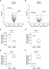
Identification of uEV-MMP-7. Volcano plots for the comparison of protein abundance in isolated uEVs of patients with rapid disease progression versus stable disease in (A) group 1 and (B) group 2. X-axis depicts vsn-normalized abundance ratio (rapidly progressive/stable), which includes a log transformation. Y-axis depicts naturally log-transformed P values of the Student t test, with cutoff at −Ln(0.05). Significantly different proteins (P < 0.05) are marked in darker gray. Abundance of biomarker candidates (C and D) MMP-7 and (E and F) CHMP4A in the MS/MS experiments of groups 1 and 2. Abundances were vsn-normalized, which includes log transformation. *P < 0.05, **P < 0.01 by the Student t test. CHMP4A, charged multivesicular body protein 4a; MMP-7, matrix metalloproteinase 7.
Figure 5
Figure 5
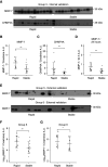
Immunoblot analysis of MMP-7 and CHMP4A in uEVs. (A) Immunoblot analysis of MMP-7 and CHMP4A in internal validation group 3 including (B and C) densitometry. (D) Daily urinary MMP-7 excretion was assessed by extrapolating uEV-MMP-7 abundance using each individual patient's 24-hour urinary creatinine excretion. (E–G) The findings were confirmed in the two external validation groups (groups 5 and 6). *P < 0.05, **P < 0.01 by the Student t test for groups 3 and 4 and the Wilcoxon rank sum test for groups 5 and 6 because of the non-normal distribution of MMP-7 abundance data.
Figure 6
Figure 6
MMP-7 abundance in whole urine. ELISA of whole-urine MMP-7 shows a nonsignificantly higher creatinine-corrected MMP-7 in patients with rapid disease progression compared with patients with stable disease in (A and B) the two internal validation groups (groups 3 and 4) and (C) one of the external validation groups (group 6).
Figure 7
Figure 7
MMP-7 in human ADPKD kidney tissue. Single-nucleus RNA sequencing of human kidneys for 20 different recognized cell types (A) identified MMP-7 for human control and ADPKD tissue when expression was projected on the cell type mapping (B) and directly compared (C) between ADPKD patient kidneys and control tissue. (D) Both the percentage of PT-2 and TAL-2 cells expressing MMP-7 and their mean expression was greater in ADPKD compared with controls. PT, proximal tubule; TAL, thick ascending limb.
Figure 8
Figure 8
Profibrotic profile of MMP-7 expressing cells. Single-nucleus RNA sequencing of human kidneys recognizes a significantly increased profibrotic expression profile in (A) MMP-7–positive cells for all cell types and those cell types with an increased MMP-7 expression, including (B) PT epithelial cells and (C) TAL cells. COL1A1, collagen type I alpha 1 chain; COL3A1, collagen type III alpha 1 chain; DCDC2, doublecortin domain containing 2; FGF2, fibroblast growth factor 2; FN1, fibronectin 1; HAVCR1, hepatitis A virus cellular receptor 1; NOTCH1; notch receptor 1; PROM1, prominin-1; SNAI1, snail family transcriptional repressor 1; TIMP1, tissue inhibitor of metalloproteinases 1; TGFB1, TGF beta 1; VCAM1, vascular cell adhesion molecule 1.

References

    1. Bergmann C, Guay-Woodford LM, Harris PC, Horie S, Peters DJM, Torres VE. Polycystic kidney disease. Nat Rev Dis Primers. 2018;4(1):50. doi:10.1038/s41572-018-0047-y - DOI - PMC - PubMed
    1. Cornec-Le Gall E, Torres VE, Harris PC. Genetic complexity of autosomal dominant polycystic kidney and liver diseases. J Am Soc Nephrol. 2018;29(1):13–23. doi:10.1681/ASN.2017050483 - DOI - PMC - PubMed
    1. Lanktree MB Guiard E Li W, et al. . Intrafamilial variability of ADPKD. Kidney Int Rep. 2019;4(7):995–1003. doi:10.1016/j.ekir.2019.04.018 - DOI - PMC - PubMed
    1. Cornec-Le Gall E, Alam A, Perrone RD. Autosomal dominant polycystic kidney disease. Lancet. 2019;393(10174):919–935. doi:10.1016/S0140-6736(18)32782-X - DOI - PubMed
    1. Gansevoort RT Arici M Benzing T, et al. . Recommendations for the use of tolvaptan in autosomal dominant polycystic kidney disease: a position statement on behalf of the ERA-EDTA Working Groups on Inherited Kidney Disorders and European Renal Best Practice. Nephrol Dial Transplant. 2016;31(3):337–348. doi:10.1093/ndt/gfv456 - DOI - PMC - PubMed

Publication types

Associated data

Grants and funding

LinkOut - more resources