. 2023 Nov 25:46:102294.
doi: 10.1016/j.jcot.2023.102294. eCollection 2023 Nov.

Locating an iliac cortical window for reduction of an acetabular dome impaction: A computed tomography and cadaveric study

Affiliations

Locating an iliac cortical window for reduction of an acetabular dome impaction: A computed tomography and cadaveric study

Rahat Jarayabhand et al. J Clin Orthop Trauma. .

Abstract

This study aimed to determine the optimal location of the iliac cortical window (ICW) for the direct reduction of acetabular dome impactions using a reference bony landmark.

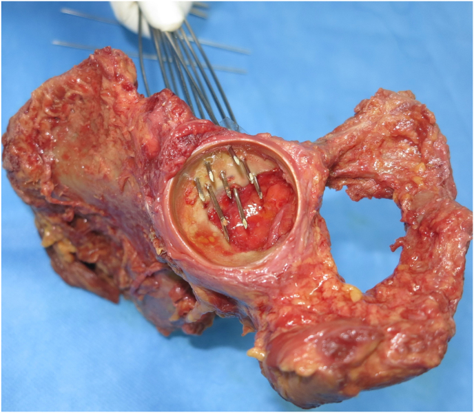
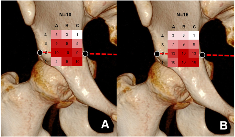
Methods: In the first part of the study, computed tomography scans of 10 normal acetabula, the femoral head weight bearing area, were projected through the superior iliac cortical surface perpendicular to the plane of the true pelvis to show the area that corresponds to the acetabular dome. A line connecting each pair of anterior inferior iliac spines (AIIS) was drawn then reflected in the superior surface of the acetabulum and a reference point (RP) was marked on the line halfway between the AIIS and the pelvic brim. A 12-point 1-cm interval grid with horizontal and vertical axes labeled A, B, C and 1 to 4, respectively, overlying the acetabular surface projection was created to identify the location of the acetabular dome. In the second part of the study, the 12-point grid was marked on eight fresh cadavers (16 acetabula) and the same acetabular dome reference point was identified. K-wires were drilled into the acetabula using a parallel drill guide at each of the twelve grid points. An arthrotomy was carried out and the locations of the K-wires which penetrated the acetabular dome were recorded.

Results: The average distance from the AIIS to the medial pelvic brim in the CT scans and cadaveric study were 47.7 and 45.9 mm, respectively. The K-wires at grid points B2 and C1 had a 100% correlation to the dome area. The A2, B1, and C2 grid points had a correlation with the dome area of >80%. The remaining grid points had joint penetrations ranging from 6.25% to 62.5%.

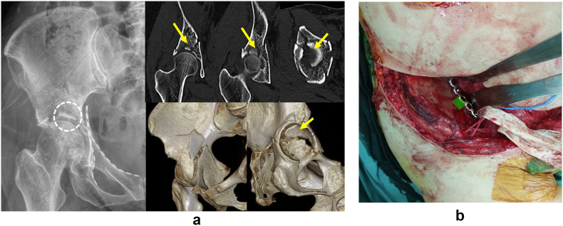
Conclusion: The proposed RP, which can be easily identified intraoperatively, and the area 1 cm2 around the RP (except in the posterior direction) can be used as reliable reference landmarks and for identification of the location of the ICW for the reduction of an acetabular dome impaction.

Keywords: Acetabular fracture; Dome impaction; Iliac cortical window; Reduction.


Conflict of interest statement

The authors declare that they have no known competing financial interests or personal relationships that could have appeared to influence the work reported in this paper.

Figures

Fig. 1
Fig. 1
An imaginary line (thick dash line) connecting both AIISs was drawn and reflected (thin dash line) on the superior cortical surface of the pelvis. The Reference Point (RP, x) was marked at the midpoint from AIIS to the medial pelvic brim.
Fig. 2
Fig. 2
Demonstration of the method to locate the bony landmark in relation to the femoral head a) A 12-point grid with 1 cm spacing was drawn across the iliac cortical surface to locate the B2 at the RP b) The image reconstruction software projected the femoral head upward perpendicular to the plane of the true pelvis on the iliac cortical surface c) The overlap area of the grid and the femoral head projection represents the area that overlays the femoral head.
Fig. 3
Fig. 3
a) Plastic bone model illustrates where the first K wire was drilled at the RP perpendicular to the plane of the true pelvis b) The first K wire was drilled at the RP via the ilioinguinal approach c) The rest of the K wires were drilled on the grid points using a parallel drill guide.
Fig. 4
Fig. 4
The K wires from the RP and the grid which penetrated into the joint.
Fig. 5
Fig. 5
Demonstration of the reference point in relation to the femoral head area. a) The number and opacity of the red color in the grids represented the femoral head projection of the dome in the CT scan b) The number and opacity of the red color in the grids represented the K wires which penetrate into the hip joint.
Fig. 6
Fig. 6
a) Preoperative radiographs and CT scans showed an anterior column posterior hemitransverse fracture with superomedial dome impaction (arrow). B) Intraoperative view of the location of the iliac cortical window (green area) from the lateral window of the ilioinguinal approach.
Fig. 7
Fig. 7
a) Fluoroscopic images demonstrating dome impaction reduction with impactor b) Follow-up radiographs at 3 years postoperatively showed a congruent hip joint with preserved joint space and no signs of posttraumatic arthritis.

References

    1. Mears D.C. Surgical treatment of acetabular fractures in elderly patients with osteoporotic bone. J Am Acad Orthop Surg. 1999;7(2):128–141. doi: 10.5435/00124635-199903000-00006. Mar-Apr. - DOI - PubMed
    1. Ferguson T.A., Patel R., Bhandari M., Matta J.M. Fractures of the acetabulum in patients aged 60 years and older: an epidemiological and radiological study. J Bone Joint Surg Br. Feb 2010;92(2):250–257. doi: 10.1302/0301-620X.92B2.22488. - DOI - PubMed
    1. Carroll E.A., Huber F.G., Goldman A.T., et al. Treatment of acetabular fractures in an older population. J Orthop Trauma. Oct 2010;24(10):637–644. doi: 10.1097/BOT.0b013e3181ceb685. - DOI - PubMed
    1. Rommens P.M., Schwab R., Handrich K., Arand C., Wagner D., Hofmann A. Open reduction and internal fixation of acetabular fractures in patients of old age. Int Orthop. Oct 2020;44(10):2123–2130. doi: 10.1007/s00264-020-04672-0. - DOI - PMC - PubMed
    1. Anglen J.O., Burd T.A., Hendricks K.J., Harrison P. The "Gull Sign": a harbinger of failure for internal fixation of geriatric acetabular fractures. J Orthop Trauma. Oct 2003;17(9):625–634. doi: 10.1097/00005131-200310000-00005. - DOI - PubMed

LinkOut - more resources