[Preprint]. 2023 Nov 4:2023.11.02.565256.
doi: 10.1101/2023.11.02.565256.

Polycomb protein binding and looping mediated by Polycomb Response Elements in the ON transcriptional state

Affiliations

Polycomb protein binding and looping mediated by Polycomb Response Elements in the ON transcriptional state

J Lesley Brown et al. bioRxiv. .

Update in

Abstract

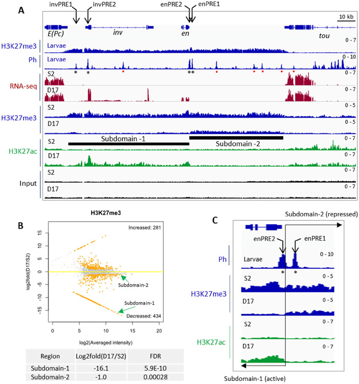
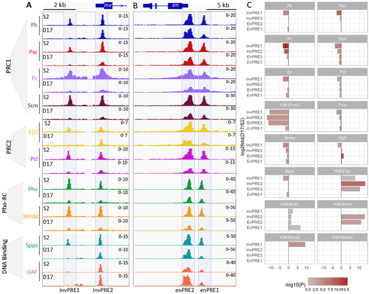
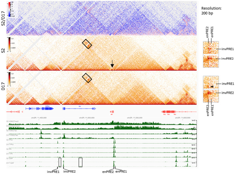
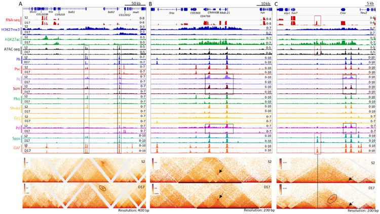
Polycomb group proteins (PcG) mediate epigenetic silencing of important developmental genes and other targets. In Drosophila, canonical PcG-target genes contain Polycomb Response Elements (PREs) that recruit PcG protein complexes including PRC2 that trimethylates H3K27 forming large H3K27me3 domains. In the OFF transcriptional state, PREs loop with each other and this looping strengthens silencing. Here we address the question of what PcG proteins bind to PREs when canonical PcG target genes are expressed, and whether PREs loop when these genes are ON. Our data show that the answer to this question is PRE-specific but general conclusions can be made. First, within a PcG-target gene, some regulatory DNA can remain covered with H3K27me3 and PcG proteins remain bound to PREs in these regions. Second, when PREs are within H3K27ac domains, PcG-binding decreases, however, this depends on the protein and PRE. The DNA binding protein GAF, and the PcG protein Ph remain at PREs even when other PcG proteins are greatly depleted. In the ON state, PREs can still loop with each other, but also form loops with presumptive enhancers. These data support the model that, in addition to their role in PcG silencing, PREs can act as "promoter-tethering elements" mediating interactions between promoter proximal PREs and distant enhancers.

Keywords: BIOLOGICAL SCIENCES; Genetics; PRE; Polycomb; chromatin loop.


Figures

Figure 1
Figure 1. Transcription, PcG binding and epigenetic patterns at the inv-en locus in S2 and D17 cells
(A) IGV tracks show the RNA-seq and ChIP-seq data at the inv-en locus. The top two tracks show the H3K27me3 and Ph occupancy from larval brains and discs. The asterisks under the Ph peaks indicate the four major PREs, and the red dots represent minor PREs. The RNA-seq and ChIP-seq (H3K27me3, H3K27ac) data for S2 and D17 cells are shown at bottom. The black bars indicate the subdomain-1 (H3K27ac covered) and subdomain-2 (H3K27me3 covered). (B) MA plot shows the alterations of H3K27me3 levels between D17 and S2 cells. The two subdomains of inv/en locus are highlighted by green arrows, and their detailed statistics are shown at bottom. (C) Zoomed in view of H3K27me3 and H3K27ac intensity over the two enPREs (asterisks). The two PREs fall into different subdomains with a precise border flanking enPRE2. The transition between the two subdomains is marked by the black line and opposing arrows.
Figure 2
Figure 2. Binding of core PcG proteins and other related factors at the four inv-en PREs in S2 and D17 cells
(A,B) IGV tracks show the occupancy of different PcG complex (PRC1, PRC2, Pho-RC) or DNA binding factors on the four major PREs in S2 and D17 cells. The PREs located at the inv (A) and en (B) regions are visualized separately. (C) Bar plots show the differences in ChIP signals for different PcG proteins and related factors at the individual inv and en PREs 1 and 2 between D17 and S2 cells.
Figure 3
Figure 3. PcG protein binding and chromatin structure over the inv-en domain in S2 and D17 cells
The heatmaps show the interaction changes over the inv-en region in S2 versus D17 cells. The top heatmap shows the differences between S2 and D17 cells, while the other two heatmaps are for S2 and D17 cells respectively. The arrow shows en is contained within its own small domain in S2 cells, whereas in the same domain as inv in D17 cells. The details are highlighted by the rectangle at right. The upper two dots are absent in D17 cells. The arrowhead on the zoomed heatmaps at the right shows an interaction which present in D17 but not S2 cells. At the bottom, the intensity for H3K27me3, H3K27ac, Pho, and Ph are shown to determine the extent of the different domains and the positions of the PREs. The rectangles highlight the D17-specific GAF binding sites. The positions of inv-en PREs are also labelled.
Figure 4
Figure 4. Transcription, PcG protein binding and chromatin structure over the bab1/bab2, mab-21 and Croc domains in S2 and D17 cells
The figures show the gene transcription, PcG protein binding and chromatin structure over three representative domains including bab1/bab2 (A), mab-21 (B) and Croc (C) in S2 and D17 cells, with top panels for the RNA-seq, ATAC-seq and ChIP-seq data, and lower panels for micro-C data. (A) The rectangles highlight presumptive PREs. The ovals highlight an area of difference between S2 and D17 cells. (B) The rectangles highlight the major differences in binding peaks for the PRC1 components Pc and Scm, and the PRC2 associated protein Pcl between S2 and D17 cells. The arrows show the interaction of the two major presumptive PREs that is present in both cell types. At this locus, the changes of the domains do not correlate with changes in PRE binding. (C) The black rectangles highlight the altered binding of Pc, Scm, and Pcl between S2 and D17 cells. The red rectangles highlight the locus with elevated binding of GAF, Spps and chromatin accessibility and a novel transcript in D17 cells. The vertical line highlights the PcG binding site with novel interaction (marked by the black oval) over the two presumptive PREs upstream of croc in D17 cells. The arrows in both panels show the interaction between the two PREs upstream of croc.

References

    1. Lewis E. B., A gene complex controlling segmentation in Drosophila. Nature 276, 565–570 (1978). - PubMed
    1. Schuettengruber B., Bourbon H. M., Di Croce L., Cavalli G., Genome Regulation by Polycomb and Trithorax: 70 Years and Counting. Cell 171, 34–57 (2017). - PubMed
    1. Kuroda M. I., Kang H., De S., Kassis J. A., Dynamic Competition of Polycomb and Trithorax in Transcriptional Programming. Annu Rev Biochem 89, 235–253 (2020). - PMC - PubMed
    1. Kassis J. A., Kennison J. A., Tamkun J. W., Polycomb and Trithorax Group Genes in Drosophila. Genetics 206, 1699–1725 (2017). - PMC - PubMed
    1. Kim J. J., Kingston R. E., Context-specific Polycomb mechanisms in development. Nat Rev Genet 10.1038/s41576-022-00499-0 (2022). - DOI - PMC - PubMed

Publication types

LinkOut - more resources