

This is a preprint.
Cerebrovascular disease drives Alzheimer plasma biomarker concentrations in adults with Down syndrome
- PMID: 38076904
- PMCID: PMC10705616
- DOI: 10.1101/2023.11.28.23298693
Cerebrovascular disease drives Alzheimer plasma biomarker concentrations in adults with Down syndrome
Update in
-
Cerebrovascular disease is associated with Alzheimer's plasma biomarker concentrations in adults with Down syndrome.Brain Commun. 2024 Sep 25;6(5):fcae331. doi: 10.1093/braincomms/fcae331. eCollection 2024. Brain Commun. 2024. PMID: 39403075 Free PMC article.
Abstract
Importance: By age 40 years over 90% of adults with Down syndrome (DS) have Alzheimer's disease (AD) pathology and most progress to dementia. Despite having few systemic vascular risk factors, individuals with DS have elevated cerebrovascular disease (CVD) markers that track with the clinical progression of AD, suggesting a role for CVD that is hypothesized to be mediated by inflammatory factors.
Objective: To examine the pathways through which small vessel CVD contributes to AD-related pathophysiology and neurodegeneration in adults with DS.
Design: Cross sectional analysis of neuroimaging, plasma, and clinical data.
Setting: Participants were enrolled in Alzheimer's Biomarker Consortium - Down Syndrome (ABC-DS), a multisite study of AD in adults with DS.
Participants: One hundred eighty-five participants (mean [SD] age=45.2 [9.3] years) with available MRI and plasma biomarker data were included. White matter hyperintensity (WMH) volumes were derived from T2-weighted FLAIR MRI scans and plasma biomarker concentrations of amyloid beta (Aβ42/Aβ40), phosphorylated tau (p-tau217), astrocytosis (glial fibrillary acidic protein, GFAP), and neurodegeneration (neurofilament light chain, NfL) were measured with ultrasensitive immunoassays.
Main outcomes and measures: We examined the bivariate relationships of WMH, Aβ42/Aβ40, p-tau217, and GFAP with age-residualized NfL across AD diagnostic groups. A series of mediation and path analyses examined causal pathways linking WMH and AD pathophysiology to promote neurodegeneration in the total sample and groups stratified by clinical diagnosis.
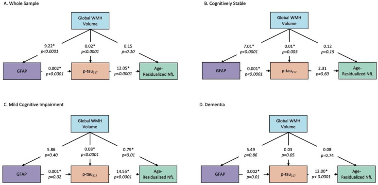
Results: There was a direct and indirect bidirectional effect through GFAP of WMH on p-tau217 concentration, which was associated with NfL concentration in the entire sample. Among cognitively stable participants, WMH was directly and indirectly, through GFAP, associated with p-tau217 concentration, and in those with MCI, there was a direct effect of WMH on p-tau217 and NfL concentrations. There were no associations of WMH with biomarker concentrations among those diagnosed with dementia.
Conclusions and relevance: The findings suggest that among individuals with DS, CVD promotes neurodegeneration by increasing astrocytosis and tau pathophysiology in the presymptomatic phases of AD. This work joins an emerging literature that implicates CVD and its interface with neuroinflammation as a core pathological feature of AD in adults with DS.
Figures


References
-
- NIA-AA Revised Criteria for Diagnosis and Staging of Alzheimer’s Disease. 2023:38. Oct 9 2023. https://alz.org/media/Documents/scientific-conferences/NIA-AA-Clinical-C....
Publication types
Grants and funding
- RF1 AG079519/AG/NIA NIH HHS/United States
- P50 AG005681/AG/NIA NIH HHS/United States
- U54 HD087011/HD/NICHD NIH HHS/United States
- P50 HD105353/HD/NICHD NIH HHS/United States
- U01 AG051412/AG/NIA NIH HHS/United States
- P50 AG005133/AG/NIA NIH HHS/United States
- UL1 TR001414/TR/NCATS NIH HHS/United States
- UL1 TR002373/TR/NCATS NIH HHS/United States
- P30 AG062715/AG/NIA NIH HHS/United States
- U54 HD090256/HD/NICHD NIH HHS/United States
- UL1 TR002345/TR/NCATS NIH HHS/United States
- UL1 TR001857/TR/NCATS NIH HHS/United States
- U24 AG021886/AG/NIA NIH HHS/United States
- P30 AG062421/AG/NIA NIH HHS/United States
- P50 AG008702/AG/NIA NIH HHS/United States
- P30 AG066519/AG/NIA NIH HHS/United States
- U19 AG068054/AG/NIA NIH HHS/United States
- R00 AG065506/AG/NIA NIH HHS/United States
- U01 AG051406/AG/NIA NIH HHS/United States
- UL1 TR001873/TR/NCATS NIH HHS/United States
LinkOut - more resources
Full Text Sources
Miscellaneous