[Preprint]. 2023 Dec 1:2023.12.01.569075.
doi: 10.1101/2023.12.01.569075.

A Mouse Model of the Protease Activated Receptor 4 (PAR4) Pro310Leu Variant has Reduced Platelet Reactivity

Affiliations

A Mouse Model of the Protease Activated Receptor 4 (PAR4) Pro310Leu Variant has Reduced Platelet Reactivity

Xu Han et al. bioRxiv. .

Update in

Abstract

Background: Protease activated receptor 4 (PAR4) mediates thrombin signaling on platelets and other cells. Our recent structural studies demonstrated a single nucleotide polymorphism in extracellular loop 3 (ECL3), PAR4-P310L (rs2227376) leads to a hypo-reactive receptor.

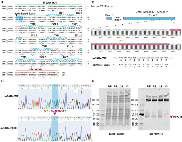
Objectives: The goal of this study was to determine how the hypo-reactive PAR4 variant in ECL3 impacts platelet function in vivo using a novel knock-in mouse model (PAR4-322L).

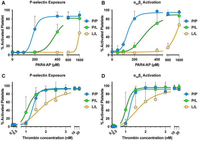
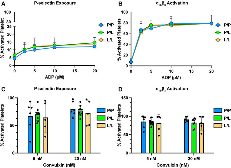
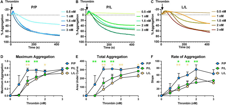
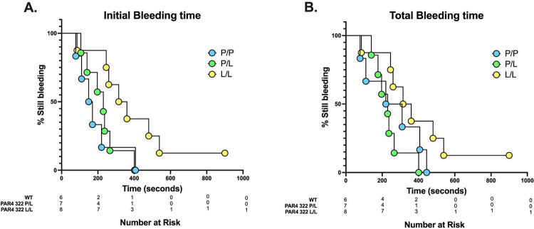
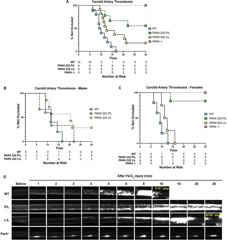
Methods: A point mutation was introduced into the PAR4 gene, F2rl3, via CRISPR/Cas9 to create PAR4-P322L, the mouse homolog to human PAR4-P310L. Platelet response to PAR4 activation peptide (AYPGKF), thrombin, ADP, and convulxin was monitored by αIIbβ3 integrin activation and P-selectin translocation using flow cytometry or platelet aggregation. In vivo responses were determined by the tail bleeding assay and the ferric chloride-induced carotid artery injury model.

Results: PAR4-P/L and PAR4-L/L platelets had a reduced response to AYPGKF and thrombin measured by P-selectin translocation or αIIbβ3 activation. The response to ADP and convulxin was unchanged among genotypes. In addition, both PAR4-P/L and PAR4-L/L platelets showed a reduced response to thrombin in aggregation studies. There was an increase in the tail bleeding time for PAR4-L/L mice. The PAR4-P/L and PAR4-L/L mice both showed an extended time to arterial thrombosis.

Conclusions: PAR4-322L significantly reduced platelet responsiveness to AYPGKF and thrombin, which is in agreement with our previous structural and cell signaling studies. In addition, PAR4-322L had prolonged arterial thrombosis time. Our mouse model provides a foundation to further evaluate the role of PAR4 in other pathophysiological contexts.

Keywords: Protease activated receptor 4; mouse model; platelet function; single nucleotide polymorphisms (SNPs); thrombin receptor.


Conflict of interest statement

Authors conflict of interest statement The authors declare no conflicts of interest.

Figures

Figure 1.
Figure 1.. Introduction of the C>T substitution in F2rl3 locus.
(A) Sequence alignment of human PAR4 and mouse PAR4. P310 at ECL3 (highlighted in blue) in humans is equal to P322 in mice. (B) gRNAs were targeted against the region of the mouse F2rl3 locus highlighted in red, while the red letters indicate the substitution target site. (C) Sanger sequencing shows the F2rl3 locus around the gRNA target site of a wildtype mouse (top) and a PAR4-P322L homozygous mouse (bottom) in which both alleles contained C>T substitutions. (D) Protein levels of PAR4 were compared across genotypes using an antibody specific for the mouse protein.
Figure 2.
Figure 2.. Platelets from PAR4-P322L mice were less responsive to PAR4 agonists.
Platelet rich plasma (PRP) from PAR4P/P, PAR4P/L, PAR4L/L littermates were stimulated with 50–1600 μM PAR4-AP, AYPGKF-NH2. (A, B). Gel-filtered platelets from PAR4P/P, PAR4P/L, PAR4L/L littermates were stimulated with 0.1 – 30 nM thrombin (C, D). The α-granule secretion (A, C) and integrin αIIbβ3 activation (B, D) were measured by flow cytometry using antibodies specific for P-Selectin and activated αΙΙbβ3. Data are means ± SD from 5 independent experiments at each concentration for A and B; Data are means from 3 independent experiments at each concentration for C and D.
Figure 3.
Figure 3.. Reactivity to non-PAR4 agonists was unaffected in platelets from PAR4-P322L mice.
Platelet rich plasma (PRP) from PAR4P/P, PAR4P/L and PAR4L/L littermates were stimulated with 2.5 – 20 μM ADP (A, B), or 5 nM or 20 nM convulxin (C, D). The α-granule secretion (A, C) and integrin αIIbβ3 activation (B, D) were measured by flow cytometry using P-selectin and activated αIIbβ3 specific antibodies. Data are means ± SD from 5 independent experiments at each concentration. Dots represent individual mice.
Figure 4.
Figure 4.. Aggregation of platelets from P322L mice had a reduced response to thrombin.
Representative tracing of thrombin-mediated platelet aggregation (A-C). Gel-filtered platelets from PAR4P/P (A), PAR4P/L (B) and PAR4L/L (C) littermates were stimulated with 0.5 – 3 nM thrombin. (D) Maximum aggregation of gel-filtered platelets from PAR4P/P, PAR4P/L and PAR4L/L were compared in response to 0.5 – 3 nM thrombin stimulation. (E) Area under curve of PAR4P/P, PAR4P/L and PAR4L/L platelet aggregation was compared in response to 0.5 – 3 nM thrombin stimulation. (F) The aggregation rate of PAR4P/P, PAR4P/L and PAR4L/L platelet aggregation was compared in response to 0.5 – 3 nM thrombin stimulation. Data are representative of 3 independent experiments. Dots represent individual mice.
Figure 5.
Figure 5.. PAR4-P322L mice had extended tail bleeding time.
Tail bleeding assay was used to evaluate the impact of the PAR4-P322L on hemostatic function. (A) Initial bleeding time was defined as the first time observing the stop of the bleeding regardless of any re-bleeding. (B) Total bleeding time was defined as the sum of bleeding times of all bleeding on/off cycles until a stable cessation occurred (no bleeding for 60 s). The experiment was terminated at 10 minutes. The data were presented as the percentage of mice that were still bleeding at a specified time point.
Figure 6.
Figure 6.. PAR4-P322L mice have longer arterial occlusion times.
Arterial thrombosis in our mice was assessed with the ferric chloride-induced carotid artery injury model. (A) The time to occlusion was visually determined as the moment blood flow stopped. The time to complete occlusion was determined in males (B) and females (C). Rhodamine 6G was used to label white blood cells and platelets in real-time over 30 minutes. Representative images are shown. (D).

References

    1. Broos K, Feys HB, De Meyer SF, Vanhoorelbeke K, Deckmyn H. Platelets at work in primary hemostasis. Blood Rev 2011; 25: 155–67. - PubMed
    1. Brass LF. Thrombin and Platelet Activation. Chest 2003; 124: 18S–25S. - PubMed
    1. Han X, Nieman MT. The domino effect triggered by the tethered ligand of the protease activated receptors. Thromb Res 2020; 196: 87–98. - PMC - PubMed
    1. Kahn ML, Nakanishi-Matsui M, Shapiro MJ, Ishihara H, Coughlin SR. Protease-activated receptors 1 and 4 mediate activation of human platelets by thrombin. J Clin Invest 1999; 103: 879–87. - PMC - PubMed
    1. Vu T-KH, Hung DT, Wheaton VI, Coughlin SR. Molecular cloning of a functional thrombin receptor reveals a novel proteolytic mechanism of receptor activation. Cell 1991; 64: 1057–68. - PubMed

Publication types