Review
. 2023 Dec 6;8(3):137-148.
doi: 10.1515/iss-2021-0045. eCollection 2023 Sep.

Virtual planning for mandible resection and reconstruction

Affiliations
Review

Virtual planning for mandible resection and reconstruction

Florian Andreas Probst et al. Innov Surg Sci. .

Abstract

In mandibular reconstruction, computer-assisted procedures, including virtual surgical planning (VSP) and additive manufacturing (AM), have become an integral part of routine clinical practice. Especially complex cases with extensive defects after ablative tumor surgery benefit from a computer-assisted approach. Various CAD/CAM-manufactured tools such as surgical guides (guides for osteotomy, resection and predrilling) support the transition from virtual planning to surgery. Patient-specific implants (PSIs) are of particular value as they facilitate both osteosynthesis and the positioning of bone elements. Computer-based approaches may be associated with higher accuracy, efficiency, and superior patient outcomes. However, certain limitations should be considered, such as additional costs or restricted availability. In the future, automation of the planning process and augmented reality techniques, as well as MRI as a non-ionizing imaging modality, have the potential to further improve the digital workflow.

Keywords: CAD/CAM; additive manufacturing; computer-assisted surgery; craniomaxillofacial surgery.


Conflict of interest statement

Competing interests: Authors state no conflict of interest.

Figures

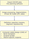
Figure 1:
Figure 1:
Steps in computer-assisted surgery.
Figure 2:
Figure 2:
CAD-process in mandibular reconstruction.
Figure 3:
Figure 3:
Ameloblastoma of the right symphyseal and body region. Mandibular resection and reconstruction with a microvascular bone flap from the iliac crest is planned. (A) Preoperative panoramic radiograph. (B) Virtual model depiction of the resection area (orange). (C) CAD with combined osteotomy/predrilling guides. (D) Planned reconstruction with ilia crest segments after opening-wedge osteotomy (the gap is later filled with cancellous bone). (E) CAD with osteotomy guide for harvesting the iliac crest transplant. (F) CAD with a patient-specific implant (PSI). (G) Harvested iliac crest flap with osteotomy guide still attached to the vascular pedicle (deep circumflex iliac artery, DCIA). (H) Postoperative panoramic radiograph.
Figure 4:
Figure 4:
Secondary mandible reconstruction with a double-barrel fibula bone flap and a patient-specific plate (PSI). (A) Virtual 3D model showing the defect area and deviation of the remaining mandible part. (B) Virtual repositioning of the mandible and depiction of the resection area (red). (C) CAD of combined osteotomy/predrilling guides. (D) Planned reconstruction with double-barrel fibula bone flap. (E) Five fibula bone segments are planned. (F) CAD of osteotomy guide for harvesting and segmentation of the fibula bone flap. (F) CAD of a patient-specific implant (PSI).
Figure 5:
Figure 5:
Transparent acrylic STL model including the remaining mandibular elements and the neomandible in the symphyseal area in a case where mandibular resection and reconstruction with a microvascular scapular bone flap is planned.
Figure 6:
Figure 6:
Lingual application of a patient-specific mandible reconstruction plate. (A) Panoramic radiograph of a patient with antiresorptive drug-induced osteonecrosis of the jaw (ARONJ) in the ramus and body region left and a fractured reconstruction plate and (B) compromised soft tissue with cutaneous fistula. (C) Virtual model depiction of the resection area (orange). (D) CAD of combined osteotomy/predrilling guides. (E) and (F) CAD of a patient-specific implant (PSI). (G) Predrilling at the symphyseal area. (H) Resection at the ramus area. (I) Fixation of lingual positioned PSI at the symphyseal area. (J) PSI attached to the mandible. (K) Postoperative panoramic radiograph.
Figure 7:
Figure 7:
Virtual surgical planning (VSP) for mandibular reconstruction with a fibula bone flap. Four blue dental implant dummies indicate the correct position of the fibula bone segments in relation to the maxillary teeth in order to facilitate the later insertion of dental implants as part of the dental rehabilitation.

References

    1. Weyh AM, Quimby A, Fernandes RP. Three-dimensional computer-assisted surgical planning and manufacturing in complex mandibular reconstruction. Atlas Oral Maxillofac Surg Clin North Am. 2020;28:145–50. doi: 10.1016/j.cxom.2020.05.007. - DOI - PubMed
    1. Ehrenfeld M, Futran ND, Manson PN, Prein J. Advanced craniomaxillofacial surgery – tumor, corrective bone surgery and trauma. Stuttgart, New York: Thieme Publishers; 2020.
    1. Fernandes RP, Yetzer JG. Reconstruction of acquired oromandibular defects. Oral Maxillofac Surg Clin. 2013;25:241–9. doi: 10.1016/j.coms.2013.02.003. - DOI - PubMed
    1. Urken ML, Weinberg H, Vickery C, Buchbinder D, Lawson W, Biller HF. Oromandibular reconstruction using microvascular composite free flaps. Report of 71 cases and a new classification scheme for bony, soft-tissue, and neurologic defects. Arch Otolaryngol Head Neck Surg. 1991;117:733–44. doi: 10.1001/archotol.1991.01870190045010. - DOI - PubMed
    1. Martola M, Lindqvist C, Hänninen H, Al-Sukhun J. Fracture of titanium plates used for mandibular reconstruction following ablative tumor surgery. J Biomed Mater Res B Appl Biomater. 2007;80:345–52. doi: 10.1002/jbm.b.30603. - DOI - PubMed

LinkOut - more resources