Review
. 2023 Dec 15;150(24):dev201786.
doi: 10.1242/dev.201786. Epub 2023 Dec 11.

Endothelin signaling in development

Affiliations
Review

Endothelin signaling in development

Stanley M Kanai et al. Development. .

Abstract

Since the discovery of endothelin 1 (EDN1) in 1988, the role of endothelin ligands and their receptors in the regulation of blood pressure in normal and disease states has been extensively studied. However, endothelin signaling also plays crucial roles in the development of neural crest cell-derived tissues. Mechanisms of endothelin action during neural crest cell maturation have been deciphered using a variety of in vivo and in vitro approaches, with these studies elucidating the basis of human syndromes involving developmental differences resulting from altered endothelin signaling. In this Review, we describe the endothelin pathway and its functions during the development of neural crest-derived tissues. We also summarize how dysregulated endothelin signaling causes developmental differences and how this knowledge may lead to potential treatments for individuals with gene variants in the endothelin pathway.

Keywords: Auriculocondylar syndrome; Cardiovascular; Craniofacial; Enteric; G protein; Hirschsprung disease; Melanocyte.


Conflict of interest statement

Competing interests The authors declare no competing or financial interests.

Figures

Fig. 1.
Fig. 1.
The endothelin signaling pathway. The endothelin ligands endothelin 1 (EDN1), EDN2 and EDN3 are initially produced as inert precursors, prepro-EDN1-3, which are processed into big-EDN1-3 by furin or a furin-like protease before being secreted. Big-EDN1-3 are then converted to mature, bioactive peptides EDN1-3 by membrane-bound metalloproteases endothelin converting enzyme 1 (ECE1) and ECE2. Binding of EDN peptides to either the endothelin receptor type A (EDNRA) or EDNRB receptors stimulate signal transduction cascades mediated by heterotrimeric G proteins. EDNRA and EDNRB couple to all four families of Gα proteins, with each family inducing distinct intracellular signaling responses. EDNRA has differential binding affinities for EDN peptides [illustrated by solid (high affinity) and hatched (low affinity) arrows], whereas EDNRB binds non-selectively to all EDN peptides. cAMP, cyclic adenosine monophosphate; CDC42, cell division control protein 42 homolog; CREB, cAMP responsive element binding protein; DAG, diacylglycerol; ERK, extracellular signal-regulated kinase; IP3, inositol triphosphate; JNK, c-Jun N-terminal kinase; PAK, P21-activated kinase; PKA, protein kinase A; PKC, protein kinase C; PLCB, phospholipase Cβ; RhoA, Ras homolog gene family member A GTPase; RhoGEF, Rho guanine nucleotide exchange factor.
Fig. 2.
Fig. 2.
Neural crest cell populations. All five subpopulations of neural crest cells (NCCs; top portion of each box) require either EDNRA (black) or EDNRB (purple) signaling for proper development of NCC-derived cells and structures (bottom portion of each box).
Fig. 3.
Fig. 3.
Signaling during lower jaw development. (A,B) Lateral (A) and ventral (B) views of an E10.5 mouse embryo schematic; purple dashed boxes in A and B illustrate the embryo area that is further expanded in the colored cartoons. Areas of color highlight expression domains of selected transcription factors required for establishing positional identity of NCCs. Expression domains for ligands Edn1 and Bmp4 are also shown. The three primary domains of the mandibular arch [proximal (P), intermediate (I) and distal (D)] are denoted. EDN1-EDNRA signaling is required for gene expression in the distal and intermediate domains (e.g. Dlx5/6 and Hand2) but not in the proximal domain (Pou3f3 and Hey1). (C,D) Schematic of mouse skulls illustrating structures dependent on EDN1-EDNRA signaling. (C) In E18.5 wild-type embryos, the mandible (md) arises from the mandibular portion of arch 1 (blue). The maxilla (mx), palatine (p) and jugal (j) bones arise from the maxillary prominence of arch 1 (orange). (D) Loss of EDN1-EDNRA signaling results in homeotic transformation of the mandible into maxilla-, palatine- and jugal-like bones. In the skull base, the pterygoid (p) and alisphenoid (as) bones are also duplicated (grey). Duplicated bones are denoted with an asterisk. Middle ear structures, including the malleus (ma), incus (in) and tympanic ring (ty), are also malformed, hypoplastic or absent. bo, basioccipital; bs, basisphenoid; eo, exoccipital; et, ethmoid; f, frontal; h, hyoid; i, incisor; ip, interparietal; la, lacrimal; n, nasal; or, orbitosphenoid; pa, parietal; pe, petrosal; pm, premaxilla; ps, presphenoid; s, stapes; so, supraoccipital; sq, squamosal; tb, temporal bone; th, thyroid cartilage; v, vomer.
Fig. 4.
Fig. 4.
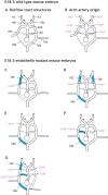
Representative aortic arch malformations in Edn1−/−, Ednra−/− or Ece1−/− mouse embryos. (A) A normally remodeled outflow tract observed in E18.5 embryos. Thin dotted lines represent normal regression of vessels during outflow tract remodeling. (B) The normally remodeled outflow tract shown in A with arch artery (aa) origins for each structure noted in pink. (C-G) Abnormal regression of arch arteries and abnormal outflow tract patterning observed in Edn1−/−, Ednra−/− and Ece1−/− mouse embryos. Blue vessels represent abnormal persistence of vessels; thick dotted lines represent abnormal regression of vessels. (C) Abnormal regression of the right arch artery 4 (raa4) and persistence of the right ductus caroticus (rdc) results in a right subclavian artery (rsa) of cervical origin. (D) Abnormal regression of right and left arch arteries 4 (raa4 and laa4) leads to persistence of right and left ductus caroticus (rdc and ldc). The right dorsal aorta (rda) also persists. (E) Abnormal regression of right arch artery 4 leads to persistence of right dorsal aorta, resulting in a right subclavian artery of dorsal aorta origin. (F) Aberrant regression of left arch artery 6 (laa6) and persistence of right arch artery 6 (raa6) and right dorsal aorta results in a right-sided aortic arch (raoa). (G) Aberrant regression of the left and right arch arteries 4, the left arch artery 6 and the left dorsal aorta (lda) and aberrant persistence of the right arch artery 6, the right ductus caroticus and the right dorsal aorta resulting in a right-sided aortic arch. da, ductus arteriosus; lsa, left subclavian artery; laa3, left arch artery 3; raa3, right arch artery 3. Adapted with permission from Yanagisawa et al. (1998a).
Fig. 5.
Fig. 5.
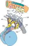
Endothelin signaling during cardiovascular development. Cardiac NCCs (blue circles) expressing Ednra arise from the cardiac neural crest (cnc) region (green) at the level of somites 1-3 (s1-s3), migrating ventrally towards the heart through the circumpharyngeal region (cpr). They subsequently surround the lumens of paired pharyngeal arch arteries (aa) 3, 4 and 6, which are lined with endothelial cells expressing Edn1. Once there, asymmetric remodeling of the arch arteries occurs, with the cardiac NCC-derived mesenchyme forming the smooth muscle cells that surround the lumen. Cardiac NCC-derived mesenchyme also give rise to the aorticopulmonary septum (aps) that divides the cardiac outflow into aortic (ao) and pulmonary (p) trunks. A subset of cardiac NCCs continue into the conotruncus (lined with Edn1-expressing endothelial cells), where they later contribute to the semilunar and atrioventricular valves. ct, conotruncus; lda, left dorsal aorta; lv, left ventricle; nt, neural tube; rda, right dorsal aorta; rv, right ventricle. Adapted with permission from Hutson and Kirby (2007).
Fig. 6.
Fig. 6.
Melanocyte and enteric nervous system development. (A) Migratory paths of trunk NCCs. In mouse and chick embryos, the melanoblast population migrates dorsolaterally between the somites and developing epidermis to colonize the dorsal surface of the embryo. Trunk NCCs that migrate ventrally between the somites and neural tube give rise to neurons and glia of the peripheral nervous system. (B) Characteristic coat pigmentation defects in Edn3 and Ednrb mutant mice. Compared with wild-type mice (top left), lethal spotting mice (ls/ls; loss of Edn3 expression) exhibit extensive white spotting (top right). Pigmented patches are typically observed on the head and hip regions. Piebald mice (s/s; reduced Ednrb expression) exhibit mild coat color spotting (bottom left). White spotting is typically observed on the shoulder and hip regions. Spotted lethal mice (sl/sl; complete loss of Ednrb expression) exhibit severe coat pigmentation defects and are almost completely white (bottom right). (C) Migratory paths of NCCs through the fetal gut of a prototypical midgestation embryo. Vagal NCCs, or enteric NCCs (ENCC), traverse long distances to colonize the entire length of the developing gut. In mice, ENCCs enter the developing intestine at the foregut (indicated by black arrow adjacent somites 1-7) around E8.5-E9.5 and migrate in a rostral-caudal direction (indicated by purple arrow in midgut). Colonization of the gut is completed by E15.5. In mouse and chick embryos, sacral NCCs enter the posterior hindgut (indicated by the black arrow adjacent the cloaca) and colonize the proximal and distal colon. Areas of high EDN3 are denoted by purple-brown shading (the stomach, cecum and cloaca). EDN3-EDNRB signaling is required for ENCCs to migrate past the ileo-cecal junction and into the proximal colon (indicated by orange circle). Regions of the gut (foregut, midgut and hindgut) are color-coded. Adapted with permission from Nagy and Goldstein (2017).

References

    1. Abu-Issa, R. and Kirby, M. L. (2007). Heart field: from mesoderm to heart tube. Annu. Rev. Cell Dev. Biol. 23, 45-68. 10.1146/annurev.cellbio.23.090506.123331 - DOI - PubMed
    1. Adameyko, I., Lallemend, F., Furlan, A., Zinin, N., Aranda, S., Kitambi, S. S., Blanchart, A., Favaro, R., Nicolis, S., Lübke, M.et al. (2012). Sox2 and Mitf cross-regulatory interactions consolidate progenitor and melanocyte lineages in the cranial neural crest. Development 139, 397-410. 10.1242/dev.065581 - DOI - PMC - PubMed
    1. Alexander, C., Zuniga, E., Blitz, I. L., Wada, N., Le Pabic, P., Javidan, Y., Zhang, T., Cho, K. W., Crump, J. G. and Schilling, T. F. (2011). Combinatorial roles for BMPs and Endothelin 1 in patterning the dorsal-ventral axis of the craniofacial skeleton. Development 138, 5135-5146. 10.1242/dev.067801 - DOI - PMC - PubMed
    1. Alexander, C., Piloto, S., Le Pabic, P. and Schilling, T. F. (2014). Wnt signaling interacts with bmp and edn1 to regulate dorsal-ventral patterning and growth of the craniofacial skeleton. PLoS Genet. 10, e1004479. 10.1371/journal.pgen.1004479 - DOI - PMC - PubMed
    1. Amiel, J., Sproat-Emison, E., Garcia-Barcelo, M., Lantieri, F., Burzynski, G., Borrego, S., Pelet, A., Arnold, S., Miao, X., Griseri, P.et al. (2008). Hirschsprung disease, associated syndromes and genetics: a review. J. Med. Genet. 45, 1-14. 10.1136/jmg.2007.053959 - DOI - PubMed

Publication types

LinkOut - more resources