. 2023 Dec;15(2):2281011.
doi: 10.1080/19490976.2023.2281011. Epub 2023 Dec 11.

Butyrate reduces adherent-invasive E. coli-evoked disruption of epithelial mitochondrial morphology and barrier function: involvement of free fatty acid receptor 3

Affiliations

Butyrate reduces adherent-invasive E. coli-evoked disruption of epithelial mitochondrial morphology and barrier function: involvement of free fatty acid receptor 3

Samira A Hamed et al. Gut Microbes. 2023 Dec.

Abstract

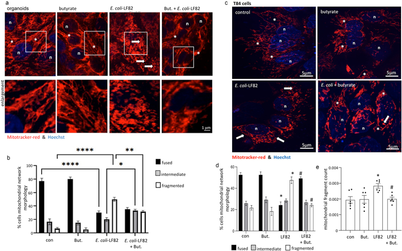
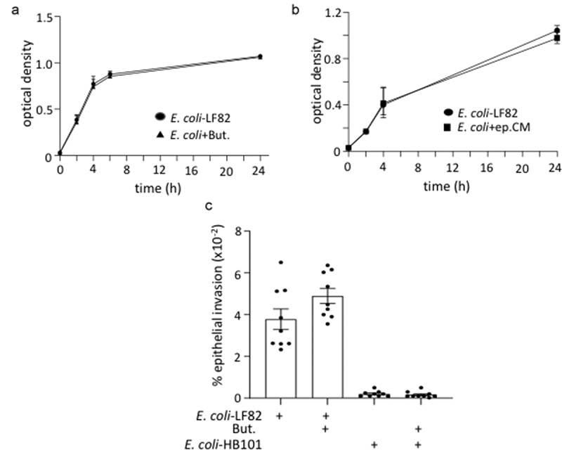
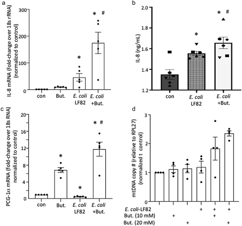
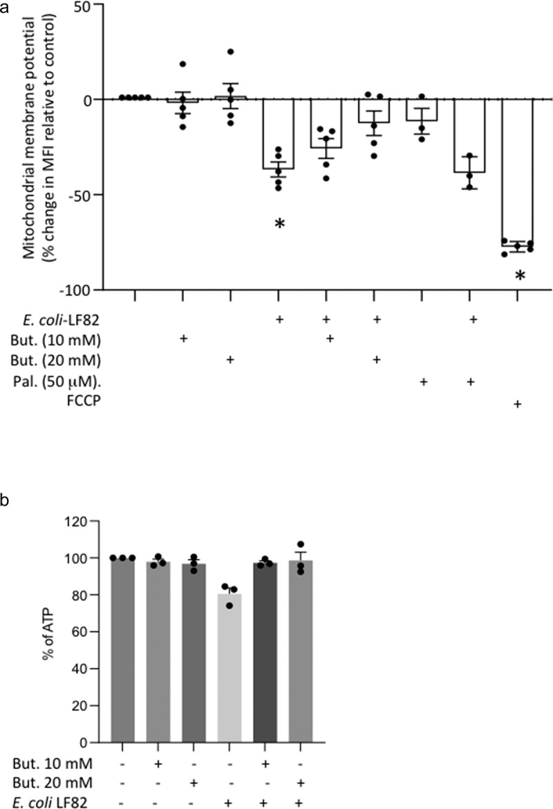
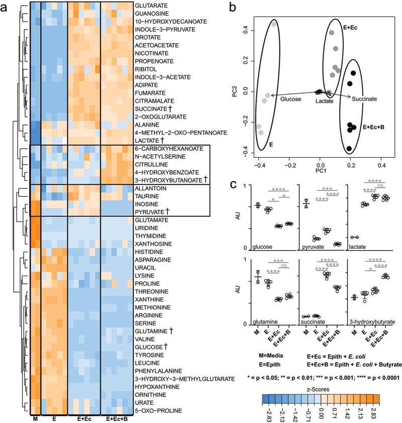
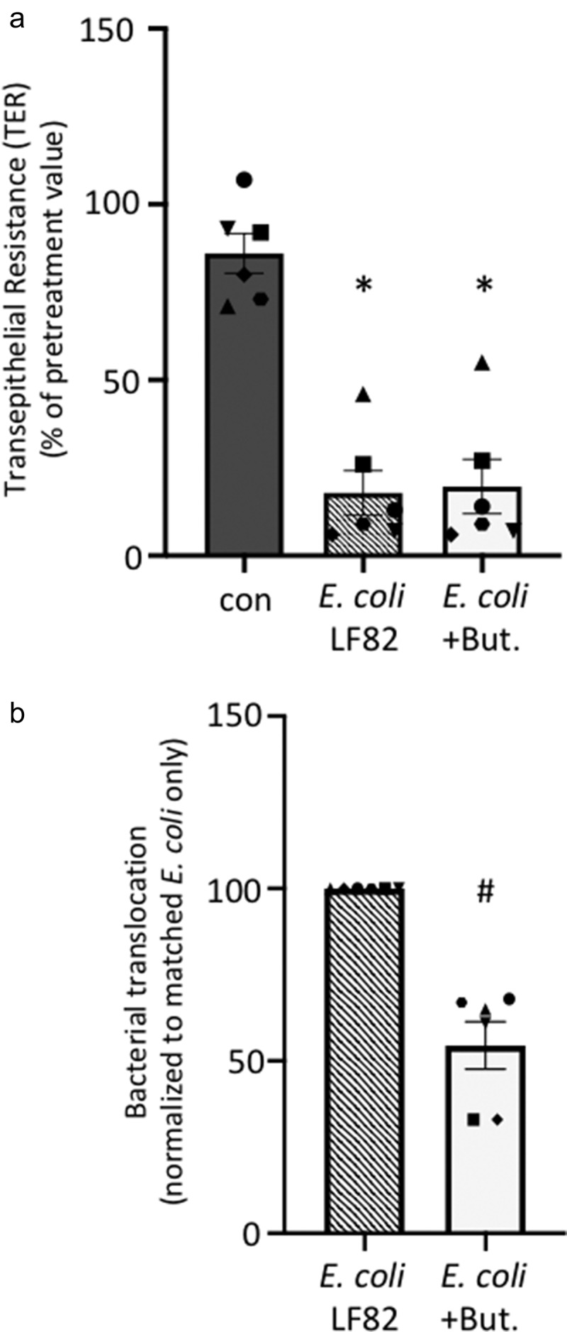
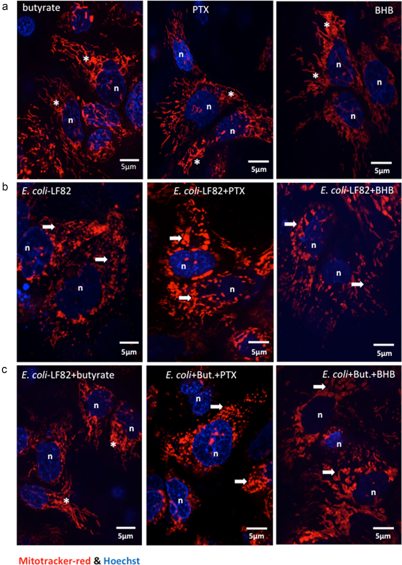
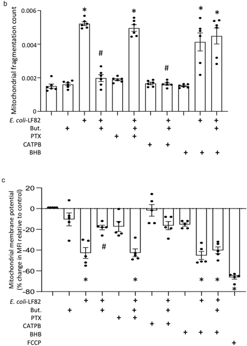
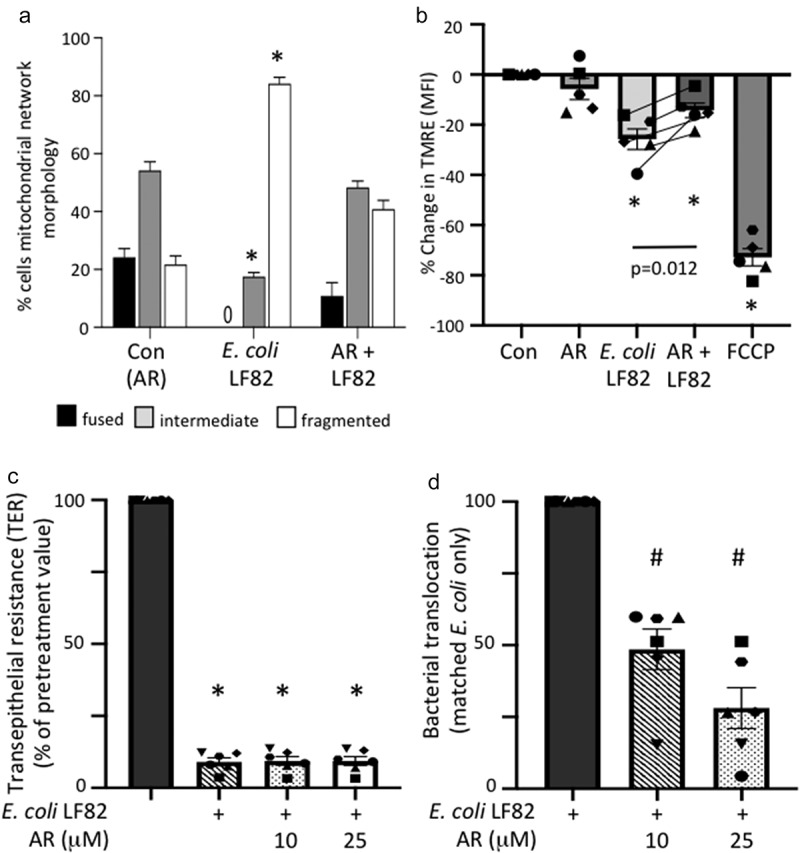
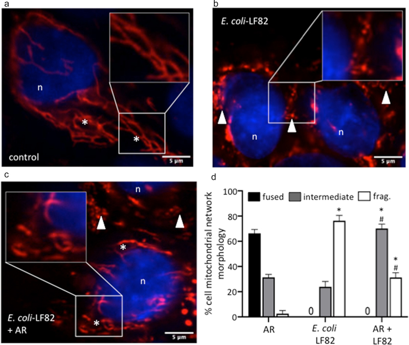
Gut bacteria provide benefits to the host and have been implicated in inflammatory bowel disease (IBD), where adherent-invasive E. coli (AIEC) pathobionts (e.g., strain LF82) are associated with Crohn's disease. E. coli-LF82 causes fragmentation of the epithelial mitochondrial network, leading to increased epithelial permeability. We hypothesized that butyrate would limit the epithelial mitochondrial disruption caused by E. coli-LF82. Human colonic organoids and the T84 epithelial cell line infected with E. coli-LF82 (MOI = 100, 4 h) showed a significant increase in mitochondrial network fission that was reduced by butyrate (10 mM) co-treatment. Butyrate reduced the loss of mitochondrial membrane potential caused by E. coli-LF82 and increased expression of PGC-1α mRNA, the master regulator of mitochondrial biogenesis. Metabolomics revealed that butyrate significantly altered E. coli-LF82 central carbon metabolism leading to diminished glucose uptake and increased succinate secretion. Correlating with preservation of mitochondrial network form/function, butyrate reduced E. coli-LF82 transcytosis across T84-cell monolayers. The use of the G-protein inhibitor, pertussis toxin, implicated GPCR signaling as critical to the effect of butyrate, and the free fatty acid receptor three (FFAR3, GPR41) agonist, AR420626, reproduced butyrate's effect in terms of ameliorating the loss of barrier function and reducing the mitochondrial fragmentation observed in E. coli-LF82 infected T84-cells and organoids. These data indicate that butyrate helps maintain epithelial mitochondrial form/function when challenged by E. coli-LF82 and that this occurs, at least in part, via FFAR3. Thus, loss of butyrate-producing bacteria in IBD in the context of pathobionts would contribute to loss of epithelial mitochondrial and barrier functions that could evoke disease and/or exaggerate a low-grade inflammation.

Keywords: Pathobiont; T84 cells; gut epithelium; mitochondrial dynamics; organoid; permeability.


Conflict of interest statement

No potential conflict of interest was reported by the author(s).

Figures

Figure 1.
Figure 1.
E. coli-LF82 evoked mitochondrial fragmentation of enteric epithelia is reduced in butyrate co-treated cells. Human colon organoids (a, b) or monolayers of the human colon-derived T84 epithelial cell line (c-e) were treated with E. coli-LF82 (108 cfu, 4 h) ± co-treatment with sodium butyrate (But., 10 mM). Representative images were collected in a random fashion by first identifying epithelia nuclei (blue, n) and then swapping the confocal laser channel to assess the mitochondrial network (Mitotracker (red)). Twenty cells per monolayer were characterized by semi-quantitative analysis (b, d) and an image-J analysis program (e) (data are mean ± SEM, from 3 organoids and 6–9 epithelial monolayers assessed in 2–3 separate experiments; *, **, **** statistically different at p < .05, p < .01 and p < .001 respectively (panel b); * and #, p < .05 compared to control uninfected cells (con) and E. coli-LF82 only infected cells, respectively (panels d, e) by two-way ANOVA followed by Tukey’s multiple comparisons test; image *, fused mitochondrial network with elongated strands; arrow, fragmented, vesiculated area of the mitochondrial network).
Figure 2.
Figure 2.
Butyrate does not kill E. coli-LF82. E. coli-LF82 (108 cfu) only (no epithelium) were cultured in antibiotic-free medium with butyrate (But., 10 mM) (a) or a 50% spent-medium from T84+butyrate+E. coli-LF82 cultures (4 h, 0.2 μm filtered (ep.CM)) (b) and optical density measured over a 24 h period (n = 3). (c) Invasion assays revealed that butyrate did not alter the ability of E. coli-LF82 to invade T84 epithelia (multiplicity of infection = 100, 4 h; non-invasive E. coli-HB101 (108 cfu) shown for comparison) (data are mean ± SEM, 9 epithelial monolayers from 3 experiments).
Figure 3.
Figure 3.
Butyrate and E. coli-LF82 increase epithelial transcription of IL-8 and PGC-1α. T84 epithelial cell monolayers exposed to E. coli-LF82 (108 cfu, 4 h) showed increased IL-8 mRNA and protein that was enhanced by co-treatment with butyrate (But., 10 mM) (a, b). Expression of the master regulator of mitochondrial synthesis, peroxisome proliferator-activated receptor gamma co-activator 1-alpha (PGC-1α), was significantly increased by butyrate, and more so in the presence of E. coli-LF82, with a trend toward increased mitochondrial DNA (mtDNA) copy number (c, d) (data are mean ± SEM; n = 5–4; * and #, p <0.05 compared to control (con) uninfected cells and E. coli-LF82 only infected cells respectively by two-way ANOVA followed by Tukey’s multiple test; mRNA target genes were compared to expression of 18s rRNA as a housekeeping gene and the data normalized against control conditions set at 1).
Figure 4.
Figure 4.
Butyrate co-treatment prevents E. coli-LF82 evoked loss of mitochondrial membrane potential and reduced intracellular ATP. (a) T84 epithelial cells were treated with E. coli-LF82 (108, cfu) ± butyrate (But.) or palmitate (Pal.) for 4 h and then membrane potential was assessed by TMRE fluorescence in a flow cytometer. A 10 min treatment with the metabolic toxin, FCCP (10 μM) was used as a positive control (n = 5, except Pal. where n = 3)). (b) ATP was measured in epithelial lysates by commercial assay in triplicate in three experiments (data are mean ± SEM; *, p < .05 compared to control uninfected cells by the Kruskal–Wallis test followed by Dunn’s multiple comparison test for normalized data; statistics not performed on data in panel B because n = 3; MFI, mean fluorescent intensity).
Figure 5.
Figure 5.
Butyrate modulates metabolism in E. coli-LF82 infected T84 epithelial cells. The metabolic activity of E. coli-LF82 infected (108 cfu, 4 h) and non-infected T84 epithelial (epith) cells ± butyrate (10 mM) was assessed by LC-MS analyses of culture media. Metabolites that were consumed or produced by cultures were identified and quantified. (a) heat-map of 52 media metabolites with signal intensities shown as z-scores (i.e., mean centered, variance stabilized signal intensities). Boxed metabolites indicate clusters of butyrate-linked metabolic perturbations. A selection of representative metabolites (†) illustrating the main metabolic patterns are shown as dot-plots in panel 5C. (b) Principal component analysis (PCA) of metabolite signals with the magnitude and direction of metabolite contributions shown as vectors (in a bi-plot). The three largest metabolic contributors to clustering are annotated above the vectors. (c) Representative metabolite levels (data show as mean ± SEM for n = 6; AU, arbitrary units) are plotted with significant differences denoted as calculated by pairwise t-test.
Figure 6.
Figure 6.
E. coli-LF82 translocation across epithelia is less pronounced in butyrate co-treated epithelia. Human colon-derived T84 cells (106) were seeded onto porous (3 μm) transwell supports and cultured until electrically confluent, typically 7 days when transepithelial resistance exceeded 750 Ohms.cm2. E. coli-LF82 (108 cfu) ± butyrate (But., 10 mM) were added to the apical surface and TER and transcytosis of the bacteria assessed 24 h later. (a) TER is presented as the change after 24 h with each monolayer being its’ own control (i.e., pretreatment value). Starting TERs in these experiments ranged from 750–2460 Ohms.cm2. (b) Bacterial transcytosis was assessed via serial dilution of culture-well basolateral medium on agar plates, with the data being converted to % transcytosis based on bacterial counts in the apical compartment. E. coli-LF82 data were normalized to 100 for comparison with E. coli+But. in the same experiment (data are mean ± SEM; each data point is an individual experiment (n = 6) in which bacterial transcytosis across 3 or 4 epithelial monolayers was averaged and are represented by a different symbol; * and #, p <.05 compared to control uninfected cells (con) and E. coli-LF82 only infected cells, respectively).
Figure 7.
Figure 7.
Pertussis toxin and β-hydroxybutyrate (BHB) implicate FFAR3 in butyrate’s maintenance of epithelial mitochondrial networks. Monolayers of the human colon-derived T84 epithelial cell line (106) were treated with E. coli-LF82 (108 cfu, 4 h) ± a co-treatment with sodium butyrate (But., 10 mM) ± an 18 h pre-treatment with pertussis toxin (PTX, 50 ng/ml) or BHB (5 mM). (a) Representative images were collected in a random fashion by first identifying epithelia nuclei (blue, n) and then swapping the confocal laser channel to assess the mitochondrial network (Mitotracker (red)). Twenty cells per monolayer were characterized for mitochondrial fragmentation count by ImageJ analysis and averaged/monolayer (b). (c) Mitochondrial membrane potential was assessed by TMRE fluorescence in a flow cytometer. A 10 min treatment with the metabolic toxin, FCCP (10 Ohms.cm2) was used as a positive control. The effect of the FFAR2 antagonist (S)-3-(2-(3-chlorophenyl) acetamido)-4-(4-(trifluoromethyl) phenyl) butanoic acid (CATPB, 10 Ohms.cm2, 30 min pre-treatment) is also shown (mean ± SEM; n = 5–6 epithelial monolayers from separate experiments in panels a and b; * and #, p <.05 compared to control uninfected cells and E. coli-LF82 only infected cells respectively by two-way ANOVA followed by Tukey’s multiple comparison test (b) or the Kruskal–Wallis test from by Dunn’s multiple comparison test; image *, fused mitochondrial network with elongated strands; arrow, fragmented, vesiculated area of the mitochondrial network; MFI, mean fluorescence intensity).
Figure 7.
Figure 7.
(continue).
Figure 8.
Figure 8.
An FFAR3 agonist improves mitochondrial network connectivity and barrier function in E. coli-LF82-infected T84 epithelial cells. Monolayers of the human colon-derived T84 epithelial cell line were treated with E. coli-LF82 (108 cfu, 4 h) ± a co-treatment with the FFAR3 agonist AR420626 (25 µM) and representative images collected in a random fashion by first identifying epithelia nuclei (blue, n) and then swapping the confocal laser channel to assess the mitochondrial network as defined by TOMM-20 immunostains. Twenty cells per monolayer were characterized by semi-quantitative assessment (a). (b) Mitochondrial membrane potential was assessed by TMRE fluorescence in a flow cytometer. A 10 min treatment with the metabolic toxin, FCCP (10 µM, n = 5 epithelial monolayers from separate experiments (indicated by different symbols)). Filter-grown T84 cell monolayers (starting transepithelial resistance (TER) range = 957–2155 Ohms.cm) were cultured with E. coli-LF82 (108 cfu) ± AR420626 and TER and transcytosis of the bacteria assessed 24 h later. (c) TER is presented as the change over 24 h with each monolayer being its’ own control (i.e., pre-treatment value). (d) Bacterial transcytosis was assessed via serial dilution of culture-well basolateral medium on agar plates, with the data being converted to % transcytosis based on bacterial counts in the apical compartment and then E. coli-LF82 was normalized to 100 for comparison with E. coli+AR420626 in the same experiment (data are mean ± SEM; each data point is an individual experiment (n = 6) in which measurements from 3 or 4 monolayers were averaged and are shown as a different symbol; * and #, p <.05 compared to control uninfected cells (con) and E. coli-LF82 only infected cells, respectively).
Figure 9.
Figure 9.
An FFAR3 agonist improves mitochondrial network connectivity in E. coli-LF82-infected human organoids. Monolayers of colonic organoids derived from two healthy controls were treated with E. coli-LF82 (MOI = 100, 4 h) ± a co-treatment with the FFAR3 agonist AR420626 (AR: 25 µM). Representative confocal images of MitoTrackerTM (red) and Hoescht (blue) co-stained organoids show the fused mitochondrial network of control organoid cells treated with the FFAR3 agonist alone (a), the puncta-like fragmented mitochondrial network of E. coli-LF82 infected organoid cells (b), and the intermediate fragments of the E. coli-LF82 infected organoids co-treated with the FFAR3 agonist (c) (n, nucleus; *, filamentous mitochondria; arrowhead, fragmented mitochondria). Twenty cells were assessed from four monolayers for semi-quantitative analysis (d). Data are mean ± SEM, * and #, p < .05 compared to control uninfected drug-treated cells (AR) and E. coli-LF82 only infected cells, respectively (two-way ANOVA followed by Tukey’s multiple comparison test) (frag., fragmented mitochondria).

References

    1. O’Riordan KJ, Collins MK, Moloney GM, Knox EG, Aburto MR, Fulling C, Morley SJ, Clarke G, Schellekens H, Cryan JF.. Short chain fatty-acids: microbial metabolites for gut-brain axis signalling. Mol Cell Endocrinol. 2022;456:111572. doi:10.1016/j.mce.2022.111572. - DOI - PubMed
    1. Skelly AN, Sato Y, Kearney S, Honda K.. Mining the microbiota for microbial and metabolite-based immunotherapies. Nat Rev Immunol. 2019;19(5):305–21. doi:10.1038/s41577-019-0144-5. - DOI - PubMed
    1. Chang PV, Hao L, Offermanns S, Medzhitov R. The microbial metabolite butyrate regulates intestinal macrophage function via histone deacetylase inhibition. Proc Natl Acad Sci (USA). 2014;111(6):2247–2252. doi:10.1073/pnas.1322269111. - DOI - PMC - PubMed
    1. Siddiqui MT, Cresci GAM. The immunomodulatory functions of butyrate. J Inflamm Res. 2021;14:6025–6041. doi:10.2147/JIR.S300989. - DOI - PMC - PubMed
    1. Rashed R, Valcheva R, Dieleman LA. Manipulation of gut microbiota as a key target for Crohn’s disease. Front Med. 2022;9:887044. doi:10.3389/fmed.2022.887044. - DOI - PMC - PubMed

Publication types

MeSH terms