. 2024 Feb 5;223(2):e202302081.
doi: 10.1083/jcb.202302081. Epub 2023 Dec 11.

BLK positively regulates TLR/IL-1R signaling by catalyzing TOLLIP phosphorylation

Affiliations

BLK positively regulates TLR/IL-1R signaling by catalyzing TOLLIP phosphorylation

Wei-Wei Li et al. J Cell Biol. .

Abstract

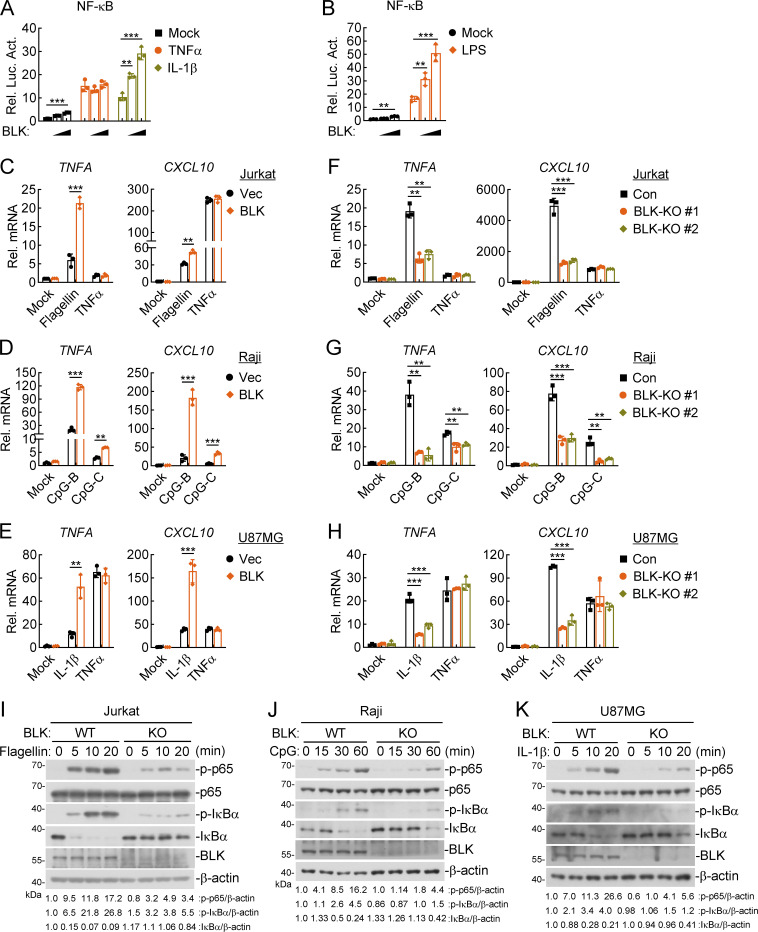
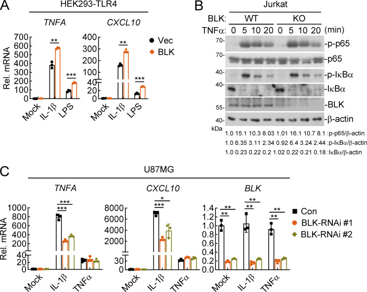
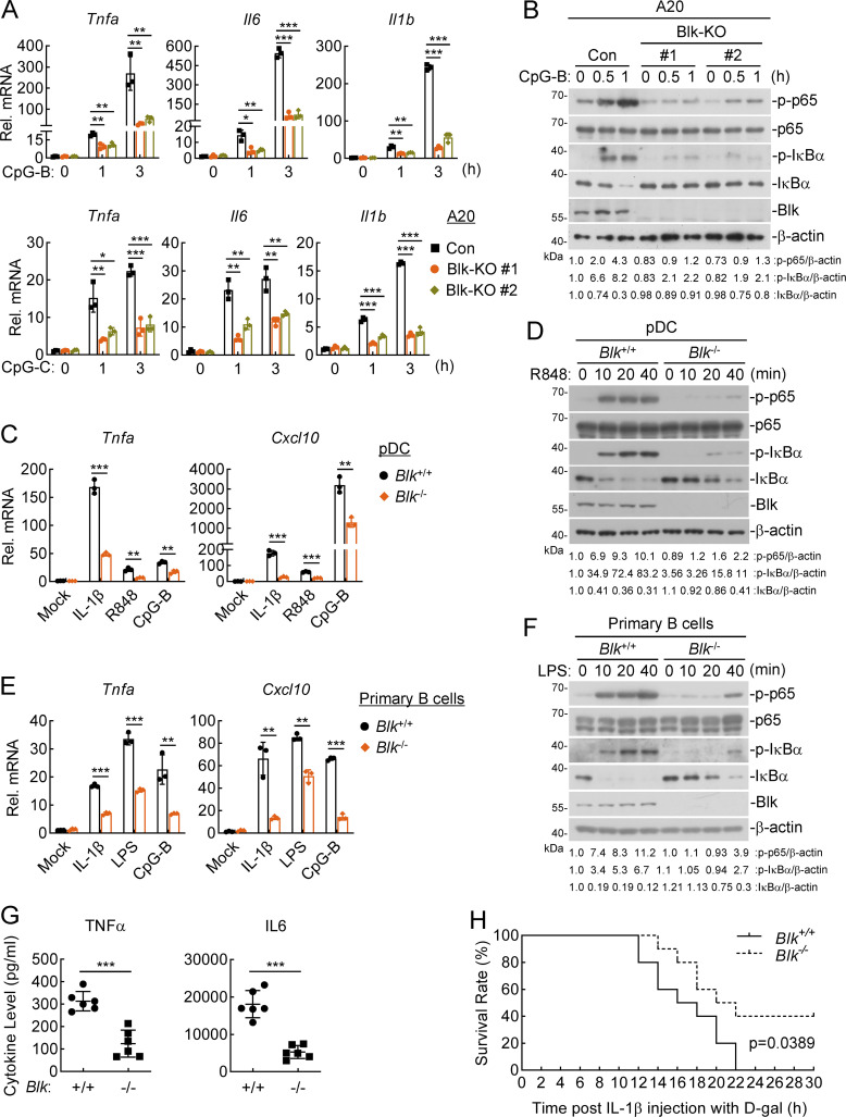
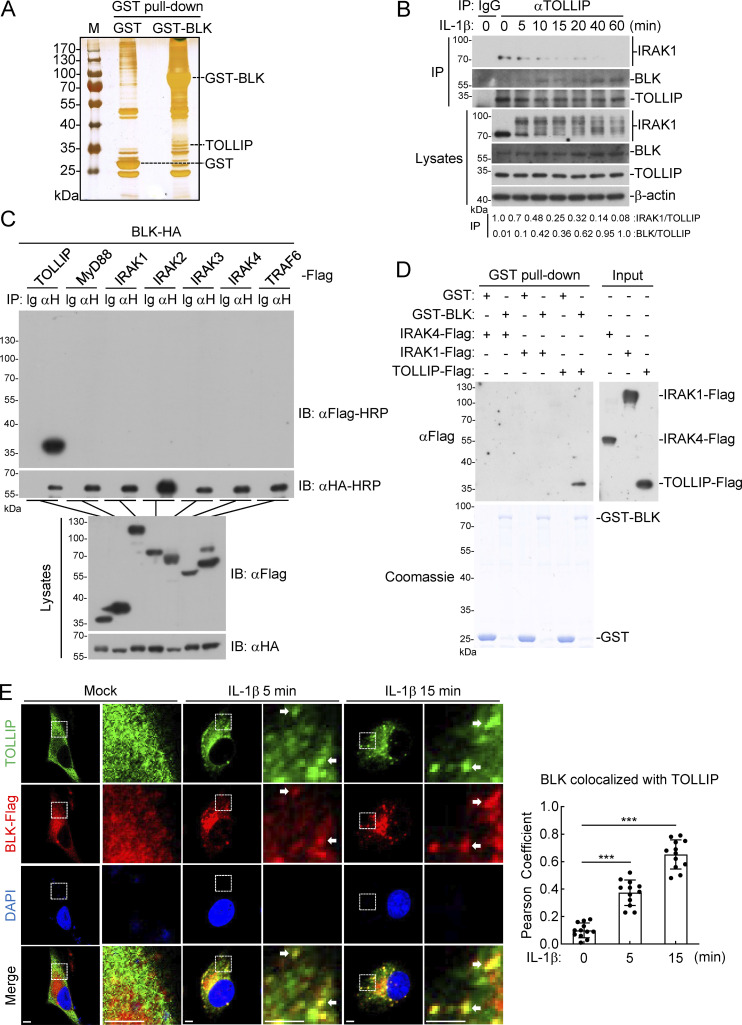
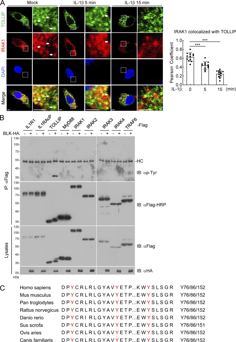
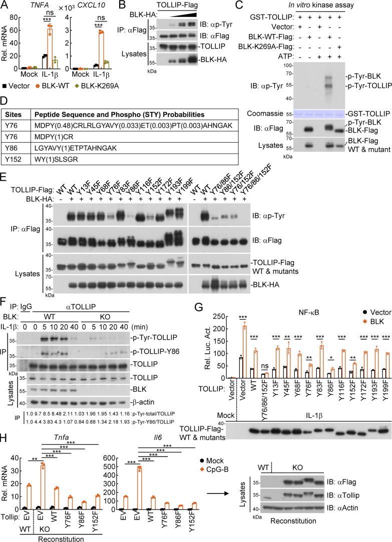
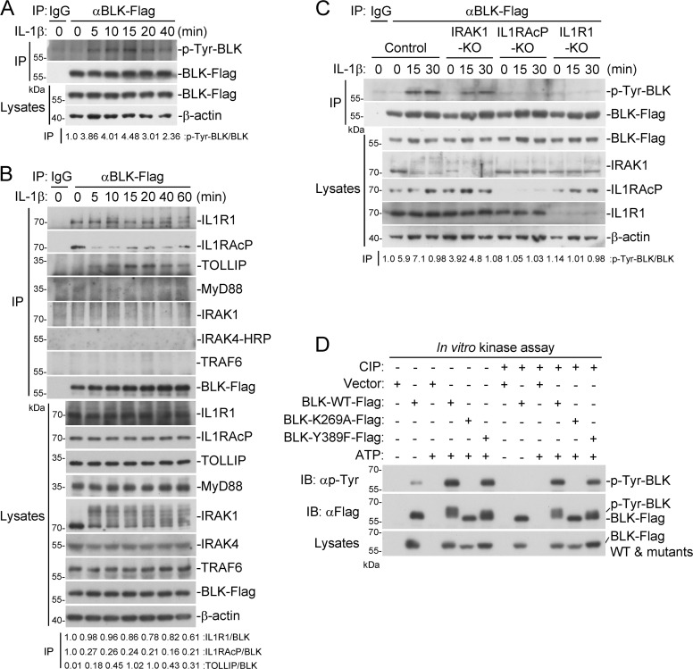
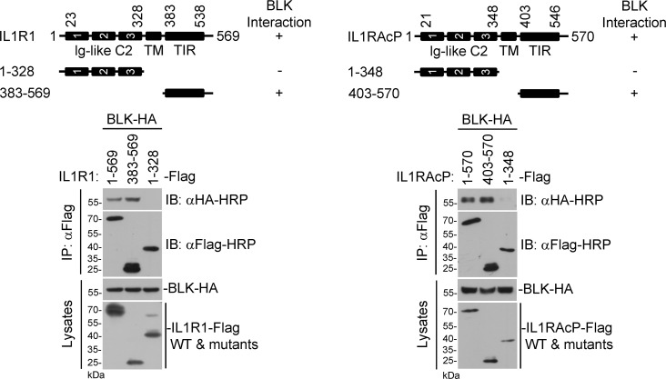
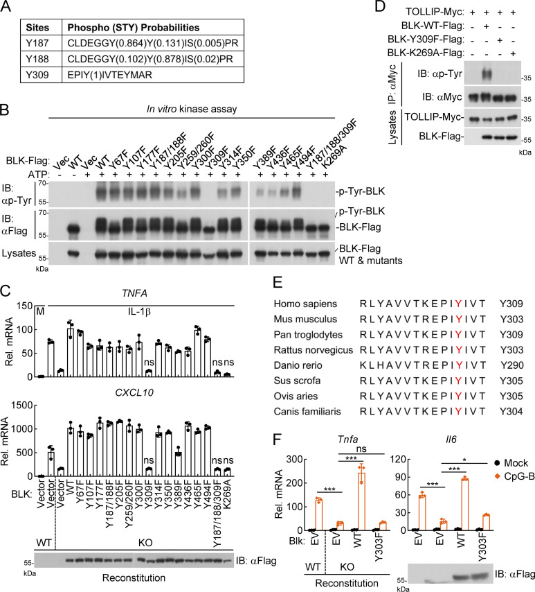
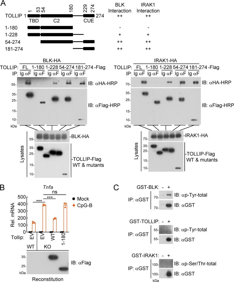
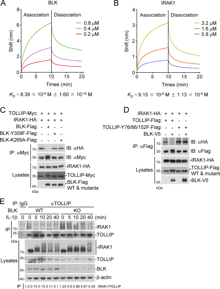
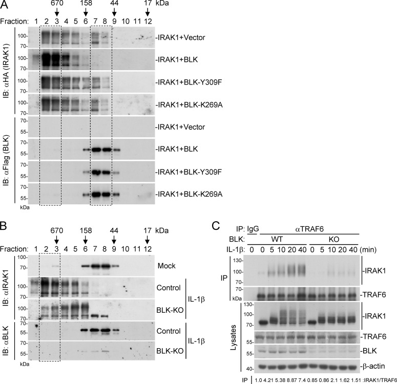
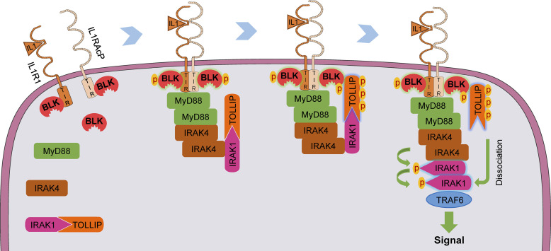
TLR/IL-1R signaling plays a critical role in sensing various harmful foreign pathogens and mounting efficient innate and adaptive immune responses, and it is tightly controlled by intracellular regulators at multiple levels. In particular, TOLLIP forms a constitutive complex with IRAK1 and sequesters it in the cytosol to maintain the kinase in an inactive conformation under unstimulated conditions. However, the underlying mechanisms by which IRAK1 dissociates from TOLLIP to activate TLR/IL-1R signaling remain obscure. Herein, we show that BLK positively regulates TLR/IL-1R-mediated inflammatory response. BLK-deficient mice produce less inflammatory cytokines and are more resistant to death upon IL-1β challenge. Mechanistically, BLK is preassociated with IL1R1 and IL1RAcP in resting cells. IL-1β stimulation induces heterodimerization of IL1R1 and IL1RAcP, which further triggers BLK autophosphorylation at Y309. Activated BLK directly phosphorylates TOLLIP at Y76/86/152 and further promotes TOLLIP dissociation from IRAK1, thereby facilitating TLR/IL-1R-mediated signal transduction. Overall, these findings highlight the importance of BLK as an active regulatory component in TLR/IL-1R signaling.


Conflict of interest statement

Disclosures: The authors declare no competing interests exist.

Figures

Figure 1.
Figure 1.
BLK positively regulates TLR/IL-1R–mediated inflammatory signaling. (A and B) Effects of BLK on TNFα-, IL-1β–, and LPS-induced activation of NF-κB. U87MG (A) or HEK293-TLR4 cells (B) (1 × 10⁵) were cotransfected with NF-κB reporter (0.002 μg), pRL-TK (Renilla luciferase) reporter (0.01 μg), and increased amounts of BLK expression plasmids (0.01, 0.02 μg) for 24 h. Cells were then left untreated or treated with TNFα (20 ng/ml), IL-1β (20 ng/ml), or LPS (100 ng/ml) for 10 h before luciferase assays. (C–E) Effects of BLK on flagellin-, TNFα-, CpG-B/C–, and IL-1β–induced transcription of downstream genes. Jurkat (C), Raji (D), or U87MG (E) cells were transduced with vector (Vec) or BLK expression plasmids by lentivirus-mediated gene transfer to establish stable cell lines. Cells (2 × 10⁵) were left untreated or treated with flagellin (0.1 μg/ml), TNFα (20 ng/ml), CpG-B/C (1 μM), or IL-1β (20 ng/ml) for 3 h before qPCR analysis. (F–H) Effects of BLK deficiency on flagellin-, TNFα-, CpG-B/C–, and IL-1β–induced transcription of downstream genes. Jurkat (F), Raji (G), or U87MG (H) cells were transduced with control (Con) or the indicated gRNA plasmids targeting BLK gene by the CRISPR/Cas9 method to establish stable cell lines. BLK-deficient and control cells (2 × 10⁵) were treated with the indicated stimuli for 3 h before qPCR analysis. (I–K) Effects of BLK deficiency on flagellin-, CpG-B–, and IL-1β–induced phosphorylation of p65 and IκBα. BLK-deficient and control Jurkat (I), Raji (J), or U87MG (K) cells (2 × 10⁵) (BLK-KO #1 plasmids were used) were left untreated or treated with flagellin (0.1 μg/ml), CpG-B (1 μM), or IL-1β (20 ng/ml) for the indicated times before immunoblot analysis. KO, knockout. Graphs show mean ± SD (n = 3 biological replicates in A and B, n = 3 technical replicates in C–H) from one representative experiment. **P < 0.01, ***P < 0.001 (unpaired, two-tailed Student’s t test). Data are representative of three independent experiments with similar results. Source data are available for this figure: SourceData F1.
Figure S1.
Figure S1.
BLK positively regulates IL-1β–, LPS-, but not TNFα-induced inflammatory responses. (A) Effects of BLK on IL-1β– and LPS-induced transcription of downstream genes. HEK293-TLR4 cells (2 × 10⁵) were transfected with BLK expression plasmids for 24 h. Cells were then left untreated or treated with IL-1β (20 ng/ml) or LPS (100 ng/ml) for 3 h before qPCR analysis. (B) Effects of BLK deficiency on TNFα-induced phosphorylation of p65 and IκBα. BLK-deficient and control Jurkat cells (2 × 10⁵; BLK-KO #1 plasmids were used) were left untreated or treated with TNFα (20 ng/ml) for the indicated times before immunoblot analysis. KO, knockout. (C) Effects of BLK knockdown on TNFα- and IL-1β–induced transcription of downstream genes. U87MG cells (2 × 10⁵) were transfected with the indicated siRNA (final concentration, 40 nM). 48 h later, cells were left untreated or treated with TNFα (20 ng/ml) or IL-1β (20 ng/ml) for 3 h before qPCR analysis. Graphs show mean ± SD (n = 3 technical replicates in A and C) from one representative experiment. *P < 0.05, **P < 0.01, ***P < 0.001 (unpaired, two-tailed Student’s t test). Data are representative of three independent experiments with similar results. Source data are available for this figure: SourceData FS1.
Figure 2.
Figure 2.
Blk deficiency attenuates TLR/IL-1R–mediated inflammatory responses in vivo. (A and B) Effects of Blk deficiency on CpG-B/C–induced transcription of downstream genes and phosphorylation of p65 and IκBα. Murine A20 cells were transduced with control (Con) or the indicated gRNA plasmids targeting Blk gene by the CRISPR/Cas9 method to establish stable cell lines. Blk-deficient and control cells (2 × 10⁵) were left untreated or treated with CpG-B/C (1 μM) for the indicated times before qPCR (A) and immunoblot (B) analyses. KO, knockout. (C and D) Effects of Blk deficiency on IL-1β–, R848-, and CpG-B–induced transcription of downstream genes and phosphorylation of p65 and IκBα. pDCs derived from the spleens of Blk+/+ and Blk−/− mice (1 × 10⁵) were stimulated with murine IL-1β (20 ng/ml), R848 (10 μg/ml), or CpG-B (1 μM) for 3 h before qPCR analysis (C), or stimulated with R848 (10 μg/ml) for the indicated times before immunoblot analysis (D). (E and F) Effects of Blk deficiency on IL-1β–, LPS- and CpG-B–induced transcription of downstream genes and phosphorylation of p65 and IκBα. Primary B cells derived from the spleens of Blk+/+ and Blk−/− mice (5 × 10⁵) were seeded in 12-well plates for 48 h in the presence of anti-lgM/IgG (5 μg/ml) and anti-CD40 antibody (1 μg/ml). Cells were then stimulated with murine IL-1β (20 ng/ml), LPS (100 ng/ml), or CpG-B (1 μM) for 3 h before qPCR analysis (E), or stimulated with LPS (100 ng/ml) for the indicated times before immunoblot analysis (F). (G) Effects of Blk deficiency on IL-1β–induced serum cytokine levels. Sex- and age-matched Blk+/+ and Blk−/− mice (n = 6 for each group) were injected i.p. with murine IL-1β (150 μg/kg) for 2 h before serum cytokines were measured by ELISA. (H) Effects of Blk deficiency on IL-1β–induced inflammatory death. Sex- and age-matched Blk+/+ and Blk−/− mice (n = 10 for each group) were injected i.p. with murine IL-1β (150 μg/kg) plus D-gal (0.5 mg/g) per mouse, and mouse survival was recorded every 1 h. Graphs show mean ± SD (n = 3 technical replicates in A, C, and E, n = 6 biological replicates in G) from one representative experiment. *P < 0.05, **P < 0.01, ***P < 0.001 (unpaired, two-tailed Student’s t test). For the mouse survival study in H, Kaplan–Meier survival curves were generated and analyzed by the log-rank test. Data are representative of at least two independent experiments with similar results. Source data are available for this figure: SourceData F2.
Figure S2.
Figure S2.
Genotyping of Blk-deficient mice. (A) Blk gene targeting strategy. Exons 3–9 and part of the intron of Blk gene were deleted by the CRISPR/Cas9 method. The arrow marks the sequence position gRNA targeted. (B) Genotyping of Blk−/− mice. PCR analysis of genomic DNA to identify wild-type, heterozygous, and homozygous mice. Source data are available for this figure: SourceData FS2.
Figure 3.
Figure 3.
BLK associates with TOLLIP. (A) Identification of the BLK interactome by silver staining and mass spectrometry methods. Silver staining showed GST-associated factors (Control) and GST-BLK–associated factors, respectively. TOLLIP was among the convincing interacting factors. (B) Endogenous interaction between TOLLIP and IRAK1 or BLK. U87MG cells (2 × 10⁷) were left untreated or treated with IL-1β (20 ng/ml) for the indicated times. Coimmunoprecipitation (IP) and immunoblot (IB) analysis were performed with the indicated antibodies. (C) BLK interacts with TOLLIP in the mammalian overexpression system. HEK293 cells (2 × 10⁶) were transfected with the indicated plasmids for 24 h. Coimmunoprecipitation and immunoblot analysis were performed with the indicated antibodies. (D) BLK directly binds to TOLLIP. Prokaryotically expressed and purified GST-BLK protein coupled to glutathione sepharose beads was incubated with purified Flag-tagged IRAK1, IRAK4, or TOLLIP proteins for 3 h at 4°C and then subjected to in vitro GST pull-down assays. (E) BLK colocalizes with TOLLIP upon IL-1β stimulation (left panel). U87MG cells stably expressing BLK (1 × 10⁵) were left untreated or treated with IL-1β (20 ng/ml) for the indicated times, then fixed with 4% PFA and stained with anti-Flag and anti-TOLLIP antibodies before confocal microscopy. The arrow marks the colocalized pixels. Scale bars, 2 μm. Quantitative analysis of colocalization of BLK with TOLLIP (right panel). Statistical analysis was based on colocalization images using ImageJ software. Graphs show mean ± SD (n = 12 cells from three individual images). ***P < 0.001 (unpaired, two-tailed Student’s t test). Data are representative of at least two independent experiments with similar results. Source data are available for this figure: SourceData F3.
Figure S3.
Figure S3.
BLK mediates tyrosine phosphorylation of TOLLIP. (A) IRAK1 colocalizes with TOLLIP under resting conditions. U87MG cells (1 × 10⁵) were left untreated or treated with IL-1β (20 ng/ml) for the indicated times, then fixed with 4% PFA, and stained with anti-IRAK1 and anti-TOLLIP antibodies before confocal microscopy (left panel). The arrow marks the colocalized pixels. Scale bars, 2 μm. Statistical analysis of colocalization of IRAK1 with TOLLIP was based on colocalization images using ImageJ software (right panel). Graphs show mean ± SD (n = 12 cells from three individual images). ***P < 0.001 (unpaired, two-tailed Student’s t test). (B) BLK mediates tyrosine phosphorylation of TOLLIP but not other adaptors. HEK293 cells (2 × 10⁶) were transfected with the indicated plasmids for 24 h. Coimmunoprecipitation (IP) and immunoblot (IB) analysis were performed with the indicated antibodies. HC, heavy chain. (C) Sequence alignment of TOLLIP from the indicated species. The sequences correspond to aa74–157 of human TOLLIP. The conserved tyrosine residues are highlighted in red. Data are representative of three independent experiments with similar results. Source data are available for this figure: SourceData FS3.
Figure 4.
Figure 4.
Phosphorylation of TOLLIP at Y76/86/152 is essential for its regulatory function in TLR/IL-1R signaling. (A) Effects of BLK and its mutant on IL-1β–induced transcription of downstream genes. U87MG cells (2 × 10⁵) were transfected with BLK or BLK(K269A) plasmids for 24 h. Cells were then left untreated or treated with IL-1β (20 ng/ml) for 3 h before qPCR analysis. (B) BLK mediates tyrosine phosphorylation of TOLLIP in a dose-dependent manner. HEK293 cells (2 × 10⁶) were transfected with the indicated plasmids for 24 h. Coimmunoprecipitation (IP) and immunoblot (IB) analysis were performed with the indicated antibodies. (C) BLK catalyzes tyrosine phosphorylation of TOLLIP in vitro. Purified Flag-tagged BLK and its mutant coupled with Protein G sepharose were subjected to in vitro kinase assays with purified GST-TOLLIP. (D) Identification of potential tyrosine phosphorylation sites of TOLLIP by mass spectrometry. The in vitro phosphorylated TOLLIP in C was subjected to mass spectrometry. The list shows the phosphorylated peptide sequences and phosphorylation (STY) probabilities. (E) BLK mediates tyrosine phosphorylation of TOLLIP at Y76, Y86, and Y152. HEK293 cells (2 × 10⁶) were transfected with the indicated plasmids for 24 h. Coimmunoprecipitation and immunoblot analysis were performed with the indicated antibodies. (F) Effects of BLK deficiency on IL-1β–triggered TOLLIP Y86 phosphorylation. BLK-deficient and control U87MG cells (2 × 10⁷) were left untreated or treated with IL-1β (20 ng/ml) for the indicated times. Coimmunoprecipitation and immunoblot analysis were performed with the indicated antibodies. KO, knockout. (G) Effects of BLK and all TOLLIP mutants co-transfection on IL-1β–triggered NF-κB activation. U87MG cells (1 × 10⁵) were cotransfected with NF-κB reporter (0.002 μg), pRL-TK reporter (0.01 μg), BLK plasmids (0.05 μg), either TOLLIP or its mutants (0.05 μg) for 24 h and then treated with IL-1β (20 ng/ml) for 10 h before luciferase assays. The lower blot shows the expression levels of TOLLIP and its mutants as detected by anti-Flag antibody. (H) Effects of reconstitution of Tollip-deficient cells with Tollip or its mutants on CpG-B–induced transcription of downstream genes. A20 cells were transduced with vector or the gRNA plasmids targeting Tollip gene by the CRISPR/Cas9 method to establish the stable cell lines with puromycin (1 μg/ml) selection. Wild-type and Tollip-deficient A20 cells were then transduced with empty vector (EV), Tollip, or its mutants by lentivirus-mediated gene transfer to establish the stable cell lines with blasticidin S (10 μg/ml) selection. Subsequently, the indicated cell lines (2 × 10⁵) were left untreated or treated with CpG-B (1 μM) for 3 h before qPCR analysis. The blots show the expression levels of Tollip and its mutants in the indicated cell lines as detected by anti-Flag or anti-Tollip antibodies, respectively. Graphs show mean ± SD (n = 3 technical replicates in A and H, n = 3 biological replicates in G) from one representative experiment. *P < 0.05, **P < 0.01, ***P < 0.001, ns, not significant (unpaired, two-tailed Student’s t test). Data are representative of three independent experiments with similar results. Source data are available for this figure: SourceData F4.
Figure 5.
Figure 5.
Preassociation of BLK with IL1R1 and IL1RAcP is necessary for BLK autophosphorylation. (A) BLK undergoes tyrosine phosphorylation following IL-1β stimulation. U87MG cells stably expressing BLK (2 × 10⁷) were left untreated or treated with IL-1β (20 ng/ml) for the indicated times. Coimmunoprecipitation (IP) and immunoblot (IB) analysis were performed with the indicated antibodies. (B) BLK is constitutively associated with IL1R1 and IL1RAcP under unstimulated conditions. U87MG cells stably expressing BLK (2 × 10⁷) were left untreated or treated with IL-1β (20 ng/ml) for the indicated times. Coimmunoprecipitation and immunoblot analysis were performed with the indicated antibodies. (C) IL1R1 or IL1RAcP deficiency abolishes IL-1β–induced BLK phosphorylation. U87MG cells were transduced with control or gRNA plasmids targeting IRAK1, IL1R1, or IL1RAcP genes by the CRISPR/Cas9 method to establish stable cell lines. The indicated cell lines (2 × 10⁵) were left untreated or treated with IL-1β (20 ng/ml) for the indicated times before immunoblot analysis. KO, knockout. (D) BLK catalyzes autophosphorylation in a kinase activity-dependent manner. Purified Flag-tagged BLK and its mutants coupled with Protein G sepharose were subjected to in vitro BLK autophosphorylation assays without or with CIP treatment. The detailed procedures are shown in Materials and methods. Data are representative of three independent experiments with similar results. Source data are available for this figure: SourceData F5.
Figure S4.
Figure S4.
BLK interacts with the cytoplasmic TIR domains of IL1R1 and IL1RAcP. Domain mapping of the interaction between BLK and IL1R1 or IL1RAcP. HEK293 cells (2 × 10⁶) were transfected with the indicated plasmids for 24 h. Coimmunoprecipitation (IP) and immunoblot (IB) analysis were performed with the indicated antibodies. −: no interaction, +: interaction. TM, transmembrane. Data are representative of three independent experiments with similar results. Source data are available for this figure: SourceData FS4.
Figure 6.
Figure 6.
Autophosphorylation of BLK at Y309 is critical for its regulation of TLR/IL-1R–mediated inflammatory signaling. (A) Identification of potential autophosphorylation sites of BLK by mass spectrometry. The in vitro phosphorylated BLK in Fig. 5 D was subjected to mass spectrometry. The list shows the phosphorylated peptide sequences and phosphorylation (STY) probabilities. (B) BLK catalyzes autophosphorylation mainly at Y309. Purified Flag-tagged BLK and its mutants coupled with Protein G sepharose were subjected to in vitro BLK autophosphorylation assays. (C) Effects of reconstitution of BLK-deficient cells with BLK and its mutants on IL-1β–induced transcription of downstream genes. BLK-deficient and control U87MG cells (2 × 10⁵) were transfected with the same amount of BLK or its mutants for 24 h. Cells were then left untreated or treated with IL-1β (20 ng/ml) for 3 h before qPCR analysis. The lower blot shows the expression levels of BLK and its mutants as detected by anti-Flag antibody. KO, knockout. (D) BLK(Y309F) fails to mediate tyrosine phosphorylation of TOLLIP. HEK293 cells (2 × 10⁶) were transfected with the indicated plasmids for 24 h. Coimmunoprecipitation (IP) and immunoblot (IB) analysis were performed with the indicated antibodies. (E) Sequence alignment of BLK from the indicated species. The sequences correspond to aa 298–312 of human BLK. The conserved tyrosine residues are highlighted in red. (F) Effects of reconstitution of Blk-deficient cells with Blk or its mutant on CpG-B–induced transcription of downstream genes. Blk-deficient A20 cells were transduced with Blk or Blk(Y303F) plasmids by lentivirus-mediated gene transfer to establish the stable cell lines with blasticidin S (10 μg/ml) selection. The indicated cell lines (2 × 10⁵) were then left untreated or treated with CpG-B (1 μM) for 3 h before qPCR analysis. The lower blot shows the expression levels of Blk and Blk(Y303F) in the indicated cell lines as detected by anti-Flag antibody. Graphs show mean ± SD (n = 3 technical replicates in C and F) from one representative experiment. *P < 0.05, ***P < 0.001, ns, not significant (unpaired, two-tailed Student’s t test). Data are representative of three independent experiments with similar results. Source data are available for this figure: SourceData F6.
Figure S5.
Figure S5.
The C-terminus (aa181–274) containing CUE domain of TOLLIP is responsible for its association with BLK and IRAK1. (A) Domain mapping of the interaction between TOLLIP and BLK or IRAK1. HEK293 cells (2 × 10⁶) were transfected with the indicated plasmids for 24 h. Coimmunoprecipitation (IP) and immunoblot (IB) analysis were performed with the indicated antibodies. −: no interaction, +: weak interaction, ++: strong interaction. FL, full length. (B) Effects of reconstitution of Tollip-deficient cells with Tollip or Tollip(1–180) on CpG-B–induced transcription of downstream genes. Wild-type and Tollip-deficient A20 cells were transduced with empty vector (EV), Tollip, or Tollip(1–180) by lentivirus-mediated gene transfer to establish the stable cell lines with blasticidin S (10 μg/ml) selection. Subsequently, the indicated cell lines (2 × 10⁵) were left untreated or treated with CpG-B (1 μM) for 3 h before qPCR analysis. The lower blot shows the expression levels of Tollip and Tollip(1–180) in the indicated cell lines as detected by anti-Flag antibody. Graphs show mean ± SD (n = 3 technical replicates) from one representative experiment. ***P < 0.001, ns, not significant (unpaired, two-tailed Student’s t test). KO, knockout. (C) Detection of the phosphorylation status of BLK, TOLLIP, and IRAK1. The purified recombinant proteins BLK, TOLLIP, and IRAK1 were subjected to coimmunoprecipitation and immunoblot analysis. Data are representative of three independent experiments with similar results. Source data are available for this figure: SourceData FS5.
Figure 7.
Figure 7.
BLK promotes TOLLIP dissociation from IRAK1 by competitively binding to TOLLIP. (A and B) TOLLIP-binding affinities of BLK and IRAK1. ForteBio Octet Red system was used to examine the binding affinities of recombinant BLK (A) and IRAK1 (B) to TOLLIP. The vertical and horizontal axes represent the light shift distance (nm) and association/dissociation times, respectively. (C) Effects of BLK and its mutants on the association of TOLLIP and IRAK1. HEK293 cells (2 × 10⁶) were transfected with the indicated plasmids for 24 h. Coimmunoprecipitation (IP) and immunoblot (IB) analysis were performed with the indicated antibodies. (D) Effects of BLK on the association of TOLLIP(Y76/86/152F) and IRAK1. HEK293 cells (2 × 10⁶) were transfected with the indicated plasmids for 24 h. Coimmunoprecipitation and immunoblot analysis were performed with the indicated antibodies. (E) Effects of BLK deficiency on IL-1β–induced dissociation of IRAK1 from TOLLIP. BLK-deficient and control U87MG cells (2 × 10⁷) (BLK-KO #1 plasmids were used) were left untreated or treated with IL-1β (20 ng/ml) for the indicated times before coimmunoprecipitation and immunoblot analysis. KO, knockout. Data are representative of three independent experiments with similar results. Source data are available for this figure: SourceData F7.
Figure 8.
Figure 8.
BLK facilitates TLR/IL-1R–mediated signal transduction. (A) Effects of BLK and its mutants on the oligomerization of IRAK1. HEK293 cells (4 × 10⁷) were transfected with IRAK1 and BLK or its mutants for 24 h. Cells were collected and lysed, followed by gel filtration. Fraction size was calibrated with the gel filtration standard (151-1901; Bio-Rad). (B) Effects of BLK deficiency on IL-1β–induced oligomerization of endogenous IRAK1. BLK-deficient and control U87MG cells (1 × 10⁸) were untreated or treated with IL-1β (20 ng/ml) for 10 min before gel filtration. Fraction size was calibrated with the gel filtration standard. (C) Effects of BLK deficiency on IL-1β–induced hyperphosphorylation of IRAK1 and subsequent recruitment of IRAK1 to TRAF6. BLK-deficient and control U87MG cells (2 × 10⁷) (BLK-KO #1 plasmids were used) were left untreated or treated with IL-1β (20 ng/ml) for the indicated times before coimmunoprecipitation (IP) and immunoblot (IB) analysis. Data are representative of three independent experiments with similar results. Source data are available for this figure: SourceData F8.
Figure 9.
Figure 9.
A schematic presentation for the role of BLK in regulating TLR/IL-1R–mediated inflammatory response. Under unstimulated conditions, BLK is constitutively associated with the cytoplasmic TIR domains of IL1R1 and IL1RAcP. Upon binding of IL-1β to IL1R1, IL1RAcP is recruited to form a high-affinity heterodimer, which results in the conformational change of TIR domains and further triggers BLK autophosphorylation. Simultaneously, the receptor complex recruits MyD88, IRAKs, and TOLLIP to form a signal transduction complex termed Myddosome. Activated BLK phosphorylates TOLLIP and further promotes TOLLIP dissociation from IRAK1 by competitively binding to the CUE domain of TOLLIP, thus enabling downstream signaling cascade.

Similar articles

Cited by

References

    1. Akerblad, P., and Sigvardsson M.. 1999. Early B cell factor is an activator of the B lymphoid kinase promoter in early B cell development. J. Immunol. 163:5453–5461. 10.4049/jimmunol.163.10.5453 - DOI - PubMed
    1. Akira, S., Takeda K., and Kaisho T.. 2001. Toll-like receptors: Critical proteins linking innate and acquired immunity. Nat. Immunol. 2:675–680. 10.1038/90609 - DOI - PubMed
    1. Akira, S., Uematsu S., and Takeuchi O.. 2006. Pathogen recognition and innate immunity. Cell. 124:783–801. 10.1016/j.cell.2006.02.015 - DOI - PubMed
    1. Barton, G.M., and Medzhitov R.. 2003. Toll-like receptor signaling pathways. Science. 300:1524–1525. 10.1126/science.1085536 - DOI - PubMed
    1. Blasius, A.L., and Beutler B.. 2010. Intracellular toll-like receptors. Immunity. 32:305–315. 10.1016/j.immuni.2010.03.012 - DOI - PubMed

Publication types