Review
. 2024 Jul;34(7):4243-4260.
doi: 10.1007/s00330-023-10413-9. Epub 2023 Dec 11.

Imaging for the assessment of the arrhythmogenic potential of mitral valve prolapse

Affiliations
Review

Imaging for the assessment of the arrhythmogenic potential of mitral valve prolapse

Antonio Esposito et al. Eur Radiol. 2024 Jul.

Abstract

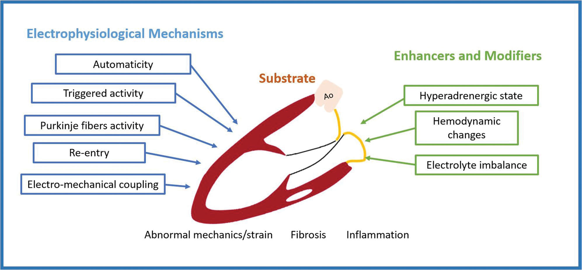
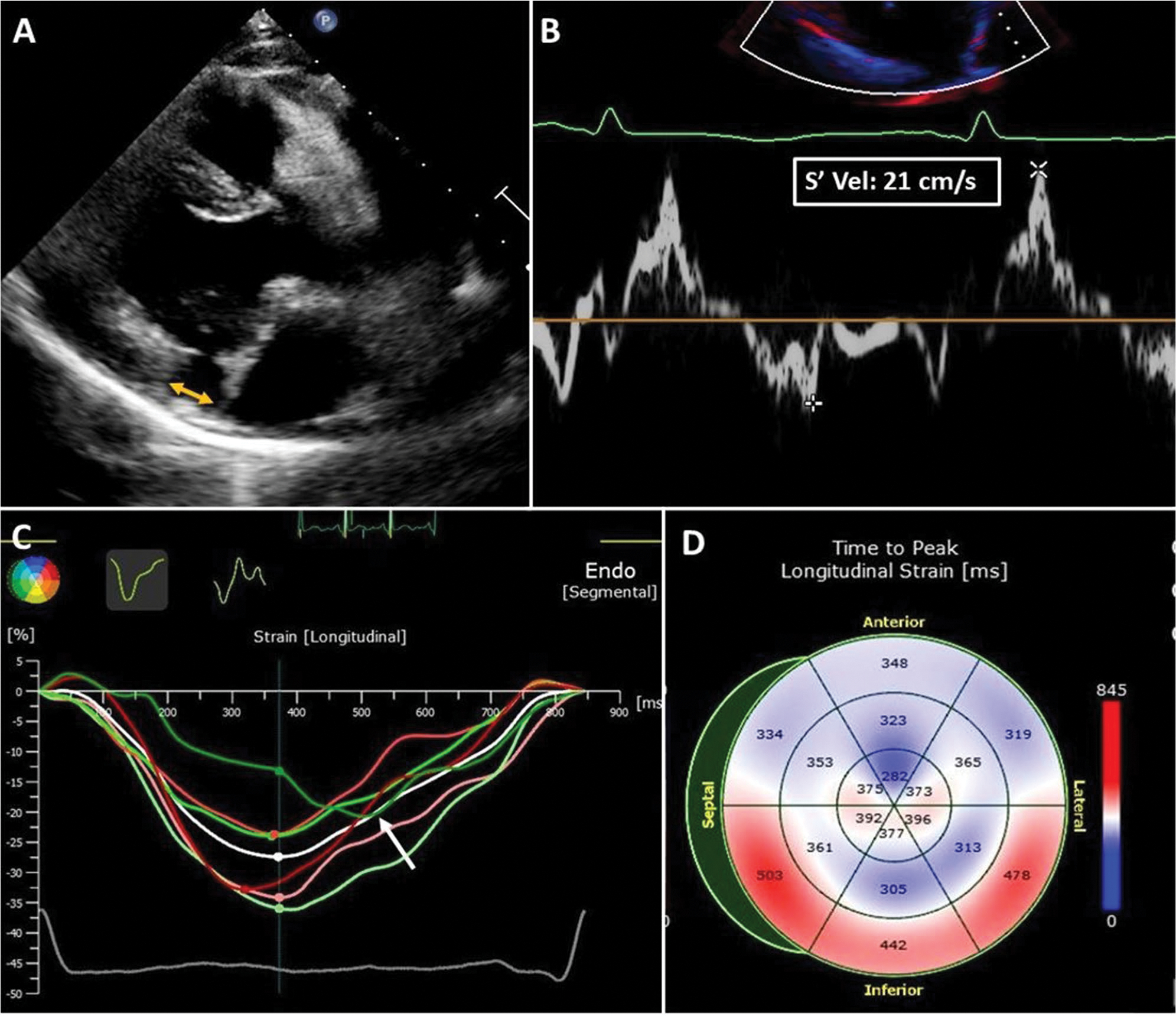
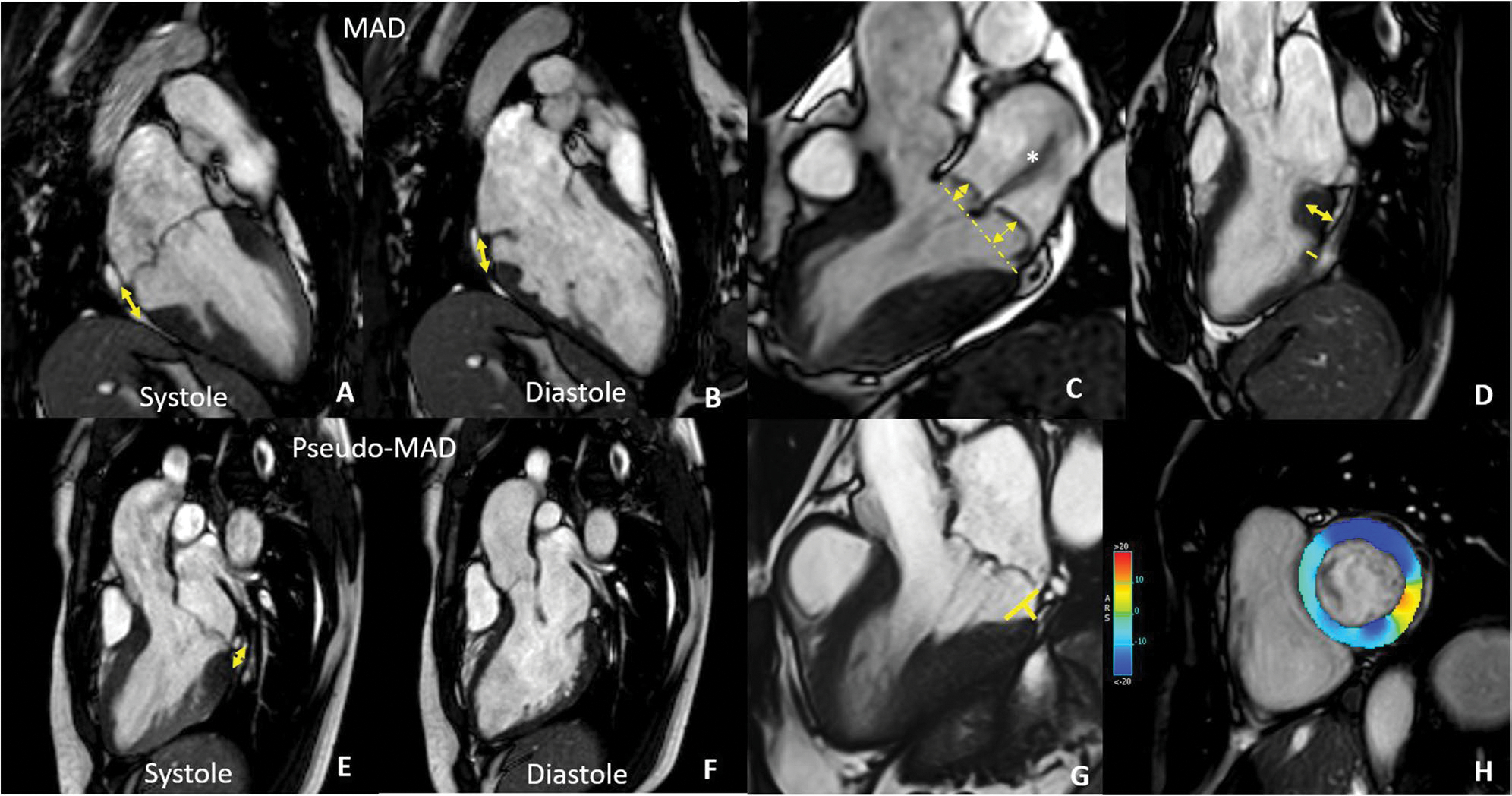
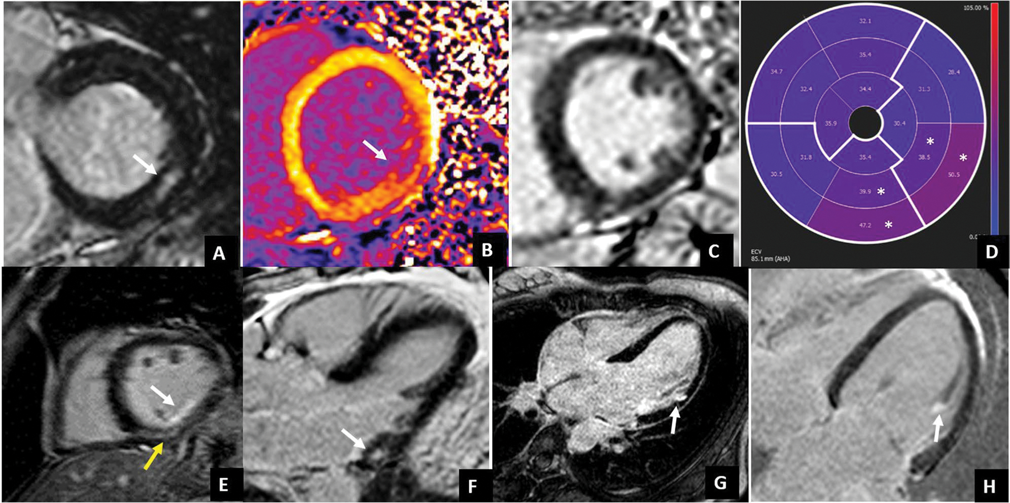
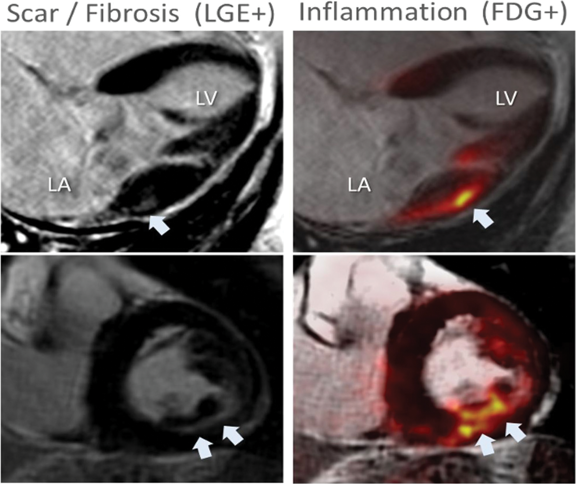
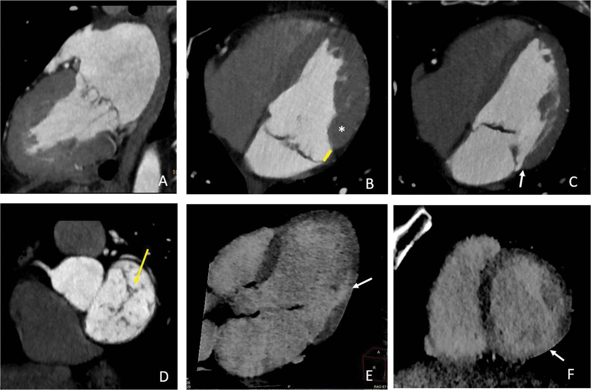
Mitral valve prolapse (MVP) is the most common valve disease in the western world and recently emerged as a possible substrate for sudden cardiac death (SCD). It is estimated an annual risk of sudden cardiac death of 0.2 to 1.9% mostly caused by complex ventricular arrhythmias (VA). Several mechanisms have been recognized as potentially responsible for arrhythmia onset in MVP, resulting from the combination of morpho-functional abnormality of the mitral valve, structural substrates (regional myocardial hypertrophy, fibrosis, Purkinje fibers activity, inflammation), and mechanical stretch. Echocardiography plays a central role in MVP diagnosis and assessment of severity of regurgitation. Several abnormalities detectable by echocardiography can be prognostic for the occurrence of VA, from morphological alteration including leaflet redundancy and thickness, mitral annular dilatation, and mitral annulus disjunction (MAD), to motion abnormalities detectable with "Pickelhaube" sign. Additionally, speckle-tracking echocardiography may identify MVP patients at higher risk for VA by detection of increased mechanical dispersion. On the other hand, cardiac magnetic resonance (CMR) has the capability to provide a comprehensive risk stratification combining the identification of morphological and motion alteration with the detection of myocardial replacement and interstitial fibrosis, making CMR an ideal method for arrhythmia risk stratification in patients with MVP. Finally, recent studies have suggested a potential role in risk stratification of new techniques such as hybrid PET-MR and late contrast enhancement CT. The purpose of this review is to provide an overview of the mitral valve prolapse syndrome with a focus on the role of imaging in arrhythmic risk stratification. CLINICAL RELEVANCE STATEMENT: Mitral valve prolapse is the most frequent valve condition potentially associated with arrhythmias. Imaging has a central role in the identification of anatomical, functional, mechanical, and structural alterations potentially associated with a higher risk of developing complex ventricular arrhythmia and sudden cardiac death. KEY POINTS: • Mitral valve prolapse is a common valve disease potentially associated with complex ventricular arrhythmia and sudden cardiac death. • The mechanism of arrhythmogenesis in mitral valve prolapse is complex and multifactorial, due to the interplay among multiple conditions including valve morphological alteration, mechanical stretch, myocardial structure remodeling with fibrosis, and inflammation. • Cardiac imaging, especially echocardiography and cardiac magnetic resonance, is crucial in the identification of several features associated with the potential risk of serious cardiac events. In particular, cardiac magnetic resonance has the advantage of being able to detect myocardial fibrosis which is currently the strongest prognosticator.

Keywords: Arrhythmogenic mitral valve prolapse; Cardiac imaging technique; Computed tomography; Magnetic resonance; Mitral valve prolapse.


Conflict of interest statement

Conflict of interest

Marco Francone is European Radiology’s section editor of Cardiac in the Scientific editorial board. He has not taken part in the review or selection process of this article.

The authors of this manuscript declare no relationships with any companies, whose products or services may be related to the subject matter of the article. Anna Palmisano is a member of the European Radiology Experimental scientific editorial board.

Figures

Fig. 1
Fig. 1
Pathophysiology of arrhythmogenic mitral valve prolapse
Fig. 2
Fig. 2
Echocardiographic biomarkers of arrhythmic MVP in a 35-year-old woman with MVP, moderate mitral regurgitation, and frequent premature ventricular contractions. A MAD. The yellow arrow shows the distance between the basal ventricular myocardium and the hinge point of the posterior leaflet in late systole. Bulging of the basal inferior-lateral myocardium, typical of the systolic curling, was also present in this patient. B Pickelhaube sign. A spiked late systolic high-velocity (21 cm/s) signal is recorded at the level of the lateral mitral annulus in a four-chamber view. C Speckle tracking imaging. Late post-systolic shortening (after aortic valve closure) of the basal inferior-lateral left ventricular wall (white arrow, green line). D Mechanical dispersion. Prolonged time-to-peak longitudinal strain is observed at the basal inferior, inferior-septal, and inferior-lateral walls and is related to myocardial periannular fibrosis
Fig. 3
Fig. 3
CMR findings in MVP. In A and B are reported systolic (A) and diastolic (B) frames of the same patient showing mitral annulus disjunction (double-headed arrow) that needs to be distinguished from pseudo-MAD (E and F) due to the juxtaposition of the posterior leaflet on the atrial wall in systole. C 3-chamber long axis showing severe bileaflet mitral valve prolapse with high prolapse volume and a huge jet of regurgitation (asterisks). D Basal LV hypertrophy with a ratio of LV thickness between basal and mid segments of the inferolateral wall > 1.5 at end-diastole. G Curling distance by tracing a line between the top of the LV I wall and the LA–MV leaflet junction, and from this line, a perpendicular line to the lower limit of the mitral annulus at end-systole. H GLS analysis showing contractility alteration of the inferolateral wall
Fig. 4
Fig. 4
LGE patterns typically associated with arrhythmic MVP. LGE usually occurs at the level of the LV inferolateral wall. Different LGE patterns have been described: non-ischemic mid-wall LGE (white arrows in A and F, yellow arrow in E), subendocardial LGE (white arrows in E and G), papillary muscle LGE (with arrow in H). Interstitial fibrosis documented by native T1 mapping and ECV values has been found to be increased not only at the site of LGE (white arrow in native T1 map in B) but also in LGE negative patients (case example in C) with diffusely high ECV values which are higher in the inferior and inferolateral mid-basal wall (white asterisks)
Fig. 5
Fig. 5
Hybrid [18F]FDG PET/MRI in a patient with MVP. An example of concordant [18F]FDG uptake and LGE in an asymptomatic patient with chronic severe degenerative mitral regurgitation and absent left ventricular remodeling (LVEF 60%, LVESD 38 mm). White arrowheads indicate areas of either LGE or FDG uptake. LA indicates left atrium; LV, left ventricle
Fig. 6
Fig. 6
Cardiac CT in a patient with arrhythmic MVP. CT images show a bileaflet multiscallop mitral valve prolapse (A) in a patient with MAD recognizable in systole (arrow in B) and diastole (arrow in C), with curling of the inferolateral mid-basal wall (white asterisks in B) and mild regurgitation due to a small coaptation defect (arrow in D). Late contrast enhancement 3-chambers long axis (E) and 2-chambers short axis (F) documented a small area of late enhancement in the mid inferolateral wall close to the posterior papillary muscle insertion point (arrows in E and F)

References

    1. Delling FN, Vasan RS (2014) Epidemiology and pathophysiology of mitral valve prolapse. Circulation. 10.1161/CIRCULATIONAHA.113.006702 - DOI - PMC - PubMed
    1. Adabifirouzjaei F, Hsiao A, DeMaria AN (2022) Mitral valve prolapse—the role of cardiac imaging modalities. Structural Heart. 10.1016/j.shj.2022.100024 - DOI - PMC - PubMed
    1. Sabbag A, Essayagh B, Barrera JDR et al. (2022) EHRA expert consensus statement on arrhythmic mitral valve prolapse and mitral annular disjunction complex in collaboration with the ESC Council on valvular heart disease and the European Association of Cardiovascular Imaging endorsed cby the Heart Rhythm Society, by the Asia Pacific Heart Rhythm Society, and by the Latin American Heart Rhythm Society. EP Europace. 10.1093/europace/euac125 - DOI - PMC - PubMed
    1. Parwani P, Avierinos J-F, Levine RA, Delling FN (2017) Mitral valve prolapse: multimodality imaging and genetic insights. Prog Cardiovasc Dis. 10.1016/j.pcad.2017.10.007 - DOI - PMC - PubMed
    1. Palmisano A, Nicoletti V, Colantoni C et al. (2021) Dynamic changes of mitral valve annulus geometry at preprocedural CT: relationship with functional classes of regurgitation. Eur Radiol Exp. 10.1186/s41747-021-00231- - DOI - PMC - PubMed

MeSH terms