. 2024 May 3;147(5):1809-1821.
doi: 10.1093/brain/awad413.

PAK2 is necessary for myelination in the peripheral nervous system

Affiliations

PAK2 is necessary for myelination in the peripheral nervous system

Bo Hu et al. Brain. .

Abstract

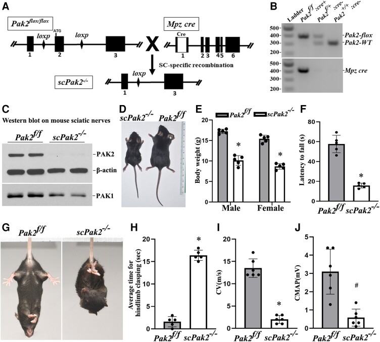
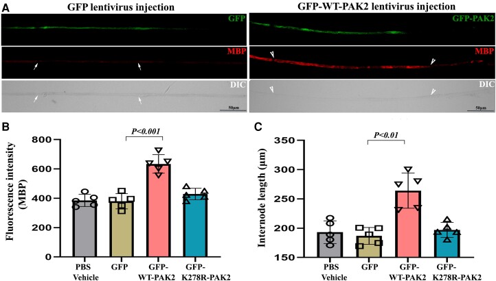
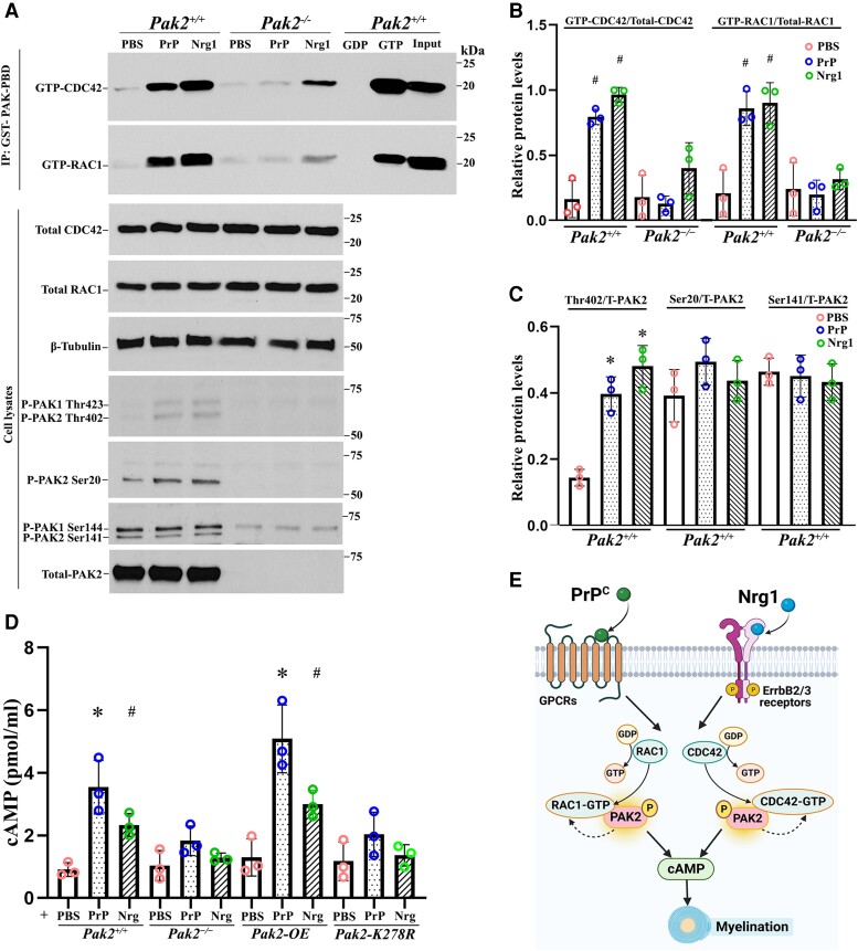
Myelination enables electrical impulses to propagate on axons at the highest speed, encoding essential life functions. The Rho family GTPases, RAC1 and CDC42, have been shown to critically regulate Schwann cell myelination. P21-activated kinase 2 (PAK2) is an effector of RAC1/CDC42, but its specific role in myelination remains undetermined. We produced a Schwann cell-specific knockout mouse of Pak2 (scPak2-/-) to evaluate PAK2's role in myelination. Deletion of Pak2, specifically in mouse Schwann cells, resulted in severe hypomyelination, slowed nerve conduction velocity and behaviour dysfunctions in the scPak2-/- peripheral nerve. Many Schwann cells in scPak2-/- sciatic nerves were arrested at the stage of axonal sorting. These abnormalities were rescued by reintroducing Pak2, but not the kinase-dead mutation of Pak2, via lentivirus delivery to scPak2-/- Schwann cells in vivo. Moreover, ablation of Pak2 in Schwann cells blocked the promyelinating effect driven by neuregulin-1, prion protein and inactivated RAC1/CDC42. Conversely, the ablation of Pak2 in neurons exhibited no phenotype. Such PAK2 activity can also be either enhanced or inhibited by different myelin lipids. We have identified a novel promyelinating factor, PAK2, that acts as a critical convergence point for multiple promyelinating signalling pathways. The promyelination by PAK2 is Schwann cell-autonomous. Myelin lipids, identified as inhibitors or activators of PAK2, may be utilized to develop therapies for repairing abnormal myelin in peripheral neuropathies.

Keywords: Pak2 knock-out mouse; GTPases Rac1/Cdc42; Schwann cells; myelin lipids; myelination; peripheral nerve.


Conflict of interest statement

The authors report no competing interests.

Figures

Figure 1
Figure 1
PAK2 is highly expressed during peripheral nerve development. (A) The expression levels of P21-activated kinases (PAKs) were analysed by western blot in the sciatic nerves of P7 wild-type (WT) mice. Each antibody was tested in duplicates of nerve lysate. PAK1 and PAK2 were detectable in mouse sciatic nerves, while PAK3, PAK4, PAK5 and PAK6 were barely detectable or undetectable. β-Actin served as a loading control. The relative protein level of the PAKs was determined by normalizing to β-actin. The top panel was from a short exposure. The bottom panel was developed by a long exposure. This experiment was performed three times using different samples. (B) Total PAK1 and PAK2 levels were measured by western blot in E18.5, P1, P7, P14, P28 and P150 wild-type mouse sciatic nerves. PAK1 and PAK2 levels were normalized against β-actin levels. PAK2 levels were significantly higher in the developing peripheral nerves (E18.5 to P14) than those in mature ones (P150). PAK1 is highly expressed in peripheral nerves during the embryonic period (E18.5) and gradually decreases during the maturation of the peripheral nerves. For each developmental stage, duplicates of nerve samples were used in each experiment. The experiment was replicated with different samples. (C) Longitudinal (iiv) and transverse (vviii) sections of 3-week-old wild-type sciatic nerves were stained with antibodies against PAK2 (red) and myelin basic protein (MBP) (green, stain myelin). The staining showed PAK2 localization predominantly in myelin. The top panels [C(i, iii, v and vii)] show the original images, while the bottom panels [C(ii, iv, vi and viii)] are the magnified view of the white boxes in the top panels. The specificity of the PAK2 antibody was confirmed through knockout mouse tissue (Supplementary Fig. 1). (D) A section of the dorsal root ganglia (DRG) from a 3-week-old wild-type mouse was reacted with PAK2 (red) and neurofilament-200 (NF, green) antibodies. PAK2 was localized in the cytoplasm of the DRG neurons. [D(ii and iv)] Magnified view of the white box. Scale bars = 10 μm.
Figure 2
Figure 2
Schwann cell-specific Pak2 knockout mouse. (A) Exon 2 in Pak2 gene was flanked by LoxP. Mpz activates Cre recombinase expression in Schwann cells at embryonic Day 14.5. Upon Cre recombination, the exon 2 of Pak2 between loxP sites was removed to produce Schwann cell-specific knockout mice (scPak2−/−). (B) Mice were genotyped by PCR. The amplified wild-type (WT) Pak2 allele is 391 bp, and the amplified Pak2 flox allele is 306 bp. Lane 1 = DNA ladder (M); Lane 2 = Pak2flox/flox:Mpz-Cre+ (scPak2−/−); Lane 3 = Pak2flox/+:Mpz-Cre (Pak2f/+); Lane 4 = Pak2wt/wt:Mpz-Cre (Pak2+/+). (C) At P21, sciatic nerves with stripped epineurium were analysed by western blot using an antibody against PAK1 and PAK2. In Pak2f/f nerves, both PAK2 and PAK1 were detectable. However, in scPak2−/− nerves, PAK2 expression was hardly detectable, while PAK1 level also exhibited a minor reduction. β-Actin was used as a loading control. The nerves of each genotype were from two different samples. (D). One-month-old Pak2f/f and scPak2−/− littermate were placed next to a centimetre measure. (E) The body size of the 1-month-old scPak2−/− mice was significantly smaller than that of their Pak2f/f littermates (P < 0.001, n = 5 in each group). (F) Rotarod analysis of motor function at 1 month of age revealed a significant reduction in scPak2−/− mice, compared to that in Pak2f/f mice (P < 0.001, n = 5 in each group). (G) Tail-suspension test showed hind paw clasping in 1-month-old scPak2−/− mice, but not in control littermates. (H) Quantification of hindlimb clasping test. Data are presented as the average time for hindlimb splaying. One-month-old scPak2−/− mice exhibited significantly increased hindlimb clasp compared to that in their Pak2f/f littermates (P < 0.001, n = 5 for each genotyping group). (I and J) Nerve conduction studies showed that conduction velocity (CV) and compound muscle action potential (CMAP) were significantly decreased in 1-month-old scPak2−/− mice compared to Pak2f/f mice (P < 0.05, n = 6 in each genotype). Mean ± SD. *P < 0.001, #P < 0.01.
Figure 3
Figure 3
Morphometric analysis on mouse sciatic nerve. (A) The semithin sections on 1-month-old scPak2−/− sciatic nerves showed a reduction in the number of myelinated nerve fibres, which appeared smaller in diameter compared to that in the Pak2f/f nerves. There were no signs of demyelination, such as myelin debris, broken myelin or excessive macrophages. (B) The morphometric analysis confirmed a significant decrease in myelinated nerve fibres of scPak2−/− nerves, compared with that in the Pak2f/f nerves (P < 0.001, n = 3 in each genotype). (C) Myelin sheath thickness was reduced in scPak2−/− sciatic nerves. (D) A plot of g-ratio against nerve fibre diameter supports the observed reduction of myelin thickness in scPak2−/− mice. (E) Electronic microscopy images revealed scPak2−/− axons either failed to be myelinated (arrowhead) or thinly myelinated (arrow). Scale bars = 2 μm.
Figure 4
Figure 4
Intraneural lentivirus delivery restores PAK2 expression in Schwann cells of scPak2−/− sciatic nerves in vivo. (A) A sciatic nerve from a 3-day-old scPak2−/− mouse was surgically exposed and locally injected with 2 μl lentivirus particles at 1010 cfu/ml of GFP-only, GFP-WT-PAK2 (wild-type PAK2) or GFP-K278R-PAK2 (kinase-dead PAK2 mutation). The contralateral sciatic nerve was injected with 2 μl PBS as a control. At P30, the sciatic nerves were dissected. After fixation, teased nerve fibres were stained with myelin basic protein (MBP) antibody and imaged. (B) The MBP fluorescence intensity of each internode within nerve fibres was quantified and the average intensity for each mouse was calculated. The intensity of MBP was significantly increased in GFP-positive fibres (between arrowheads) from scPak2−/− nerves transfected with GFP-WT-Pak2 lentivirus, but not GFP-positive fibres from scPak2−/− nerves transfected with GFP-K278R-Pak2 lentivirus, compared with fibres (between arrows) in scPak2−/− nerve fibres transfected with GFP-only lentivirus (mean ± SD, P < 0.001, n = 10–20 fibres per mouse, n = 5 mice per group). (C) Internodal lengths of each nerve fibre were quantified. The average internodal length increased by 40% in scPak2−/− nerves treated with GFP-WT-Pak2 lentivirus (264.2 ± 30 μm for internodes in scPak2−/− nerves injected with GFP-WT-Pak2 lentivirus versus 186.9 ± 14 μm for internodes in scPak2−/− nerve fibres injected with GFP-only lentivirus, mean ± SD, P < 0.01, 10–20 fibres per mouse, n = 5 mice per group). This rescue was not observed in nerve fibres injected with kinase-dead Pak2 mutation. DIC = differential interference contrast; GFP = green fluorescent protein; SD = standard deviation.
Figure 5
Figure 5
PAK2 is essential for Nrg1- or PrPC-driven CDC42/RAC1 activation and cAMP levels. (A) Endogenous RAC1 and CDC42 activity were measured using an affinity-precipitation assay with recombinant GST-PAK-PBD. Western blots were performed with antibodies for total RAC1, total CDC42, total PAK2 and phosphorylated-PAK2 (Thr402, Ser20 and Ser141) in Pak2+/+ and Pak2−/− Schwann cells stimulated with prion protein (PrPC) and Nrg1. (B) The levels of GTP-CDC42 and GTP-RAC1 were normalized against total CDC42 and total RAC1, respectively. After 30 min of exposure to PrPC or Nrg1, GTP-CDC42, and GTP-RAC1 levels significantly increased in Pak2+/+ Schwann cells, compared with PBS-treated Schwann cells (#P < 0.05). Pak2−/− Schwann cells had no or minimal response to the stimulation. (C) The level of Thr402, but not Ser20 and Ser 141, significantly increased in PrPC or Nrg1-induced Pak2+/+ Schwann cells, compared with those in the vehicle (*P < 0.01). Total and phosphorylated-PAK2 proteins were undetectable in Pak2−/− Schwann cells. (D) Human WT-PAK2 and kinase-dead PAK2-K278R plasmids were transfected into Pak2−/− Schwann cells to obtain Pak2-OE and Pak2-K278R cell clones. Pak2+/+, Pak2−/−, Pak2-OE and Pak2-K278R Schwann cells were treated with PBS, PrPC or Nrg1 for 30 min. The intracellular cyclic adenosine monophosphate (cAMP) concentrations were measured by direct cAMP ELISA assay in cell lysates. PrPC and Nrg1 significantly increased cAMP levels in Pak2+/+ and Pak2-OE Schwann cells, but not Pak2−/− and Pak2-K278R Schwann cells, compared with that in PBS (mean ± SD, *P < 0.01, #P < 0.05). (E) A proposed model highlights PAK2 serves as a crucial convergence point for axon-derived Nrg1 and PrPC promyelination pathways, subsequently facilitating the activation of RAC1/CDC42 and cAMP level within Schwann cells. The curved arrows indicate the proposed feedback stimulation from PAK2 to CDC42/RAC1. SD = standard deviation; WT = wild-type.
Figure 6
Figure 6
Neuronal-specific Pak2 conditional knockout mice. (A) Pak2f/f mice were crossed with Syncre mice to generate a conditional neuronal knockout (nPak2−/−). The genotype of conditional knockouts was identified by PCR. (B) A western blot analysis indicated that PAK2 level was undetectable in neuron cells from nPak2−/− dorsal root ganglia (DRGs). The neuron cells of each genotype were derived from two separate samples. (C) Semi-thin sections of sciatic nerves showed myelinated nerve fibres were similar between 6-month-old Pak2f/f and nPak2−/− mice. Scale bars = 10 µm. (DG) Morphometric evaluations revealed no difference in axon radius, axon density, myelin thickness and the g-ratio between the sciatic nerves of 6-month-old Pak2f/f and nPak2−/− mice (P > 0.05; n = 4 for both groups). (H) Nerve conduction studies showed no significant difference in conduction velocity (CV) between 6-month-old Pak2f/f and nPak2−/− mice (P > 0.05, n = 8 in each genotype). (I) Compound muscle action potential (CMAP) revealed normal values in mutants. Mean ± SD. SD = standard deviation.
Figure 7
Figure 7
Comparative analysis of PAK1 and PAK2 in Nrg1- or PrPC-driven CDC42/RAC1 activation and protein structure analysis. (A and B) The activity of CDC42 and RAC1 were evaluated through an affinity-precipitation assay using recombinant GST-PAK-PBD. Western blot analyses were conducted using antibodies against RAC1, CDC42, PAK2 and PAK1 in Schwann cells of wild-type (WT), Pak1−/− and Pak2−/−, which were stimulated with prion protein (PrPC) or Nrg1. Upon stimulation with PrPC or Nrg1, GTP-CDC42 and GTP-RAC1 levels significantly increased in wild-type Schwann cells when compared to the Schwann cells exposed to PBS (#P < 0.05). On the contrary, Pak2−/− Schwann cells showed minimal or no response to the stimulation. (CF) AlphaFold-Multimer models of full-length PAK1 and PAK2 with CDC42 and RAC1. (C) PAK1/CDC42. (D) PAK1/RAC1. (E) PAK2/CDC42. (F) PAK2/RAC1. Regions with pLDDT (a confidence measure produced by AlphaFold-Multimer per residue) <50 are not shown. These regions are intrinsically disordered regions. The domains are coloured as follows: PAK kinase domain (yellow); PAK PBD regions (magenta); CDC42/RAC1 (blue); CDC42/RAC1 Switch 2 loop/helix (green); GTP from PDB: 5CJP (white spheres); Mg2+ ion (grey sphere); residue differences between PAK1 and PAK2 in the PBD region (orange spheres).
Figure 8
Figure 8
PAK2 activity is regulated by myelin lipids in Schwann cells. (A) Subconfluent Pak2+/+ and Pak2−/− Schwann cell cultures grown on poly-L-lysine (PLL)-coated wells were starved and stimulated with the indicated lipids (100 μM) or vehicle for 30 min. Cells were lysed in RIPA buffer and the supernatants (soluble fraction) were collected. Total PAK2 and its phosphorylation levels were determined by western blots using the indicated antibodies in Pak2+/+ Schwann cells, but not in Pak2−/− Schwann cells. β-Tubulin was used as loading control. (B) The pellets were solubilized by sonication in RIPA lysis buffer containing 1% NP40 and the supernatants (insoluble fraction) were obtained. Western blot of phosphorylated and total PAK2 was performed in the insoluble fraction. The phosphorylated PAK2 proteins (Ser20 and Ser141) were not detectable in the insoluble fraction. GAPDH was used as loading control. (C) Densitometric analysis: the Thr402 level, but not Ser20, Ser141 and total PAK2, significantly increased in the soluble fraction of Pak2+/+ Schwann cells after treatment with cholesteryl oleate, L-α-phosphatidylinositol (PI), C6 ceramide or ganglioside GM3, compared to that in vehicles. Conversely, Thr402 level was decreased by sphingosine and PI(3,5)P2. *P < 0.01. (D) In the insoluble fraction of Pak2+/+ Schwann cells, the Thr402 PAK2 level showed a significant increase following sphingosine treatment compared to that by vehicle. Total PAK2 levels were elevated when treated with sphingosine and N-Hexanoyl-D-sphingosine. *P < 0.01. C6 Ceramide = N-Hexanoyl-D-sphingosine; Chol = cholesterol; CholE = cholesteryl oleate; Gal Cer = Galactosyl(β) Ceramide; GM3 = Ganglioside GM3; KO = Pak2−/− Schwann cells; PS = L-α-phosphatidylserine; SM = sphingomyelin; SPH = sphingosine; WT = Pak2+/+ Schwann cells.

References

    1. Salzer JL. Schwann cell myelination. Cold Spring Harb Perspect Biol. 2015;7:a020529. - PMC - PubMed
    1. Newbern J, Birchmeier C. Nrg1/ErbB signaling networks in Schwann cell development and myelination. Semin Cell Dev Biol. 2010;21:922–928. - PMC - PubMed
    1. Mogha A, D'Rozario M, Monk KR. G protein-coupled receptors in myelinating Glia. Trends Pharmacol Sci. 2016;37:977–987. - PMC - PubMed
    1. Küffer A, Lakkaraju AK, Mogha A, et al. The prion protein is an agonistic ligand of the G protein-coupled receptor Adgrg6. Nature. 2016;536:464–468. - PMC - PubMed
    1. Bacallao K, Monje PV. Requirement of cAMP signaling for Schwann cell differentiation restricts the onset of myelination. PLoS One. 2015;10:e0116948. - PMC - PubMed

Publication types