. 2023 Dec 11;11(12):e007502.
doi: 10.1136/jitc-2023-007502.

Knockout of the inhibitory receptor TIGIT enhances the antitumor response of ex vivo expanded NK cells and prevents fratricide with therapeutic Fc-active TIGIT antibodies

Affiliations

Knockout of the inhibitory receptor TIGIT enhances the antitumor response of ex vivo expanded NK cells and prevents fratricide with therapeutic Fc-active TIGIT antibodies

Md Faqrul Hasan et al. J Immunother Cancer. .

Abstract

Background: Inhibitory receptor T-cell Immunoreceptor with Ig and ITIM domains (TIGIT) expressed by Natural Killer (NK) and T cells regulates cancer immunity and has been touted as the next frontier in the development of cancer immunotherapeutics. Although early results of anti-TIGIT and its combinations with antiprogrammed death-ligand 1 were highly exciting, results from an interim analysis of phase III trials are disappointing. With mixed results, there is a need to understand the effects of therapeutic anti-TIGIT on the TIGIT+ immune cells to support its clinical use. Most of the TIGIT antibodies in development have an Fc-active domain, which binds to Fc receptors on effector cells. In mouse models, Fc-active anti-TIGIT induced superior immunity, while Fc receptor engagement was required for its efficacy. NK-cell depletion compromised the antitumor immunity of anti-TIGIT indicating the essential role of NK cells in the efficacy of anti-TIGIT. Since NK cells express TIGIT and Fc-receptor CD16, Fc-active anti-TIGIT may deplete NK cells via fratricide, which has not been studied.

Methods: CRISPR-Cas9-based TIGIT knockout (KO) was performed in expanded NK cells. Phenotypic and transcriptomic properties of TIGIT KO and wild-type (WT) NK cells were compared with flow cytometry, CyTOF, and RNA sequencing. The effect of TIGIT KO on NK-cell cytotoxicity was determined by calcein-AM release and live cell imaging-based cytotoxicity assays. The metabolic properties of TIGIT KO and WT NK cells were compared with a Seahorse analyzer. The effect of the Fc-component of anti-TIGIT on NK-cell fratricide was determined by co-culturing WT and TIGIT KO NK cells with Fc-active and Fc-inactive anti-TIGIT.

Results: TIGIT KO increased the cytotoxicity of NK cells against multiple cancer cell lines including spheroids. TIGIT KO NK cells upregulated mTOR complex 1 (mTORC1) signaling and had better metabolic fitness with an increased basal glycolytic rate when co-cultured with cancer cells compared with WT NK cells. Importantly, TIGIT KO prevented NK-cell fratricide when combined with Fc-active anti-TIGIT.

Conclusions: TIGIT KO in ex vivo expanded NK cells increased their cytotoxicity and metabolic fitness and prevented NK-cell fratricide when combined with Fc-active anti-TIGIT antibodies. These fratricide-resistant TIGIT KO NK cells have therapeutic potential alone or in combination with Fc-active anti-TIGIT antibodies to enhance their efficacy.

Keywords: cell engineering; immune checkpoint inhibitors; immunotherapy, adoptive.


Conflict of interest statement

Competing interests: AJC: licensed IP to, consultancy and research support from Kiadis Pharma, a Sanofi company; JLO: licensed IP to, consultancy with Kiadis Pharma, a Sanofi company. MFH, TJC-P: licensed IP to Kiadis Pharma, a Sanofi company; DL: scientific advisory board of and consults for Avidicure; consultancy, licensing, and royalty fees from Kiadis Pharma, a Sanofi Company; IP interests related to NK-cell therapy.

Figures

Figure 1
Figure 1
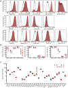
TIGIT can be knocked out in ex vivo expanded NK cells. Representative histograms from one donor demonstrate TIGIT KO (red) PM21-NK cells (A) or FC-NK cells (B) have minimal TIGIT expression compared with WT (black, gray fill) exNK cells. Isotype or unstained control are shown in gray. (C) Summary data from multiple donors (n=6–15 donors, each average of duplicates) show TIGIT KO efficiency in PM21-NK cells (circles) and FC-NK cells (triangles). (D) An example graph of NK-cell fold-change over time for one donor comparing expansion-matched, donor-matched non-electroporated PM21-NK cells (gray squares), PM21-NK cells electroporated with Cas9 only (WT, black circles), or TIGIT-specific Cas9 RNP (KO, red triangles). (E) There was no significant difference in fold-expansion of TIGIT KO (red) and WT exNK cells (black) on day 14 of culture for PM21-based expansion (circles) (n=12 donors) or for FC-based expansion (triangles) (n=3 donors). (F) Similarly, there was no difference in NK-cell number on day 14 between donor and postelectroporation seeding density-matched WT (black triangles) and TIGIT KO (red triangles) FC-NK cells (n=6 donors). Data are presented as histograms or scatter dot plots with mean and error bars representing SD. Statistical significance was determined by multiple paired t-tests. ExNK, ex vivo expanded NK; KO, knockout; NK, natural killer; TIGIT, T-cell Immunoreceptor with Ig and ITIM domains; WT, wild-type.
Figure 2
Figure 2
TIGIT KO does not change exNK-cell phenotype based on donor-matched comparisons. (A) Representative flow cytometry histograms are shown overlaying isotype control (black line), WT PM21-NK cells (black outline, gray fill), and TIGIT KO PM21-NK cells (red outline, red fill). (B) TIGIT KO (red triangles) did not significantly change the expression of any NK-cell surface receptors tested compared with WT PM21-NK cells (black circles) (n=3 donors, in duplicate). (C) CyTOF median metal intensities for NK cell markers comparing WT (circles) and TIGIT KO (triangles) FC-NK cells, with (open symbol) and without (closed symbol) exposure to the SJGBM-2 tumor cell line (n=2 donors, with four experimental conditions for each, WT and TIGIT KO NK cells). TIGIT expression was significantly decreased in TIGIT KO NK cells across all conditions (p<0.05), while no significant change in intensity was observed across other markers tested. ExNK, ex vivo expanded NK; KO, knockout; NK, natural killer; TIGIT, T-cell Immunoreceptor with Ig and ITIM domains; WT, wild-type. *P<0.05.
Figure 3
Figure 3
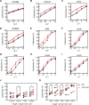
TIGIT KO enhances exNK-cell cytotoxicity against pediatric and lung cancer cell lines. The cytotoxicity of WT exNK cells and TIGIT KO exNK cells were determined against a panel of pediatric and lung cancer cell lines. Multiple E:T ratios were used to determine dose-dependent cytotoxicity curves. While no improvement in the killing of CHLA90 (A) was observed, TIGIT KO (red triangles) enhanced FC-NK-cell cytotoxicity against CHLA10 (B), RH30 (C), SJBM2 (D), and PM21-NK-cell cytotoxicity against A549 (E), H1299 (F), H358 (G), H1650 (H), and H1975 (I) compared with their respective WT exNK cells (black circles) (A–I are representative plots from a single donor). (J) Across multiple donors at a 1.25:1 E:T ratio, a significant increase in cytotoxicity was observed for CHLA10 and SJBM2, while RH30 trended toward increased cytotoxicity and no difference was seen for CHLA90 cells (n=6, each average of duplicates). (K) Cytotoxicity was also significantly increased for A549, H1299, H358, and H1650 (n=4 donors, average of triplicates, 1:1 E:T, t=48 h). ExNK, ex vivo expanded NK; KO, knockout; NK, natural killer; TIGIT, T-cell Immunoreceptor with Ig and ITIM domains; WT, wild-type. E:T, effector/target; KO, knockout; NK, natural killer; TIGIT, T-cell Immunoreceptor with Ig and ITIM domains; WT, wild-type. *P<0.05.
Figure 4
Figure 4
TIGIT KO enhances exNK cell cytotoxicity against tumor spheroids. TIGIT KO exNK-cell (PM21-NK cells) cytotoxicity against cancer spheroids was compared with WT exNK cells using kinetic live-cell imaging cytotoxicity assays. (A) Example images of untreated spheroids, with WT exNK cells, and with TIGIT KO exNK cells are shown for each cell line at 72 h post-NK-cell addition. (B) TIGIT KO exNK cells had increased cytotoxicity against all cell lines (n=3–4 donors, average of quadruplicates), with statistical significance reached for A549, H1650, and SK-N-AS. (C) TIGIT KO exNK cells had decreased t1/2 compared with WT exNK cells against all cell lines with statistical significance reached for all except H1650 (n=3–4 donors, average of quadruplicates). (D) EC50 was also decreased for TIGIT KO exNK cells compared with WT exNK cells against all cell lines, except those highly susceptible to NK cell killing, H1650 and H1975 (n=3–4 donors, average of quadruplicates). Each cancer spheroid had individual susceptibility to exNK cells, therefore the number of NK cells added, and time of analyses were used as follows: for cytotoxicity, 3333 exNK cells for A549, and H1650 while 10 000 exNK cells for H1975 and SK-N-AS, and 30 000 NK cells for H1299 and H358 spheroids. Dose-dependent cytotoxicity curves used to determine EC50 were determined at 72 h for H1299, H358, and H1975 and at 48 h for A549, H1650, and SK-N-AS. Data are presented as scatter dot plots with donor-pair lines. Statistical significance was determined by multiple paired t-tests. ExNK, ex vivo expanded NK; KO, knockout; NK, natural killer; TIGIT, T-cell Immunoreceptor with Ig and ITIM domains; WT, wild-type. *P<0.05, **p<0.01.
Figure 5
Figure 5
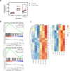
TIGIT KO exNK cells have increased basal glycolysis rate compared with WT exNK cells when exposed to K562-PVR+ cells. (A) WT exNK cells (PM21-NK cells) and TIGIT KO exNK cells were stimulated with either cytokines (IL-12/IL-15/IL-18) or PVR expressing K562 cells (K562-PVR+ cells) for 24 h and compared with unstimulated exNK cells for basal glycolysis levels (n=6 donors, in duplicate). K562-PVR+ cell stimulation significantly increased basal glycolytic rate in TIGIT KO exNK cells (red triangles) compared with WT exNK cells (black circle). Data are presented as scatter plots with donor-pair lines. Statistical significance was determined by multiple paired t-tests. RNA-sequencing analysis with Gene Set Enrichment Analysis confirmed the upregulation of mTORC1 signaling and glycolysis hallmark gene sets in TIGIT KO exNK cells compared with WT exNK cells. Enrichment plots (B) and heatmaps with dysregulated genes (false discovery rate (FDR)<0.1) (C) are shown for these gene sets. ExNK, ex vivo expanded NK; KO, knockout; NK, natural killer; TIGIT, T-cell Immunoreceptor with Ig and ITIM domains; WT, wild-type. *P<0.05.
Figure 6
Figure 6
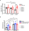
TIGIT KO prevents Fc-active anti-TIGIT driven exNK-cell fratricide and prevents the decrease in exNK-cell cytotoxicity when combined with Fc-active anti-TIGIT. A total of 10 000 WT exNK cells (PM21-NK cells) and TIGIT KO exNK cells were co-cultured with Fc-inactive anti-TIGIT or isotype control or Fc-active anti-TIGIT antibodies (10 μg/mL) for 24 h with unexposed exNK cells as a control. (A) Relative viable NK cells in antibody-exposed exNK-cell cultures were determined relative to unexposed exNK cells for each of the WT (black) and TIGIT KO (red) groups for 5 NK-cell donors, in triplicate (average for each donor represented as circles, squares, diamond, hexagon, or triangles in each group). Fc-active anti-TIGIT induced significant fratricide against exNK cells but not against TIGIT KO exNK cells. WT exNK cells and TIGIT KO exNK cells were co-cultured with A549 spheroids in the presence of Fc-inactive or Fc-active anti-TIGIT and cytotoxicity was determined. (B) WT exNK cells showed significantly lower cytotoxicity against A549 spheroids in the presence of Fc-active (red) compared with Fc-inactive anti-TIGIT (blue), whereas TIGIT KO exNK cells alone had increased cytotoxicity compared with WT exNK cells (black), and there was no significant change in cytotoxicity in presence of Fc-inactive or Fc-active anti-TIGIT (n=3 donors, each in triplicate and averages represented as circles, squares, or triangles in each group). Data are presented as scatter dot plots with error bars representing SD. Statistical significance was determined by nested one-way analysis of variance with multiple comparisons and Fisher’s Least Significant Difference (LSD) test post-hoc analysis. *P<0.05, **p<0.01, ****p<0.0001.

References

    1. Chiang EY, Mellman I. TIGIT-CD226-PVR axis: advancing immune checkpoint blockade for cancer immunotherapy. J Immunother Cancer 2022;10:e004711. 10.1136/jitc-2022-004711 - DOI - PMC - PubMed
    1. Ge Z, Peppelenbosch MP, Sprengers D, et al. . TIGIT, the next step towards successful combination immune checkpoint therapy in cancer. Front Immunol 2021;12:1–13. 10.3389/fimmu.2021.699895 - DOI - PMC - PubMed
    1. Sanchez-Correa B, Valhondo I, Hassouneh F, et al. . DNAM-1 and the TIGIT/PVRIG/TACTILE axis: novel immune checkpoints for natural killer cell-based cancer immunotherapy. Cancers (Basel) 2019;11:877. 10.3390/cancers11060877 - DOI - PMC - PubMed
    1. Manieri NA, Chiang EY, Grogan JL. TIGIT: a key inhibitor of the cancer immunity cycle. Trends Immunol 2017;38:20–8. 10.1016/j.it.2016.10.002 - DOI - PubMed
    1. Zhang Q, Bi J, Zheng X, et al. . Blockade of the checkpoint receptor TIGIT prevents NK cell exhaustion and elicits potent anti-tumor immunity. Nat Immunol 2018;19:723–32. 10.1038/s41590-018-0132-0 - DOI - PubMed

Publication types

Substances