. 2023 Dec 8;13(1):21916.
doi: 10.1038/s41598-023-49136-8.

Gankyrin inhibits ferroptosis through the p53/SLC7A11/GPX4 axis in triple-negative breast cancer cells

Affiliations

Gankyrin inhibits ferroptosis through the p53/SLC7A11/GPX4 axis in triple-negative breast cancer cells

Ming Lei et al. Sci Rep. .

Abstract

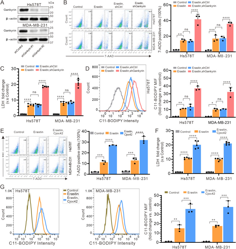
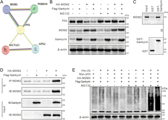
Gankyrin is found in high levels in triple-negative breast cancer (TNBC) and has been established to form a complex with the E3 ubiquitin ligase MDM2 and p53, resulting in the degradation of p53 in hepatocarcinoma cells. Therefore, this study sought to determine whether gankyrin could inhibit ferroptosis through this mechanism in TNBC cells. The expression of gankyrin was investigated in relation to the prognosis of TNBC using bioinformatics. Co-immunoprecipitation and GST pull-down assays were then conducted to determine the presence of a gankyrin and MDM2 complex. RT-qPCR and immunoblotting were used to examine molecules related to ferroptosis, such as gankyrin, p53, MDM2, SLC7A11, and GPX4. Additionally, cell death was evaluated using flow cytometry detection of 7-AAD and a lactate dehydrogenase release assay, as well as lipid peroxide C11-BODIPY. Results showed that the expression of gankyrin is significantly higher in TNBC tissues and cell lines, and is associated with a poor prognosis for patients. Subsequent studies revealed that inhibiting gankyrin activity triggered ferroptosis in TNBC cells. Additionally, silencing gankyrin caused an increase in the expression of the p53 protein, without altering its mRNA expression. Co-immunoprecipitation and GST pull-down experiments indicated that gankyrin and MDM2 form a complex. In mouse embryonic fibroblasts lacking both MDM2 and p53, this gankyrin/MDM2 complex was observed to ubiquitinate p53, thus raising the expression of molecules inhibited by ferroptosis, such as SLC7A11 and GPX4. Furthermore, silencing gankyrin in TNBC cells disrupted the formation of the gankyrin/MDM2 complex, hindered the degradation of p53, increased SLC7A11 expression, impeded cysteine uptake, and decreased GPX4 production. Our findings suggest that TNBC cells are able to prevent cell ferroptosis through the gankyrin/p53/SLC7A11/GPX4 signaling pathway, indicating that gankyrin may be a useful biomarker for predicting TNBC prognosis or a potential therapeutic target.


Conflict of interest statement

The authors declare no competing interests.

Figures

Figure 1
Figure 1
The expression of gankyrin is up-regulated in TNBC tissues and cells, and is negatively correlated with the patient’s prognosis. (A) Analysis of the GEPIA database indicates significantly higher levels of gankyrin in TNBC tumors compared to normal tissues. (B) The mRNA expression of gankyrin in various TNBC cell lines, including MCF-10A, MDA-MB-231, HCC-1937, MDA-MB-468, BT20, and Hs578T. (C) Protein mapping data highlights the differential expression of gankyrin in TNBC (tumor) and normal tissues. (D) The expression of gankyrin protein in a normal breast cell line and multiple TNBC cell lines as described in (B). (E) The GEPIA database demonstrates a contrast in overall survival rates between TNBC patients with high and low gankyrin expression. The data presented represents the mean ± standard deviation of three independent replicates and was analyzed using one-way univariate analysis of variance with multiple comparisons. Statistical significance was determined as *** < 0.001 and **** < 0.0001.
Figure 2
Figure 2
Overexpression of Gankyrin suppresses ferroptosis in TNBC cells. (A) Gankyrin protein expression in Hs578T and MDA-MB-231 cells transfected with shControl, shGankyrin #1, and shGankyrin #2 vectors, respectively. (B) Flow cytometry analysis of the proportion of 7-AAD-positive cells in Hs578T and MDA-MB-231 cells. (C) Fold changes in lactate dehydrogenase (LDH) release compared to the control. (D) Flow cytometry detection of lipid peroxides C11-BODIPY. (EG) LDH release (E), proportion of 7-AAD-positive cells (F), and lipid peroxides C11-BODIPY (G) in Hs578T and MDA-MB-231 cells after gankyrin inhibition by cjoc42. Data represent the means ± standard deviations of three independent replicates and were analyzed using one-way univariate analysis of variance with multiple comparisons. Statistical significance was denoted as *** < 0.001 and **** < 0.0001.
Figure 3
Figure 3
Effect of gankyrin on p53 protein expression in TNBC cells. (A) Levels of p53 protein expression in Hs578T and MDA-MB-231 cells after transfection with the designated siRNA interfering vectors. (B) Fluorescence intensity of p53 in Hs578T and MDA-MB-231 cells detected by Flow cytometry. (C) Expression of TP53 mRNA in Hs578T and MDA-MB-231 cells was detected by conventional RT-PCR. (D) Expression of TP53 mRNA in Hs578T and MDA-MB-231 cells was quantified by RT-qPCR. The results are presented as the mean ± standard deviation of three independent replicates and were analyzed using a one-way univariate analysis of variance with multiple comparisons. Statistical significance was determined as follows: ** < 0.01, *** < 0.001, **** < 0.0001, and ns indicating no statistical difference.
Figure 4
Figure 4
Involvement of p53 expression in the inhibition of ferroptosis. Hs578T and MDA-MB-231 cells treated with erastin were transfected with the shCtrl empty or shGankyrin expression vector for 24 h, followed by a 2-h incubation with or without the p53 inhibitor PFT-α (15 μM). (A) Fold changes in LDH release (vs. the shCtrl) were measured. (B) Proportion of 7-AAD positive cells in each group was analyzed by flow cytometry. (C) Lipid oxidates C11-BODIPY were analyzed by flow cytometry. The data presented represent the mean ± standard deviation of three independent replicates and were analyzed using one-way univariate analysis of variance with multiple comparisons. Statistical significance was determined as follows: ** < 0.01, *** < 0.001, **** < 0.0001, and “ns” indicating no statistical difference.
Figure 5
Figure 5
Gankyrin and MDM2 form a complex to promote ubiquitin-mediated degradation of p53. (A) STRING analysis reveals significant interactions among gankyrin, MDM2, p53, SLC7A11, and GPX4 proteins. (B) Expression levels of p53, MDM2, and gankyrin were assessed in HEK293T cells with a MDM2 knockout, transfected with MDM2 and gankyrin plasmids. (C) GST pull-down assay demonstrates the binding of GST-gankyrin to 35S-labeled MDM2 protein. (D) Immunoblotting using anti-gankyrin and anti-MDM2 antibodies confirms the interaction between gankyrin and MDM2 in the MDM2 knockout HEK293T cells. (E) Changes in p53 ubiquitination levels were measured by transfecting different combinations of Ub, p53, MDM2, and gankyrin plasmids in MDM2 and p53 double knockout MEF cells.
Figure 6
Figure 6
Role of PSMD10 in inhibiting ferroptosis in TNBC cells through the p53/SLC7A11/GPX4 pathway. (A) Protein expression of p53, SLC7A11, GPX4, MDM2, and gankyrin were examined in HEK293T cells expressing MDM2 with a HA-MDM2 vector. (B) Ubiquitination level of p53 (left panel) and the protein expression levels of p53, SLC7A11, GPX4, and gankyrin were assessed in Hs578T cells (right panel). (C) Hs578T and MDA-MB-231 cells were transfected with shGankyrin or shCtrl, along with a HA-MDM2 plasmid transfection, and the relative RNA expression levels of TP53 and SLC7A11 were measured. (D) Fold change in cystine uptake level compared to the shCtrl was determined. (E) Relative RNA expression level of GPX4 compared to the shCtrl was examined by RT-qPCR. (F) Fold change in LDH release compared to the control was measured. (G and H) Percentage of 7-AAD-positive dead cells (G) and the fold change in lipid peroxide C11-BODIPY compared to the control (H) were analyzed by flow cytometry. The data represent the mean ± SD from three independent experiments and were analyzed using two-way ANOVA followed by Tukey’s post-hoc multiple comparison analysis. Statistical significance is denoted as * < 0.05, ** < 0.01, *** < 0.001, **** < 0.0001. NS indicates no significance.

References

    1. Ding Y, et al. Identification of a small molecule as inducer of ferroptosis and apoptosis through ubiquitination of GPX4 in triple negative breast cancer cells. J. Hematol. Oncol. 2021;14:19. doi: 10.1186/s13045-020-01016-8. - DOI - PMC - PubMed
    1. Agostinetto E, et al. Progress and pitfalls in the use of immunotherapy for patients with triple negative breast cancer. Expert Opin. Investig. Drugs. 2022;31:567–591. doi: 10.1080/13543784.2022.2049232. - DOI - PubMed
    1. Agostinetto E, Eiger D, Punie K, de Azambuja E. Emerging therapeutics for patients with triple-negative breast cancer. Curr. Oncol. Rep. 2021;23:57. doi: 10.1007/s11912-021-01038-6. - DOI - PubMed
    1. Bergin, A. R. T. & Loi, S. Triple-negative breast cancer: Recent treatment advances. F1000Res10.12688/f1000research.18888.1 (2019). - PMC - PubMed
    1. Zou Y, et al. N6-methyladenosine regulated FGFR4 attenuates ferroptotic cell death in recalcitrant HER2-positive breast cancer. Nat. Commun. 2022;13:2672. doi: 10.1038/s41467-022-30217-7. - DOI - PMC - PubMed

MeSH terms

Substances