Review
. 2024 Apr;25(4):309-332.
doi: 10.1038/s41580-023-00683-y. Epub 2023 Dec 11.

Energy-driven genome regulation by ATP-dependent chromatin remodellers

Affiliations
Review

Energy-driven genome regulation by ATP-dependent chromatin remodellers

Sebastian Eustermann et al. Nat Rev Mol Cell Biol. 2024 Apr.

Abstract

The packaging of DNA into chromatin in eukaryotes regulates gene transcription, DNA replication and DNA repair. ATP-dependent chromatin remodelling enzymes (re)arrange nucleosomes at the first level of chromatin organization. Their Snf2-type motor ATPases alter histone-DNA interactions through a common DNA translocation mechanism. Whether remodeller activities mainly catalyse nucleosome dynamics or accurately co-determine nucleosome organization remained unclear. In this Review, we discuss the emerging mechanisms of chromatin remodelling: dynamic remodeller architectures and their interactions, the inner workings of the ATPase cycle, allosteric regulation and pathological dysregulation. Recent mechanistic insights argue for a decisive role of remodellers in the energy-driven self-organization of chromatin, which enables both stability and plasticity of genome regulation - for example, during development and stress. Different remodellers, such as members of the SWI/SNF, ISWI, CHD and INO80 families, process (epi)genetic information through specific mechanisms into distinct functional outputs. Combinatorial assembly of remodellers and their interplay with histone modifications, histone variants, DNA sequence or DNA-bound transcription factors regulate nucleosome mobilization or eviction or histone exchange. Such input-output relationships determine specific nucleosome positions and compositions with distinct DNA accessibilities and mediate differential genome regulation. Finally, remodeller genes are often mutated in diseases characterized by genome dysregulation, notably in cancer, and we discuss their physiological relevance.


Conflict of interest statement

Conflict of interest

The authors declare no competing interests.

Figures

Figure 1.
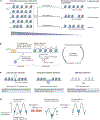
Figure 1.. Active chromatin self-organization and genome regulation.
An active interplay between chromatin organization and genome regulation establishes cellular stability and plasticity. (a) Energy-driven molecular machinery shapes the multi-level self-organisation of chromatin within the nucleus of a eukaryotic cell. At the first level of organization, ATP-dependent chromatin remodellers organize the positioning and composition of nucleosomes along the DNA. ATP-driven chromatin self-organization on this level also includes the activities of DNA topoisomerase 2 (TOP2), as well as indirectly DNA methyltransferases (not shown), and histone post-translational modifiers such as acetyl-CoA-dependent histone acetyl transferases (HAT), as the precursor metabolites used by these enzymes are synthesized in ATP-dependent reactions. ATP-dependent chromatin-loop extrusion by cohesin and related complexes, and possibly liquid–liquid phase separation (LLPS) can contribute to higher levels of chromatin organization. The resulting chromatin regulates transcription, DNA replication and DNA repair. On the right, a stereotypical organisation of a genic nucleosome array (+1, +2, … nucleosomes) downstream of a nucleosome depleted promoter is shown as a simplified example of how the first level of chromatin regulates transcription by RNA polymerase II (Pol II). (b) Schematic illustrating how remodellers couple the chemical energy of ATP hydrolysis to mechanical movements of their two motor ATPase lobes (red) relative to each other. If the motor ATPase of a remodeller is bound to a nucleosomal substrate, such movements can result in DNA translocation and lead, for example, to repositioning (sliding) of nucleosomes so that a transcription factor binding site (yellow) is wrapped around the histone octamer and is not accessible to its cognate transcription factor (TF). (c) Schematics illustrating some aspects of the composition and information content of the first level of chromatin organization, either (left) of a nucleosome core particle (NCP; PDB 1KX5), or (right) of oligo-nucleosomes (PDB 7PET) including linker histones and non-histone factors. Remodellers have key roles in determining the positioning and composition of nucleosomes, which in turn regulates access of transcription factors to their DNA binding motifs (in yellow) or interactions with other factors. Determination of nucleosome positions amounts to determining the length of linker DNA, which directly influences the 3D orientation of NCPs relative to each other and likely also higher order chromatin-fiber packing.
Figure 2.
Figure 2.. Nucleosome organization by remodellers.
a) Different remodellers (remodeller Amut represents a dysregulated disease version of remodeller A) generate a subset of specific nucleosome organizations that correspond to functional states (right) from all possible nucleosome organizations (left). This lowers the entropy of the system. Different nucleosome organizations are symbolized by different nucleosome positions, but may also differ otherwise, e.g. in histone modifications or variants. b) Left: At the 5’ ends of active genes, cooperation of different remodeller-specific activities generates a stereotypic nucleosome organization that is a prerequisite for transcription (Animation 1). The RSC or SWI/SNF remodeller disassembles nucleosomes from the promoter region, and INO80, ISW1a or ISW2 set the position of the first (+1) nucleosome, to which a nucleosome array at the gene body (+2, +3 … nucleosomes) is phased. The +1 nucleosome position is determined either by INO80 according to sequence features (not shown), or by INO80, ISW1a or ISW2 in relation to a DNA-binding barrier factor. Regular spacing in the genic array is generated mainly by ISW1a or Chd1. The remodeller SWR1 exchanges H2A–H2B dimers with H2A.Z–H2B variant dimers, especially at the +1 nucleosome. c) Different remodellers are involved in organizing nucleosomes at the indicated functional genomic sites, so that various machineries can function at these sites. Some yeast remodellers known to function at DNA damage repair sites are listed, whereas in human cells the homologous remodellers (e.g., BAF, PBAF; Supplementary Table 1), but also remodellers without yeast counterpart, such as ALC1, are involved in DNA repair. The resulting nucleosome organization at repair sites is not clear yet (stippled nucleosomes). d) Putative energy diagram for nucleosome remodelling. Left: the exemplary nucleosome organizations 1, 2 and 3 (as in panel a) in the absence of remodellers have similar free energy (G) levels, occur to similar degree at equilibrium (similar blue fill) and are kinetically trapped owing to high activation energy barriers (dashed double-headed arrow; ΔG). Middle: Binding and ATP-dependent remodelling (curved dashed arrows) by remodeller A (as in panel a) affects the energy landscape for all nucleosome organizations (turning 1, 2, 3 into 1*, 2*, 3*) and lowers the activation energy on the paths to nucleosome organization 2*, so that organization 2* becomes energetically more favorable (lower position along G axis) and more common (increased prevalence symbolized by increased blue fill). Right: Following remodelling and dissociation of remodeller A, initial energetics are restored, but organization 2 remains stably populated in a kinetic trap. Organization 2 could even correspond to a higher energy level relative to organizations 1 and 3 in the absence of the remodeller and still remain stable owing to the kinetic trap, as long as 2* is energetically favored during remodelling. CTCF: CCCTC binding factor; DSB: DNA double-strand break; ORC, origin recognition complex.
Figure 3.
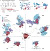
Figure 3.. Assemblies and architectures of chromatin remodellers.
a) (top) Phylogenetic tree of nucleoside 5′-triphosphate (NTP)-dependent helicases and translocases, focusing on Snf2-type ATPases. Marked in bold font in the third row from the top are Snf2-type motor ATPases of yeast chromatin remodellers. (bottom) Schematic representation of a Snf2-type motor ATPase domain with flanking regions and protein-specific structural elements. b) The modularity of multi-subunit remodeller assembly. The flanking regions of a Snf2-type motor ATPase (red) act as a scaffold for the modular assembly of additional scaffold subunits (blue) and regulatory subunits (green). Remodellers can contain different variants of subunits, which assemble in a combinatorial manner. In multi-cellular eukaryotes, these subunits are often expressed in tissue-specific and cell-type-specific manners. c) Different Snf2-type ATPases (listed in the inner circle) evolved from a common evolutionary ancestor (center; PDB 6IRO) and can be classified into the four main families (quadrants): INO80, ISWI, CHD and SWI/SNF. These ATPases assemble often into multi-subunit remodeller complexes, but some can also function as monomers (outer circle; Supplementary Table 1). Representative structures of remodellers determined to date are shown. Names of human remodellers are given, but if the homologous yeast remodeller structure is shown, the yeast name is given in parentheses. From top right going clockwise: INO80 of the fungus Chaetomium thermophilum (PDB: 8AV6, 8A5P), SWR1 from Saccharomyces cerevisiae (PDB 6GEN; the human complex is Snf2-related CBP activator (SRCAP)), Isw1 (ATPase only) from S. cerevisiae (PDB 6K1P), CHD4 (ATPase only) from Homo sapiens (PDB 6RYR), Chd1 from S. cerevisiae (PDB 7TN2), PBAF from H. sapiens (PDB 7VDV) and BAF from H. sapiens (PDB 6LTJ).
Figure 4.
Figure 4.. Nucleosome binding and DNA translocation mechanism of Snf2-type motor ATPases.
a) Top: Primary structural organization of Snf2-type motor ATPases. It features superfamily 2 (SF2)-conserved RecA1 and RecA2 core domains along with remodeller-specific protrusion 1 and protrusion 2 elements. RecA1 and protrusion 1 form the N-lobe, and RecA2 and protrusion 2 form the C-lobe. The C-terminal brace element forms a helix that spans across the lobes and couples the motions of the two lobes by connecting between protrusion 1 and protrusion 2. AutoN is an auto-regulatory element that can stabilize the inactive conformation by binding to protrusion 1 at the top of the N-lobe, and is homologous to the post-HSA domain of INO80 and SWI/SNF remodellers, which is in the C-terminal direction to the HAS domain (Supplementary Figure 1). Additional elements can include a long insertion loop (in the INO80 family). Bottom: A cryo-EM structure of yeast Isw1 motor ATPase in complex with a nucleosome core particle (NCP) (PDB 6IRO) is shown in surface representation (left, two orientations) and close-up cartoon representation (right). Super helical locations (SHLs) are numbered relative to the direction of DNA translocation: SHL0 is at the dyad, negative SHL numbers are towards the entry DNA and positive numbers are towards the exit DNA. The N-lobe of the motor domain sits between gyres and forms its primary contact with the minor groove at SHL-2 and secondary contacts at SHL+6, while the C-lobe makes only primary contacts with SHL-2. ATP-binding and hydrolysis takes place at the centrally located active site, between the two lobes, and alters the conformation of the ATPase domain.. b) Model for an ATP-driven conformational cycle of a Snf2-type ATPase, leading to directional translocation of or on DNA. Snf2-type ATPases can adopt an (allosterically) inhibited apo state in the absence of DNA substrate. DNA translocation proceeds through formation of a transient A-form DNA following ATP hydrolysis. ATP hydrolysis and release of inorganic phosphate (Pi) triggers a movement of the C-lobe leading to insertion of the gating helix into the minor groove and a 3’→5’ motion of the tracking strand. Binding of the next ATP molecule is proposed to switch the DNA back into B-form, in which the guide strand followed the tracking strand, resulting in a rotational DNA translocation of 1 bp. This rotational translocation can also lead to DNA overwinding and underwinding at the DNA entry and exit directions, respectively. Note that this step is not yet supported by structural data and consecutive cycles of ATP binding and hydrolysis might be required before DNA translocation occurs. ATP-hydrolysis can also trigger the release of DNA from the motor domain. A- and B-form DNA refers to the stretch of DNA bound by the motor ATPase lobes.
Figure 5.
Figure 5.. Structural mechanisms and principles of remodelling.
a) Current models of remodelling by an individual Snf2-type motor ATPase domain (left) and by a multi-subunit remodeller complex (right). Crystal structures of nucleosome core particles (NCPs) with different DNA sequences suggest that nucleosomes can accommodate an additional DNA base pair through a twist defect,,. Rotational DNA translocation by an individual motor ATPase domain is proposed to generate such twist defects in a directional manner and, thereby, catalyzes a sliding reaction through twist defect diffusion around the (histone) substrate. A motor–rotor–stator–grip mechanism was proposed for multi-subunit remodellers,. A grip or valve element is mechanically coupled to the motor ATPase domain through a stator element, thereby holding the motor in place and applying a rotational force onto the DNA rotor for consecutive rounds of ATP hydrolysis and translocation. The strain can be either propagated beyond the grip valve, e.g. through twist defect diffusion, resulting in nucleosome sliding, or it may accumulate or break histone–DNA contacts (red stars), which may result in histone exchange or ejection. The choice between such different remodelling outcomes and also between remodelling or non-productive DNA back-slippage may be allosterically regulated by nucleosomal and other features (FIG. 6b). b) Schematics of different motor and grip binding configurations of multi-subunit remodeller complexes (monomeric Chd1 and Isw1 without a grip element are shown for comparison). The motor and grip elements maintain a spacing of three DNA turns (three SHL steps) between them, even if binding to a hexasome. c) Motor–rotor–stator–grip mechanism of INO80 (PDB 6FML) and SWI/SNF (PDB 6UXW) remodellers and of the RNA polymerase II pre-initiation complex (Pol II PIC) (PDB 5OQM). The direction of rotational DNA translocation leads to DNA underwinding downstream of the Snf2-type motor ATPase (FIG. 4b), which is consistent with destabilization of histone–DNA contacts by remodellers and with promoter DNA opening in context of Pol II. Binding of the H2A–H2B acidic patch, at the entry or exit DNA, contributes to anchoring the grip valve on the nucleosome. d) Upon loss of one H2A–H2B dimer and hexasome formation, which may happen during the indicated processes, INO80 switches its binding configuration through a 145 degrees spin rotation and recognizes an exposed surface of the H3–H4 tetramer and the now-longer linker DNA,. e) Cryo-EM structures suggest that the yeast Snf2-type motor ATPase Mot1 (FIG. 3a) catalyzes the ejection of TATA binding protein (TBP) from DNA through a two-step mechanism similar to nucleosome remodelling. ATP hydrolysis triggers a conformational change of the motor ATPase, formation of a DNA strain and a pivot movement of the TBP-binding N-terminal HEAT repeat domain (NTD) of Mot1, which functions as grip and stator (NTD grip stator). Mot1 structural elements analogous to AutoN and gating helix are involved, too (AutoN-like protrusion coupling and gating helix insertion, respectively). TBP is dislodged from DNA and subsequently captured by a Mot1 hook. In a second step, hydrolysis of another ATP may be required to eject TBP-bound Mot1 from DNA. This mechanism may resemble other remodelling reactions that lack processive DNA translocation, such as the SWR1-catalyzed histone exchange.
Figure 6.
Figure 6.. Three stages of nucleosome remodelling and an updated hourglass model.
a) Hit-and-run model for remodeller activity, comprising the three stages of recruitment, remodelling and release. The indicated approximate duration of remodelling by the bound remodeller and 3D target search by the unbound remodeller were determined in yeast using live single molecule fluorescence imaging. A bound transcription factor may be involved in remodeller recruitment or may regulate remodelling activity. b) Updated hourglass model. Various chromatin remodellers (center) always contain an Snf2-type motor ATPase (red), which catalyzes the actual DNA translocation reaction, and may vary through combinatorial assembly (FIG. 3b). The remodellers may recognise various chromatin features (top) comprising genetic (DNA sequence) or epigenetic (all other) features, which may lead to context-dependent regulation of remodeller recruitment, remodelling activity and release (panel a). These genetic and epigenetic features of chromatin are integrated (information processing) through remodeller-specific mechanisms to affect the allosteric regulation of ATP-dependent DNA translocation and its remodelling outcome (bottom). Nucleosome sliding refers to the mobilization of histone octamers along the DNA in general, whereas nucleosome positioning leads to particular histone octamer positioning relative to the DNA sequence (vertical stippled line). Nucleosome spacing activity equalizes and sets the lengths of linkers in the nucleosome arrays, not necessarily in relation to the DNA sequence. Remodellers may catalyze the exchange of histones (dimeric blocks) with histone variants of with (un)modified histones. Losing an H2A–H2B dimer results in hexasome formation, which may be a byproduct of histone exchange or of transcription or other processes through nucleosomes (not shown). Partial histone loss also results from the collision or close stacking of nucleosomes, which may be generated or resolved by some remodellers. Only remodellers of the SWI/SNF family are able to completely disassemble nucleosomes, whereas ISWI family and CHD family remodellers support nucleosome assembly together with the deposition of histones on DNA by histone chaperones. This depiction of the hourglass model shows the combined input–output relationships of all remodellers; an individual remodeller will only enact a subset of these. PTMs, post-translational modifications.
Figure 7.
Figure 7.. Examples of input–output relationships provided by remodellers.
a) The sliding and disassembly activities by the yeast RSC complex (PDB 6TDA) are regulated through poly(dA:dT) sequences and its recruitment to acetylated histones. b) Upon sensing DNA lesions, poly(ADP-ribose) polymerase 1 (PARP1) is allosterically activated and modifies its target proteins, including histones, with poly(ADP-ribose) (PAR) chains. The autoinhibition of the ALC1 remodeller (PDB 7ENN) is allosterically relieved by binding poly(ADP-ribose) through its macrodomain, leading to nucleosome sliding. c) Yeast INO80 (PDBs 6FML, 5NBN, 8AV6, 8A5P) translates DNA mechanics features such as helix flexibility into forming stable nucleosome positions. The core module and the actin related protein (Arp) module of the complex recognize regions of intrinsic DNA flexibility or rigidity, respectively. Recognition of such features can act synergistically with the effects of barrier factors such as Reb1, which may allosterically inhibit the sliding activity. d) Yeast ISW1a (PDB 7X3T) binds a di-nucleosome so that its motor ATPase domain is on the mobile nucleosome and its ruler domain is on the neighboring, static nucleosome. The interactions of the ruler with the static nucleosome and with the motor ATPase domain may regulate nucleosome sliding so that linker DNA length is adjusted..Motor ATPases are in red.
Figure 8.
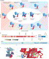
Figure 8.. Cancer mutations affect chromatin remodellers in various ways.
a) Shown in the center are the first two stages of nucleosome remodelling as depicted in FIG. 6a, but the nucleosome recruitment step is subdivided into nucleosome selection and closer engagement with nucleosomal epitopes. These stages pertain to all remodellers, but are depicted here for the human SWI/SNF-family remodeller BAF (PDB 6LTJ). Mechanistically, different mutations in BAF subunits, numbered 1–8, affect either nucleosome selection, nucleosome engagement or nucleosome remodelling. Presented from top left in clockwise direction are the four major types of mutations: missense and in-frame, abundance (remodeller expression levels), nonsense or frameshift, and gene fusion (through chromosomal translocation). Mutations 1–8 are similarly labelled in panels b & c. b) Schematics of three human BAF complex subunits with relevant domains and cancer mutations marked. Per-residue mutation frequencies (data obtained from the COSMIC database) for missense and in-frame mutations are indicated above the domain schematics and colored according to a white-gold-black heat scale; nonsense or frameshift mutation frequencies are indicated below the domain schematics in white-green-black heat scale. Mutation frequency values are scaled from white to colored bars with indicated values between 0 and N, where N is a selected per-residue mutation count that allows highlighting of sites of high mutation frequency. For sites with mutation frequencies greater than N, black bars are used. c) Human PBAF structure (PDB 7VDV) with a zoom-in view of the ATPase motor domain, which highlights the sites of specific mutations in the 3 and 4 regions. The circled 3 shows how R885 contacts the DNA backbone of the nucleosome, indicating that mutations in this residue interfere with nucleosome binding. The circled 4 shows how R1192 contacts BeFx, a mimic of the ATP γ-phosphate, indicating that mutations in this residue interfere with ATP hydrolysis. ADP, adenosine diphosphate; ARID, AT-rich interaction domain; BeFx, beryllium fluoride; BrD, bromodomain; BRK, BRM and KIS; HSA, helicase-SANT-associated; PBAF, polybromo-associated BAF; QLQ, glutamine – leucine – glutamine; SnAC, Snf2 ATP coupling; SNF5, sucrose non-fermenting 5.

References

    1. Misteli T The Self-Organizing Genome: Principles of Genome Architecture and Function. Cell 183, 28–45, doi: 10.1016/j.cell.2020.09.014 (2020). - DOI - PMC - PubMed
    1. Davidson IF & Peters JM Genome folding through loop extrusion by SMC complexes. Nature reviews. Molecular cell biology 22, 445–464, doi: 10.1038/s41580-021-00349-7 (2021). - DOI - PubMed
    1. Luger K, Mader AW, Richmond RK, Sargent DF & Richmond TJ Crystal structure of the nucleosome core particle at 2.8 A resolution. Nature 389, 251–260, doi: 10.1038/38444 (1997). - DOI - PubMed
    1. Clapier CR, Iwasa J, Cairns BR & Peterson CL Mechanisms of action and regulation of ATP-dependent chromatin-remodelling complexes. Nature reviews. Molecular cell biology 18, 407–422, doi: 10.1038/nrm.2017.26 (2017). - DOI - PMC - PubMed
    1. Martire S & Banaszynski LA The roles of histone variants in fine-tuning chromatin organization and function. Nature reviews. Molecular cell biology 21, 522–541, doi: 10.1038/s41580-020-0262-8 (2020). - DOI - PMC - PubMed

LinkOut - more resources