. 2023 Dec 11;13(1):21997.
doi: 10.1038/s41598-023-48919-3.

An α-chain modification rivals the effect of fetal hemoglobin in retarding the rate of sickle cell fiber formation

Affiliations

An α-chain modification rivals the effect of fetal hemoglobin in retarding the rate of sickle cell fiber formation

Eli H Worth et al. Sci Rep. .

Abstract

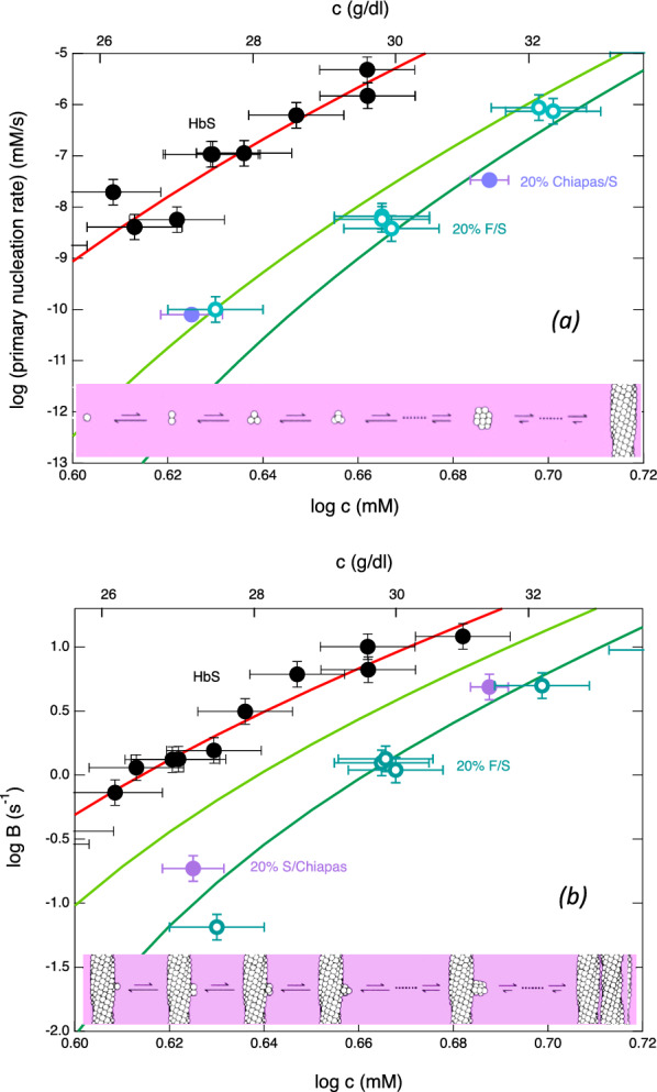
Adults with sickle cell disease bear a mutation in the β-globin gene, leading to the expression of sickle hemoglobin (HbS; α2βS2). Adults also possess the gene for γ-globin, which is a component of fetal hemoglobin (HbF, α2γ2); however, γ-chain expression normally ceases after birth. As HbF does not form the fibers that cause the disease, pharmacological and gene-modifying interventions have attempted to either reactivate expression of the γ chain or introduce a gene encoding a modified β chain having γ-like character. Here, we show that a single-site modification on the α chain, αPro114Arg, retards fiber formation as effectively as HbF. Because this addition to the repertoire of anti-sickling approaches acts independently of other modifications, it could be coupled with other therapies to significantly enhance their effectiveness.


Conflict of interest statement

The authors declare no competing interests.

Figures

Figure 1
Figure 1
Nucleation kinetics as a function of total Hb concentration. (a) Primary (homogeneous) nucleation rate. The top (solid black) points show pure HbS, while the open turquoise circles show 20% HbF mixed with 80% HbS. The filled violet points show 20% HbS Chiapas mixed with 80% HbS. Vertical error bars show the uncertainty to the fits of the distributions, which typically involve over 100 points and yield the rate. Horizontal error bars result from measuring small volumes. The solid red line shows the theory developed to describe nucleation; the lowest (dark green) line shows that theory adjusted for no copolymerization of hybrids (i.e., the effect of dilution alone). The middle line (light green) shows the theory if the hybrids have a probability of 10% to join the nucleus. (b) Exponential growth rate as a function of total Hb concentration. Secondary nucleation leads to exponential growth, so that the observed exponential factor (B) is dominated by that heterogeneous process. The symbols and lines are as in panel (a), except that the vertical error bars come from the fits to the exponential. Because the exponential factor B is related to the square root of the nucleation rate, its scale is smaller than the graph in panel (a).
Figure 2
Figure 2
Polymer structure. (a) The hemoglobin tetramer, containing 2 α chains (yellow and orange) and 2 β beta chains (cyan and blue). Tetramers form double strands (shown in panel (b)) that twist into polymers (panel c). In panel (c), each sphere represents a hemoglobin tetramer. The HbS β6V mutation docks into an adjacent β subunit pocket, forming lateral contacts, as shown by the yellow arrows. HbF lacks both donor and receptor; HbβT87Q lacks the donor and puts a bulky substitution in the receptor. α114 lies in an axial contact and is shown by the magenta circles. In a tetramer, residue 114 in either α chain packs against an adjacent molecule, so perturbing either inhibits effectively.
Figure 3
Figure 3
Coomassie-stained SDS-PAGE gel showing purified HbS Chiapas. Two closely spaced bands can be seen for the alpha and beta chains. The positions of molecular-weight markers are shown at left.

References

    1. Maitta RW, Reeves HM, Fontaine MJ. Editorial: Developments in sickle cell disease therapy and potentials for gene therapy. Front. Med. (Lausanne) 2023;10:1208192. doi: 10.3389/fmed.2023.1208192. - DOI - PMC - PubMed
    1. White SL, Hart K, Kohn DB. Diverse approaches to gene therapy of sickle cell disease. Annu. Rev. Med. 2023;74:473–487. doi: 10.1146/annurev-med-042921-021707. - DOI - PMC - PubMed
    1. Eaton WA, Hofrichter J. Sickle cell hemoglobin polymerization. Adv. Protein Chem. 1990;40:63–280. doi: 10.1016/S0065-3233(08)60287-9. - DOI - PubMed
    1. Mayuranathan T, et al. Potent and uniform fetal hemoglobin induction via base editing. Nat. Genet. 2023;55:1210–1220. doi: 10.1038/s41588-023-01434-7. - DOI - PMC - PubMed
    1. Kanter J, et al. Lovo-cel gene therapy for sickle cell disease: Treatment process evolution and outcomes in the initial groups of the HGB-206 study. Am. J. Hematol. 2023;98:11–22. doi: 10.1002/ajh.26741. - DOI - PMC - PubMed