CXCR4-directed PET/CT with [68 Ga]Ga-pentixafor in solid tumors-a comprehensive analysis of imaging findings and comparison with histopathology
- PMID: 38082196
- PMCID: PMC10957681
- DOI: 10.1007/s00259-023-06547-z
CXCR4-directed PET/CT with [68 Ga]Ga-pentixafor in solid tumors-a comprehensive analysis of imaging findings and comparison with histopathology
Abstract
Background: C-X-C motif chemokine receptor 4 (CXCR4) is overexpressed in various solid cancers and can be targeted by CXCR4-directed molecular imaging. We aimed to characterize the in-vivo CXCR4 expression in patients affected with solid tumors, along with a comparison to ex-vivo findings.
Methods: A total 142 patients with 23 different histologically proven solid tumors were imaged with CXCR4-directed PET/CT using [68 Ga]Ga-pentixafor (total number of scans, 152). A semi-quantitative analysis of the CXCR4-positive tumor burden including maximum standardized uptake values (SUVmax) and target-to-background ratios (TBR) using blood pool was conducted. In addition, we performed histopathological staining to determine the immuno-reactive score (IRS) from patients' tumor tissue and investigated possible correlations with SUVmax (by providing Spearman's rho ρ). Based on imaging, we also assessed the eligibility for CXCR4-targeted radioligand therapy or non-radioactive CXCR4 inhibitory treatment (defined as more than five CXCR4-avid target lesions [TL] with SUVmax above 10).
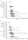
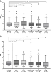
Results: One hundred three of 152 (67.8%) scans showed discernible uptake above blood pool (TBR > 1) in 462 lesions (52 primary tumors and 410 metastases). Median TBR was 4.4 (1.05-24.98), thereby indicating high image contrast. The highest SUVmax was observed in ovarian cancer, followed by small cell lung cancer, desmoplastic small round cell tumor, and adrenocortical carcinoma. When comparing radiotracer accumulation between primary tumors and metastases for the entire cohort, comparable SUVmax was recorded (P > 0.999), except for pulmonal findings (P = 0.013), indicative for uniform CXCR4 expression among TL. For higher IRS, a weak, but statistically significant correlation with increased SUVmax was observed (ρ = 0.328; P = 0.018). In 42/103 (40.8%) scans, more than five TL were recorded, with 12/42 (28.6%) exhibiting SUVmax above 10, suggesting eligibility for CXCR4-targeted treatment in this subcohort.
Conclusions: In a whole-body tumor read-out, a substantial portion of prevalent solid tumors demonstrated increased and uniform [68 Ga]Ga-pentixafor uptake, along with high image contrast. We also observed a respective link between in- and ex-vivo CXCR4 expression, suggesting high specificity of the PET agent. Last, a fraction of patients with [68 Ga]Ga-pentixafor-positive tumor burden were rendered potentially suitable for CXCR4-directed therapy.
Keywords: C-X-C motif chemokine receptor 4; CXCR4; PET/CT; Radioligand therapy; Solid tumors; Theranostics; [68 Ga]Ga-pentixafor.
© 2023. The Author(s).
Conflict of interest statement
RAW and AKB have received speaker honoraria from PentixaPharm and are involved in [68 Ga]Ga-pentixafor PET imaging in marginal zone lymphoma (LYMFOR). AKB is a member of the advisory board of PentixaPharm. All authors declare that there is no conflict of interest as well as consent for scientific analysis and publication.
Figures


References
MeSH terms
Substances
Grants and funding
LinkOut - more resources
Full Text Sources
Medical

