. 2023 Dec 11;24(1):309.
doi: 10.1186/s12931-023-02605-0.

Alpha-defensins inhibit ERK/STAT3 signaling during monocyte-macrophage differentiation and impede macrophage function

Affiliations

Alpha-defensins inhibit ERK/STAT3 signaling during monocyte-macrophage differentiation and impede macrophage function

Jungnam Lee et al. Respir Res. .

Abstract

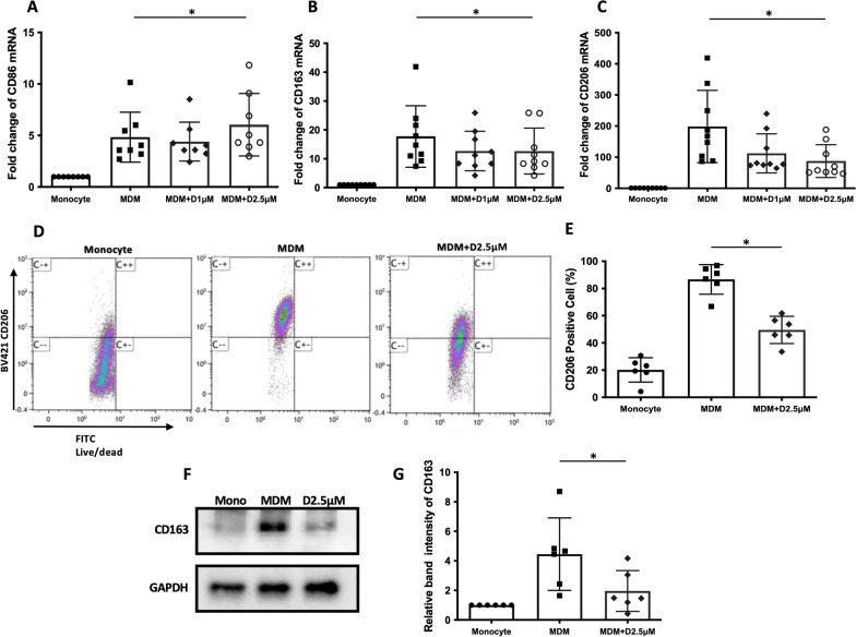
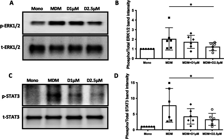
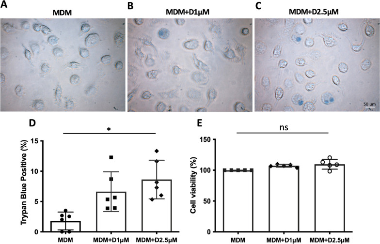
Alpha-1-antitrypsin deficiency (AATD) is a genetic disorder associated with a 5-tenfold decrease in lung levels of alpha-1-antitrypsin (AAT) and an increased risk for obstructive lung disease. α-defensins are cationic broad-spectrum cytotoxic and pro-inflammatory peptides found in the azurophilic granules of neutrophils. The concentration of α-defensins is less than 30 nM in the bronchoalveolar lavage fluid of healthy controls but is up to 6 μM in AATD individuals with significant lung function impairment. Alveolar macrophages are generally classified into pro-inflammatory (M1) or anti-inflammatory (M2) subsets that play distinct roles in the initiation and resolution of inflammation. Therefore, monocyte-macrophage differentiation should be tightly controlled to maintain lung integrity. In this study, we determined the effect of α-defensins on monocyte-macrophage differentiation and identified the molecular mechanism of this effect. The results of this study demonstrate that 2.5 μM of α-defensins inhibit the phosphorylation of ERK1/2 and STAT3 and suppress the expression of M2 macrophage markers, CD163 and CD206. In addition, a scratch assay shows that the high concentration of α-defensins inhibits cell movement by ~ 50%, and the phagocytosis assay using flow cytometry shows that α-defensins significantly reduce the bacterial phagocytosis rate of monocyte-derived macrophages (MDMs). To examine whether exogenous AAT is able to alleviate the inhibitory effect of α-defensins on macrophage function, we incubated MDMs with AAT prior to α-defensin treatment and demonstrate that AAT improves the migratory ability and phagocytic ability of MDMs compared with MDMs incubated only with α-defensins. Taken together, this study suggests that a high concentration of α-defensins inhibits the activation of ERK/STAT3 signaling, negatively regulates the expression of M2 macrophage markers, and impairs innate immune function of macrophages.

Keywords: AAT deficiency (AATD); Alpha-1-antitrysin (AAT); CD163; CD206; ERK1/2; STAT3; α-defensin.


Conflict of interest statement

The authors have declared that no conflict of interest exists.

Figures

Fig. 1
Fig. 1
The effect of α-defensins on macrophage differentiation. MDMs were differentiated using either M-CSF or GM-CSF and incubated with 2.5 µM of α-defensins for 16 h. AC The expression levels of M1 macrophage markers, CD64, CD80, and CD86, were compared among three different groups: monocyte, M-CSF-treated monocyte (M-CSF), and M-CSF and α-defensin-treated monocyte (M + D). Their relative expression is represented by fold change. DF The expression levels of M2 macrophage markers, CD163, CD204, and CD206, were also compared among the groups (monocyte, M-CSF, and M + D). GI The expression levels of M1 macrophage markers were compared among three different groups: monocyte, GM-CSF-treated monocyte (GM-CSF), and GM-CSF and α-defensin-treated monocyte (GM + D). Their relative expression is represented by fold change. JL The expression levels of M2 macrophage markers were also compared among the groups (monocyte, GM-CSF, and GM + D). Statistical analysis was conducted using Wilcoxon test. Statistical significance is denoted by (*) (p-value < 0.05)
Fig. 2
Fig. 2
The effect of α-defensins on M-CSF and GM-CSF-induced macrophages. MDMs were differentiated using both M-CSF and GM-CSF and incubated with two different concentrations of α-defensins, 1 µM and 2.5 µM, for 16 h. A The expression level of CD86 was compared between MDM controls and α-defensin-treated MDMs. B, C The expression levels of CD163 and CD206 were compared between the two MDM groups. The relative expression of the macrophage markers is represented by fold change. Statistical analysis was conducted using One-way ANOVA. Statistical significance is denoted by (*) (p-value < 0.05). The protein levels of the M2 macrophage markers were also examined in α-defensin-treated MDMs. D Cell surface distribution of CD206 was examined using flow cytometry. E The percentage of CD206-positive cells was compared between MDM controls and α-defensin-treated MDMs. F The protein level of CD163 was examined using a Western blot assay. G The protein band intensities were measured using NIH ImageJ software and compared between MDM controls and α-defensin-treated MDMs. Statistical analysis was conducted using Wilcoxon test. Statistical significance is denoted by (*) (p-value < 0.05)
Fig. 3
Fig. 3
The phosphorylation of ERK1/2 and STAT3 inhibited by α-defensins. Total proteins were isolated from monocytes, MDM controls, and α-defensin-treated MDMs. A Equal amounts of total proteins of the four different samples were analyzed via SDS-PAGE of phosphorylated ERK1/2 and total ERK1/2. C Equal amounts of total proteins of the four different samples were analyzed via SDS-PAGE of phosphorylated STAT3 and total STAT3. B and D The protein band intensities of the phosphorylated ERK1/2 and phosphorylated STAT3 were measured using NIH ImageJ software and compared among the samples. Statistical analysis was conducted using Wilcoxon test. Statistical significance is denoted by (*) (p-value < 0.05)
Fig. 4
Fig. 4
Cell viability of α-defensin-treated MDMs. MDMs were incubated with 1 µM or 2.5 µM of α-defensins for 16 h. AC MDM controls and α-defensin-treated MDMs were stained with 0.1% of trypan blue for three minutes, and the images of trypan blue-stained MDMs were captured using a light microscope. D The percentage of trypan blue-positive cells was calculated and compared between samples. Statistical analysis was conducted using One-way ANOVA. Statistical significance is denoted by (*) (p-value < 0.05). E Cell viability of each MDM sample was measured using an MTT assay. Statistical analysis was conducted using One-way ANOVA, and (ns) indicates no statistical difference among the samples
Fig. 5
Fig. 5
The effect of α-defensins on the migratory ability of MDMs. At day 7 of MDM differentiation, the scratch was performed. MDMs were incubated with or without α-defensins for 16 h. A The images of MDM controls and α-defensin-treated MDMs were taken using a light microscope; bar 200 µm. B The cells that moved into the scratched area were counted and compared among the three different MDM groups. C MDMs were incubated with 2.5 µM or 10 µM of AAT. After the incubation with AAT for one hour, MDMs were treated with 2.5 µM of α-defensins for 16 h. The images of the three different MDM groups (2.5 µM of α-defensin-treated MDMs, 2.5 µM of α-defensin and 2.5 µM of AAT-treated MDMs, and 2.5 µM of α-defensin and 10 µM of AAT-treated MDMs) were taken using a light microscope; bar 200 µm. D The cells that moved into the scratched area were counted and compared among the three different MDM groups. Statistical analysis was conducted using One-way ANOVA. Statistical significance is denoted by (*) (p-value < 0.05)
Fig. 6
Fig. 6
The bacterial phagocytosis of α-defensin-treated MDMs. MDMs were treated with 1 µM or 2.5 µM of α-defensins for 16 h and incubated with Heat-killed Staphylococcus aureus-conjugated with Alexa Fluor 488 at a multiplicity of infection (MOI) of 10 for one hour. A MDMs phagocytosing bacteria are visualized using a fluorescence microscope; bar 50 μm. Red and green indicate the host cell membrane and digested bacteria, respectively. B Green fluorescent intensity was normalized to the number of MDMs and compared among the different MDM groups. More than 10,000 cells, originating from six separate experiments, were evaluated for each MDM group. Statistical analysis was conducted using One-way ANOVA. Statistical significance is denoted by (*) (p-value < 0.05)
Fig. 7
Fig. 7
The phagocytic ability of α-defensin-treated MDMs improved by the AAT treatment. To examine the effect of exogenous AAT on α-defensin-induced injury to the MDM phagocytosis, six different samples were prepared, and without control (No phagocytosis), five samples were incubated with Heat-killed Staphylococcus aureus-conjugated with Alexa Fluor 488 at a multiplicity of infection (MOI) of 10 for one hour. The five different samples were monocytes, MDM controls, 2.5 µM of α-defensin-treated MDMs, 2.5 µM of α-defensin and 2.5 µM of AAT-treated MDMs, and 2.5 µM of α-defensin and 10 µM of AAT-treated MDMs. A The percentage of phagocytosis-positive cells was measured using flow cytometry. B To examine the effect of α-defensins on MDM phagocytosis, phagocytosis rates were compared among the four different MDM groups. The phagocytosis rate of MDM controls was set to 100%, and phagocytosis rates of the other MDM groups were normalized to the phagocytosis rate of MDM controls. Statistical analysis was conducted using One-way ANOVA. Statistical significance is denoted by (*) (p-value < 0.05)

References

    1. Ganz T, Selsted ME, Szklarek D, Harwig SS, Daher K, Bainton DF, et al. Defensins. Natural peptide antibiotics of human neutrophils. J Clin Invest. 1985;76(4):1427–1435. doi: 10.1172/JCI112120. - DOI - PMC - PubMed
    1. Ganz T. Defensins: antimicrobial peptides of innate immunity. Nat Rev Immunol. 2003;3(9):710–720. doi: 10.1038/nri1180. - DOI - PubMed
    1. Ganz T. Antimicrobial polypeptides. J Leukoc Biol. 2004;75(1):34–38. doi: 10.1189/jlb.0403150. - DOI - PubMed
    1. van Wetering S, Sterk PJ, Rabe KF, Hiemstra PS. Defensins: key players or bystanders in infection, injury, and repair in the lung? J Allergy Clin Immunol. 1999;104(6):1131–1138. doi: 10.1016/s0091-6749(99)70004-7. - DOI - PubMed
    1. Raj PA, Dentino AR. Current status of defensins and their role in innate and adaptive immunity. FEMS Microbiol Lett. 2002;206(1):9–18. doi: 10.1111/j.1574-6968.2002.tb10979.x. - DOI - PubMed

MeSH terms