. 2023 Dec 11;21(1):900.
doi: 10.1186/s12967-023-04613-6.

Bufalin targeting CAMKK2 inhibits the occurrence and development of intrahepatic cholangiocarcinoma through Wnt/β-catenin signal pathway

Affiliations

Bufalin targeting CAMKK2 inhibits the occurrence and development of intrahepatic cholangiocarcinoma through Wnt/β-catenin signal pathway

Huhu Zhang et al. J Transl Med. .

Abstract

Background: Intrahepatic cholangiocarcinoma (ICC) accounts for about 15% of primary liver cancer, and the incidence rate has been rising in recent years. Surgical resection is the best treatment for ICC, but the 5-year survival rate is less than 30%. ICC signature genes are crucial for the early diagnosis of ICC, so it is especially important to find its signature genes and therapeutic drug. Here, we studied that bufalin targeting CAMKK2 promotes mitochondrial dysfunction and inhibits the occurrence and metastasis of intrahepatic cholangiocarcinoma through Wnt/β-catenin signal pathway.

Methods: IC50 of bufalin in ICC cells was determined by CCK8 and invasive and migratory abilities were verified by wound healing, cell cloning, transwell and Western blot. IF and IHC verified the expression of CAMKK2 between ICC patients and normal subjects. BLI and pull-down demonstrated the binding ability of bufalin and CAMKK2. Bioinformatics predicted whether CAMKK2 was related to the Wnt/β-catenin pathway. SKL2001, an activator of β-catenin, verified whether bufalin acted through this pathway. In vitro and in vivo experiments verified whether overexpression of CAMKK2 affects the proliferative and migratory effects of ICC. Transmission electron microscopy verified mitochondrial integrity. Associated Ca2+ levels verified the biological effects of ANXA2 on ICC.

Results: It was found that bufalin inhibited the proliferation and migration of ICC, and CAMKK2 was highly expressed in ICC, and its high expression was positively correlated with poor prognosis.CAMKK2 is a direct target of bufalin, and is associated with the Wnt/β-catenin signaling pathway, which was dose-dependently decreased after bufalin treatment. In vitro and in vivo experiments verified that CAMKK2 overexpression promoted ICC proliferation and migration, and bufalin reversed this effect. CAMKK2 was associated with Ca2+, and changes in Ca2+ content induced changes in the protein content of ANXA2, which was dose-dependently decreasing in cytoplasmic ANXA2 and dose-dependently increasing in mitochondrial ANXA2 after bufalin treatment. In CAMKK2 overexpressing cells, ANXA2 was knocked down, and we found that reversal of CAMKK2 overexpression-induced enhancement of ICC proliferation and migration after siANXA2.

Conclusions: Our results suggest that bufalin targeting CAMKK2 promotes mitochondrial dysfunction and inhibits the proliferation and migration of intrahepatic cholangiocarcinoma through Wnt/β-catenin signal pathway. Thus, bufalin, as a drug, may also be used for cancer therapy in ICC in the future.

Keywords: ANXA2; Bufalin; CAMKK2; Ca2+; Intrahepatic cholangiocarcinoma; Migration; Mitochondrial dysfunction; Proliferation.


Conflict of interest statement

The authors declare that they have no competing interests.

Figures

Fig. 1
Fig. 1
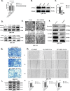
Bufalin inhibited ICC cell viability and metastasis. A-B CCK8 assay. In L02, HCCC-9810, RBE, and QBC-939 cells, bufalin resulted in a significant dose-dependent reduction in cell viability compared to controls. Bufalin had no significant effect on the activity of L02 cells. C Cell cloning of HCCC-9810, RBE, and QBC-939 cells exposed to bufalin at different concentrations and controls was analyzed. D Statistical analysis of cell proliferation capacity in cell cloning experiments. E Transwell was analyzed in HCCC-9810, RBE, and QBC-939 cells exposed to bufalin for different time or control. F Statistical analysis of the cell migration in the scratch wound healing assays. G Wound healing was analyzed in HCCC-9810, RBE, and QBC-939 cells exposed to bufalin for different time or control. H Statistical analysis of the cell migration in the scratch wound healing assays. I Western blot analysis of expression of metastasis related proteins N-cadherin and Vimentin in HCCC-9810, RBE, and QBC-939 cells exposed to bufalin or control. J The quantification of western blot. Protein levels were normalized to tubulin. All results were presented as the mean ± SD (n = 3). *p < 0.05, **p < 0.01
Fig. 2
Fig. 2
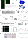
CAMKK2 is a direct interacting protein of bufalin and highly expressed in ICC. A Representative proteome microarrays results. Bufalin–protein interaction was detected (bule boxes) between bufalin and CAMKK2 by red fluorescence. Representative images of protein microarray showed positive control (blue arrow), negative control (yellow arrow) and CAMKK2 spot (red arrow) on the enlarged images. B Molecular docking predicted the potential binding sites of bufalin and CAMKK2. C Western blot analysis of CAMKK2 expression in HCCC-9810, RBE, and QBC-939 cells treated with different concentrations bufalin. D The quantification of western blot. Protein levels were normalized to tubulin. E Bufalin is labeled with biotin, pull down and verify that CAMKK2 is the target of bufalin. F CAMKK2 has stronger binding force than ATP1A1. G Pan-cancer analysis CAMMK2 was highly expressed in ICC, with red representing tumors and blue representing normal tissues. H Western blot analysis of CAMKK2 expression in HCCC-9810, RBE, L02 and QBC-939 cells. I The quantification of western blot (H). J The expression and cell localization of CAMKK2 in HCCC-9810, RBE, L02 and QBC-939 cells were analyzed by IF. K The expression level of CAMKK2 in tumor tissues and normal tissues. L mRNA expression levels of CAMKK2 in tumor tissue and normal tissue. M In the TCGA database, the top 6 signaling pathways related to CAMKK2 were enriched. All results were presented as the mean ± SD (n = 3). *p < 0.05, **p < 0.01
Fig. 3
Fig. 3
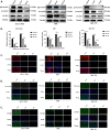
Bufalin inhibited Wnt/β-catenin signal pathway in ICC cells. A Western blot analysis of expression of proteins related to Wnt/β-catenin signal pathway in HCCC-9810, RBE and QBC-939 cells treated with bufalin or control. B The quantification of western blot. Protein levels were normalized to tubulin. C-E Cell localization and expression levels of β-catenin, CCND1 and p-β-catenin. All results were presented as the mean ± SD (n = 3). *p < 0.05, **p < 0.01
Fig. 4
Fig. 4
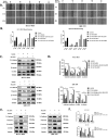
SKL2001 reversed the inhibitory effect of bufalin on the proliferation and migration of ICC. A In HCCC-9810 and QBC-939 cells, SKL2001 was activated after treatment with bufalin to detect the migration ability of the cells. After activation of SKL-2001, the cells were treated with bufalin to detect their ability to migrate. B above S indicates treatment with bufalin first, and S above B indicates treatment with advanced SKL-2001 activator. B Statistical analysis of (A) wound healing experiment. C In HCCC-9810 and QBC-939 cells, SKL2001 was activated after treatment with bufalin, and the expression levels of vimentin, N-cadherin, β-catenin and CCND1 were detected. D Statistical analysis of protein level of C. E The expression levels of vimentin, N-cadherin, β-catenin and p-β-catenin were detected in HCCC-9810 and QBC-939 cells after activation of SKL-2001 and treated with bufalin. F Statistical analysis of protein level of E. All results were presented as the mean ± SD (n = 3). *p < 0.05, **p < 0.01
Fig. 5
Fig. 5
Bufalin reversed the metastatic promotion induced by CAMKK2 in vitro. A Western blot analysis of expression of Flag-CAMKK2 protein in HCCC-9810 and QBC-939 cells overexpressing CAMKK2 or vector. B CCK8 assay in HCCC-9810 and QBC-939 cells overexpressing CAMKK2 exposed to bufalin or vector. C Would healing in HCCC-9810 and QBC-939 cells overexpressing CAMKK2 exposed to bufalin or vector. D Statistical analysis of the cell migration in the wound healing assays. E Transwell in HCCC-9810 and QBC-939 cells overexpressing CAMKK2 exposed to bufalin or vector. F Statistical analysis of the cell migration in Transwell. G Western blot analysis of expression of metastasis related proteins N-cadherin and Vimentin in HCCC-9810 and QBC-939 cells. H The quantification of western blot (F). Protein levels were normalized to tubulin. All results were presented as the mean ± SD (n = 3). * p < 0.05, **p < 0.01
Fig. 6
Fig. 6
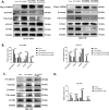
Bufalin inhibits CAMKK2-induced Wnt/β-catenin signaling pathway in vitro and vivo. A Western blot analysis of expression of proteins related to Wnt/β-catenin signal pathway in HCCC-9810 and QBC-939 cells overexpressing CAMKK2 or not, exposed to bufalin or not. B The quantification of western blot (A). Protein levels were normalized to tubulin. C Western blot analysis of expression of proteins related to vimentin, N-cadherin and Wnt/β-catenin signal pathway in solid tumors. D The quantification of western blot (C). Protein levels were normalized to tubulin. All results were presented as the mean ± SD (n = 3). * p < 0.05, **p < 0.01
Fig. 7
Fig. 7
Bufalin reversed tumor‑promoting effect induced by CAMKK2 in vivo. A Tumor-bearing mice and the dissected tumors were photographed and shown. B A ruler was used to indicate the size of the tumors. C Statistics and comparison of tumor volume in overexpression group, bufalin treatment group and vector. D HE staining was used to detect the tumors. E HE staining of main organs in nude mice. Representative HE staining photos of heart, liver, spleen, lung and kidney of each group of nude mice. Bufalin had no obvious side effects on heart, liver, spleen, lung and kidney of nude mice. Protein levels were normalized to tubulin. All results were presented as the mean ± SD (n = 3). * p < 0.05, **p < 0.01
Fig. 8
Fig. 8
Bufalin inhibits CAMKK2 by inhibiting ICC proliferation and migration via ANXA2 and promoting mitochondrial dysfunction. A Ca2+ detection showed that after overexpression of CAMKK2, Ca2+ content increased, and after treatment with bufalin, Ca2+ content decreased. B Western blot analysis of expression of metastasis related proteins ANXA2 in QBC-939 cells exposed to bufalin or control. C The quantification of western blot. Protein levels were normalized to tubulin. D Mitochondrial protein and cytoplasmic protein were isolated, and the expression of ANXA2 in mitochondria and cytoplasm was detected. E Mitochondrial morphological changes after overexpression of CAMKK2 and treatment with bufalin were detected by transmission electron microscopy. F Expression of Wnt/ β-catenin signaling pathway related proteins after ANXA2 is knocked down in QBC-939 cells. G-H After overexpression of CAMKK2 in QBC-939 cells, ANXA2 is knocked down and the cell migration ability changes. I After overexpression of CAMKK2, ANXA2 is knocked down and Wnt/ β-catenin signaling pathway related proteins are expressed in QBC-939 cells. J Statistical analysis of protein level of I. All results were presented as the mean ± SD (n = 3). ns: p ≥ 0.05, *p < 0.05, **p < 0.01
Fig. 9
Fig. 9
Bufalin targeting CAMKK2 affects ANXA2's inhibition of ICC occurrence and metastasis and promotion of mitochondrial dysfunction via the Wnt/ β-catenin signaling pathway. Bufalin down-regulates the expression of CAMKK2 in ICC, and then down-regulates ANXA2, reversing the activation of Wnt/β-catenin signaling pathway induced by CAMKK2/ANXA2, inhibiting the occurrence and metastasis of ICC, and promoting mitochondrial dysfunction. The figure is drawn in Figdraw software (www.figdraw.com)

References

    1. Shirvani H, Ghanavi J, Aliabadi A, Mousavinasab F, Talebi M, Majidpoor J, Najafi S, Miryounesi SM, Aghaei Zarch SM. MiR-211 plays a dual role in cancer development: from tumor suppressor to tumor enhancer. Cell Signal. 2023;101:110504. doi: 10.1016/j.cellsig.2022.110504. - DOI - PubMed
    1. Bahari Khasraghi L, Nouri M, Vazirzadeh M, Hashemipour N, Talebi M, Aghaei Zarch F, Majidpoor J, Kalhor K, Farnia P, Najafi S, Aghaei Zarch SM. MicroRNA-206 in human cancer: mechanistic and clinical perspectives. Cell Signal. 2023;101:110525. doi: 10.1016/j.cellsig.2022.110525. - DOI - PubMed
    1. Fattahi M, Shahrabi S, Saadatpour F, Rezaee D, Beyglu Z, Delavari S, Amrolahi A, Ahmadi S, Bagheri-Mohammadi S, Noori E, Majidpoor J, Nouri S, Aghaei-Zarch SM, Falahi S, Najafi S, Le BN. microRNA-382 as a tumor suppressor? Roles in tumorigenesis and clinical significance. Int J Biol Macromol. 2023;250:125863. doi: 10.1016/j.ijbiomac.2023.125863. - DOI - PubMed
    1. Faramin Lashkarian M, Hashemipour N, Niaraki N, Soghala S, Moradi A, Sarhangi S, Hatami M, Aghaei-Zarch F, Khosravifar M, Mohammadzadeh A, Najafi S, Majidpoor J, Farnia P, Aghaei-Zarch SM. MicroRNA-122 in human cancers: from mechanistic to clinical perspectives. Cancer Cell Int. 2023;23(1):29. doi: 10.1186/s12935-023-02868-z. - DOI - PMC - PubMed
    1. Moris D, Palta M, Kim C, Allen PJ, Morse MA, Lidsky ME. Advances in the treatment of intrahepatic cholangiocarcinoma: an overview of the current and future therapeutic landscape for clinicians. CA Cancer J Clin. 2023;73(2):198–222. doi: 10.3322/caac.21759. - DOI - PubMed

Publication types

MeSH terms