. 2024 Feb;115(2):385-400.
doi: 10.1111/cas.16038. Epub 2023 Dec 11.

Histone methyltransferase SUV420H1/KMT5B contributes to poor prognosis in hepatocellular carcinoma

Affiliations

Histone methyltransferase SUV420H1/KMT5B contributes to poor prognosis in hepatocellular carcinoma

Hirotaka Kato et al. Cancer Sci. 2024 Feb.

Abstract

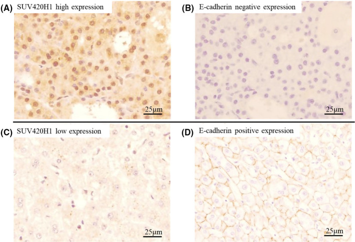
Hepatocellular carcinoma (HCC) has a high rate of recurrence and poor prognosis, even after curative surgery. Multikinase inhibitors have been applied for HCC patients, but their effect has been restricted. This study aims to clarify the clinical impact of SUV420H1/KMT5B, one of the methyltransferases for histone H4 at lysine 20, and elucidate the novel mechanisms of HCC progression. We retrospectively investigated SUV420H1 expression using HCC clinical tissue samples employing immunohistochemical analysis (n = 350). We then performed loss-of-function analysis of SUV420H1 with cell cycle analysis, migration assay, invasion assay and RNA sequence for Gene Ontology (GO) pathway analysis in vitro, and animal experiments with xenograft mice in vivo. The SUV420H1-high-score group (n = 154) had significantly poorer prognosis for both 5-year overall and 2-year/5-year disease-free survival than the SUV420H1-low-score group (n = 196) (p < 0.001 and p < 0.05, respectively). The SUV420H1-high-score group had pathologically larger tumor size, more tumors, poorer differentiation, and more positive vascular invasion than the SUV420H1-low-score group. Multivariate analysis demonstrated that SUV420H1 high score was the poorest independent factor for overall survival. SUV420H1 knockdown could suppress cell cycle from G1 to S phase and cell invasion. GO pathway analysis showed that SUV420H1 contributed to cell proliferation, cell invasion, and/or metastasis. Overexpression of SUV420H1 clinically contributed to poor prognosis in HCC, and the inhibition of SUV420H1 could repress tumor progression and invasion both in vitro and in vivo; thus, further analyses of SUV420H1 are necessary for the discovery of future molecularly targeted drugs.

Keywords: SUV420H1/KMT5B; hepatocellular carcinoma; methylation; methyltransferase.


Conflict of interest statement

Ryuji Hamamoto is an editorial board member of Cancer Science. Ryuji Hamamoto declares conflict of interest for the present article. The other authors declare no conflict of interest for the present article.

Figures

FIGURE 1
FIGURE 1
Immunohistochemical analysis using hepatocellular carcinoma (HCC) and corresponding background (non‐tumor) liver. Immunohistochemical classification based on the staining pattern according to immunoreactive scoring system in tumor and nontumor tissues.
FIGURE 2
FIGURE 2
Disease‐free and overall survival for hepatocellular carcinoma (HCC) between the SUV420H1‐high‐score and ‐low‐score groups. (A) Disease‐free survival curve in the SUV420H1‐high‐score and ‐low‐score groups. (B) Overall survival curve in the SUV420H1‐high‐score and ‐low‐score groups. p‐Values were calculated using the log‐rank test.
FIGURE 3
FIGURE 3
Cell progression analysis using small interfering RNA (siRNA) specific for SUV420H1 in Huh‐7. (A) The relative mRNA expression levels by quantitative real‐time polymerase chain reaction in siEGFP and siSUV420H1. (B, C) Immunocytochemistry and cell proliferation analysis in siEGFP and siSUV420H1. (D, E) Cell cycle analysis with flow cytometry and proportion of cell cycle phase in siEGFP and siSUV420H1. Results are presented as mean ± SEM. *p < 0.05, ***p < 0.001. All the experiments were done in triplicate.
FIGURE 4
FIGURE 4
Cell migration and cell invasion analysis using small interfering RNA (siRNA) specific for SUV420H1 in Huh‐7. (A, B) Gap area at 0, 12, and 24 h in wound‐healing assay using siEGFP and siSUV420H1. (C, D) Cell number ratio that invaded into membrane in cell invasion analysis using siEGFP and siSUV420H1. Results are presented as mean ± SEM. *p < 0.05, **p < 0.01. All the experiments were done in triplicate.
FIGURE 5
FIGURE 5
Pathways associated with SUV420H1 using Gene Ontology (GO) analysis. (A) GO terms involving SUV420H1 in Huh‐7 cell line that were derived neither from hepatitis B virus (HBV) nor C virus (HCV) (nBnC) hepatocellular carcinoma (HCC). GO enriched in siSUV420H1‐transfected cells, compared with siEGFP‐transfected cells, are indicated. (B) GO terms involving SUV420H1 in SNU475 cell line of HBV‐derived HCC. Gene ontologies enriched in siSUV420H1‐transfected cells, compared with siEGFP‐transfected cells, are indicated. (C) Common GO terms in Huh‐7 and SNU475 cell lines.
FIGURE 6
FIGURE 6
Immunohistochemistry with anti‐E‐cadherin antibody to evaluate the relationship between SUV420H1 and E‐cadherin using hepatocellular carcinoma (HCC)‐resected specimens. (A, B) The relationship between SUV420H1 high expression and E‐cadherin expression. (C, D) The relationship between SUV420H1 low expression and E‐cadherin expression.
FIGURE 7
FIGURE 7
In vivo examination employing xenograft hepatocellular carcinoma (HCC) tumors using CRISPR/Cas9 specific for SUV420H1 in Huh‐7. (A) Immunofluorescence for SUV420H1 and HDR genes cotransfected into Huh‐7 cell nucleus. (B) The relative mRNA expression level in CRISPR/Cas9 control and CRISPR/Cas9 SUV420H1 KO. (C) The changeover time of tumor progression in CRISPR/Cas9 control and CRISPR/Cas9 SUV420H1 KO. (D) Photographs of subcutaneous tumor and macroscopic specimens on day 28. Scale bars, 30 μm. DAPI, diamidino‐2‐phenylindole; GFP, green fluorescent protein; HDR, homology‐directed repair. Results are presented as mean ± SEM. *p < 0.05, ***p < 0.001. All the experiments were done in triplicate.

References

    1. Lee SC, Tan HT, Chung MC. Prognostic biomarkers for prediction of recurrence of hepatocellular carcinoma: current status and future prospects. World J Gastroenterol. 2014;20(12):3112‐3124. - PMC - PubMed
    1. Llovet JM, Ricci S, Mazzaferro V, et al. Sorafenib in advanced hepatocellular carcinoma. N Engl J Med. 2008;359(4):378‐390. - PubMed
    1. Cheng AL, Kang YK, Chen Z, et al. Efficacy and safety of sorafenib in patients in the Asia‐Pacific region with advanced hepatocellular carcinoma: a phase III randomised, double‐blind, placebo‐controlled trial. Lancet Oncol. 2009;10(1):25‐34. - PubMed
    1. Bruix J, Qin S, Merle P, et al. Regorafenib for patients with hepatocellular carcinoma who progressed on sorafenib treatment (RESORCE): a randomized, double‐blind, placebo‐controlled, phase 3 trials. Lancet. 2017;389(10064):56‐66. - PubMed
    1. Kudo M, Finn RS, Qin S, et al. Lenvatinib versus sorafenib in first‐line treatment of patients with unresectable hepatocellular carcinoma: a randomised phase 3 non‐inferiority trial. Lancet. 2018;391(10126):1163‐1173. - PubMed