

Characterizing close-focus lenses for microendoscopy
- PMID: 38084130
- PMCID: PMC10712292
- DOI: 10.1117/1.jom.3.1.011003
Characterizing close-focus lenses for microendoscopy
Abstract
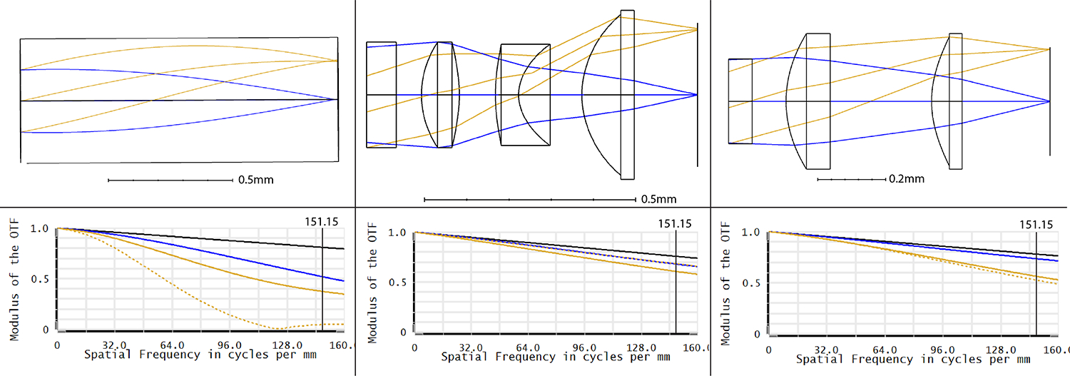
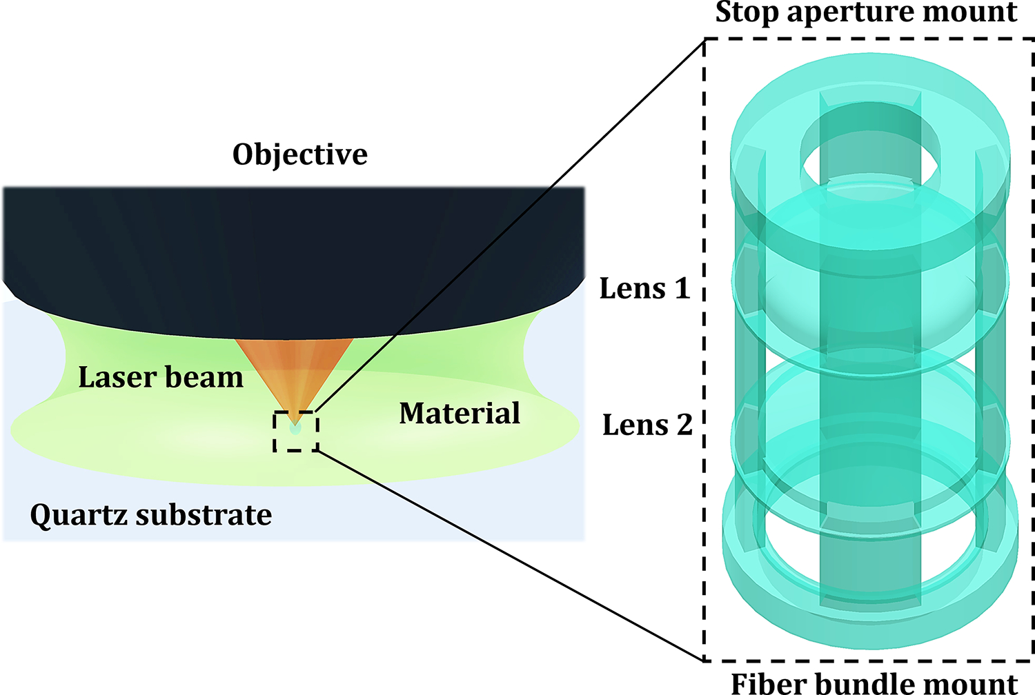
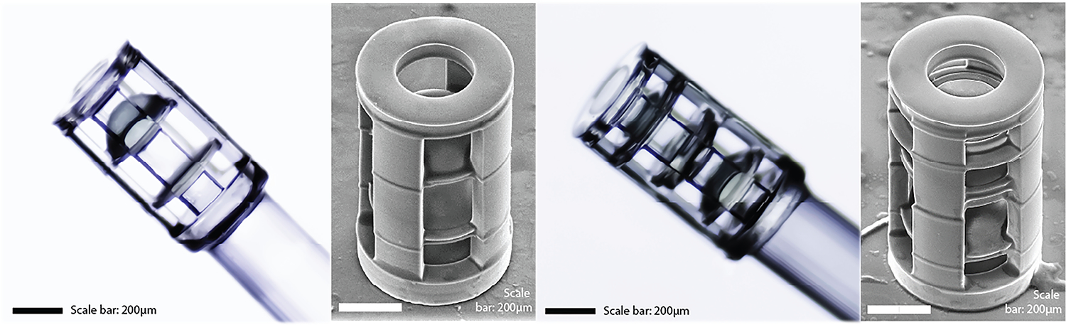
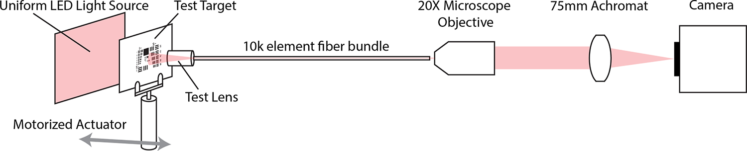
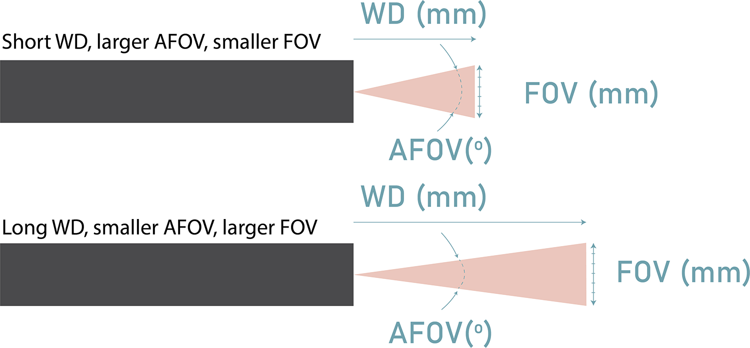
Microendoscopes are commonly used in small lumens in the body, for which a focus near to the distal tip and ability to operate in an aqueous environment are paramount for navigation and disease detection. Commercially available distal optic systems below 1mm in diameter are severely limited, and custom micro lenses are generally very expensive. Gradient index of refraction (GRIN) singlets are available in small diameters but have limited optical performance adjustability. Three-dimensional (3D) printed monolithic optical systems are an emerging option that may be suitable for enabling high performance, close-focus imaging. In this manuscript, we compared the optical performance of three custom distal optic systems; a custom-pitch GRIN singlet, 3D-printed monolithic doublet, and 3D-printed monolithic triplet, with a nominal working distance (WD) of 1.5mm, 0.5mm and 0.4mm in 0.9% saline. These short WDs are ideal for microendoscopy in collapsed or flushed lumens such as pancreatic duct or fallopian tube. The GRIN singlet had performance limited only by the fiber bundle relay over 0.9mm to 1.6 mm depth of field (DOF). The 3D printed doublet was able to achieve a comparable DOF of 0.71mm, while the 3D printed triplet suffered the most limited DOF of 0.55mm. 3D printing enables flexible design of monolithic multi-element systems with aspheric surfaces of very short WDs and relative ease of integration.
Keywords: 3D printing; Endoscopy; Lens design; microendoscope; multi-modal imaging.
Conflict of interest statement
Disclosures The authors declare that there are no conflicts of interest related to this article.
Figures
References
-
- Kahi C, Pohl H, Myers L, et al., “Colonoscopy and colorectal cancer mortality in the veter-an’s affairs health care system: a case-control study,” Ann Intern Med. 168(7), 481–8 (2018). - PubMed
-
- Kano A, Rouse AR, Kroto SM, Gmitro AF, “Microendoscopes for imaging of the pancreas,” Proceedings of SPIE - The International Society for Optical Engineering, 5318, 50–58 (2004). 10.1117/12.529310 - DOI
-
- Kerin J, Daykhovsky L, Segalowitz J, Surrey E, et al., “Falloposcopy: a microendoscopic technique for visual exploration of the human fallopian tube from the uterotubal ostium to the fimbria using a transvaginal approach,” Fertil Steril, 54(3):390–400 (1990). doi: 10.1016/s0015-0282(16)53750-9. - DOI - PubMed
Grants and funding
LinkOut - more resources
Full Text Sources