. 2024 Jan 25;52(2):856-871.
doi: 10.1093/nar/gkad1150.

A prophage encoded ribosomal RNA methyltransferase regulates the virulence of Shiga-toxin-producing Escherichia coli (STEC)

Affiliations

A prophage encoded ribosomal RNA methyltransferase regulates the virulence of Shiga-toxin-producing Escherichia coli (STEC)

Chen Gong et al. Nucleic Acids Res. .

Abstract

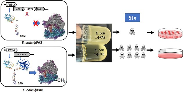
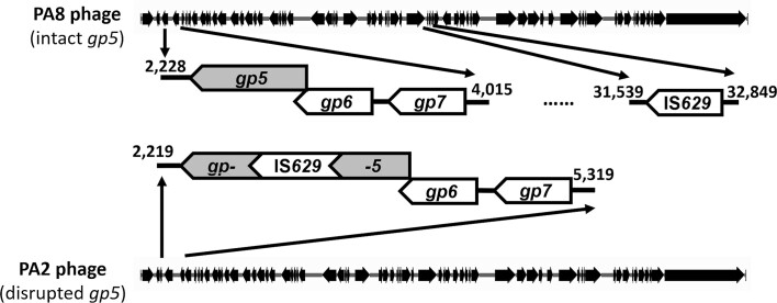
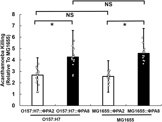
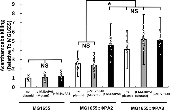
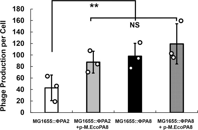
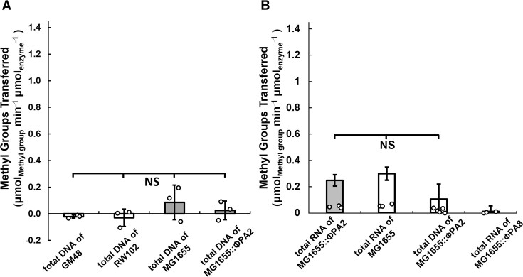
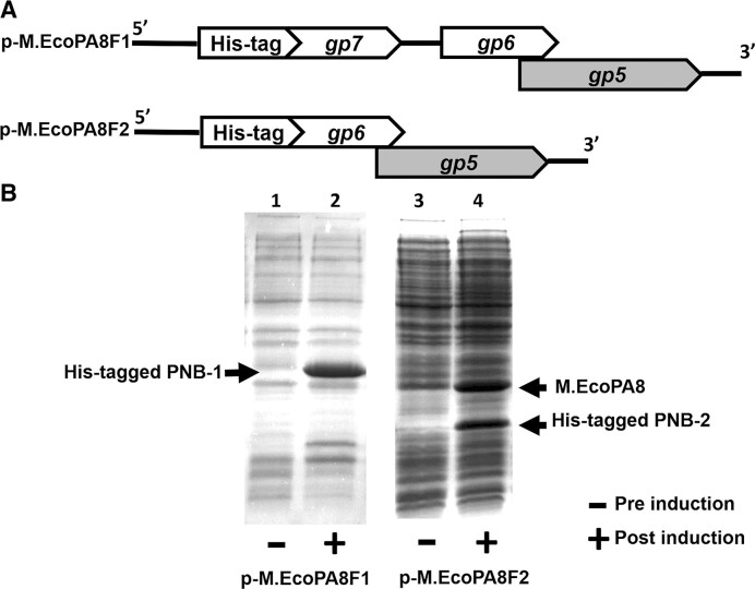
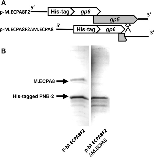
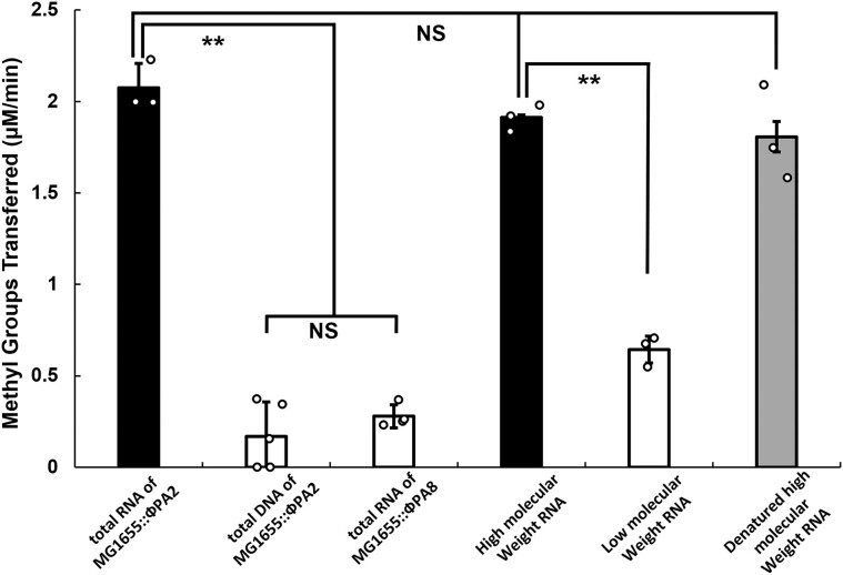
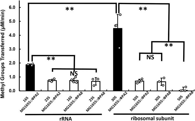
Shiga toxin (Stx) released by Shiga toxin producing Escherichia coli (STEC) causes life-threatening illness. Its production and release require induction of Stx-encoding prophage resident within the STEC genome. We identified two different STEC strains, PA2 and PA8, bearing Stx-encoding prophage whose sequences primarily differ by the position of an IS629 insertion element, yet differ in their abilities to kill eukaryotic cells and whose prophages differ in their spontaneous induction frequencies. The IS629 element in ϕPA2, disrupts an ORF predicted to encode a DNA adenine methyltransferase, whereas in ϕPA8, this element lies in an intergenic region. Introducing a plasmid expressing the methyltransferase gene product into ϕPA2 bearing-strains increases both the prophage spontaneous induction frequency and virulence to those exhibited by ϕPA8 bearing-strains. However, a plasmid bearing mutations predicted to disrupt the putative active site of the methyltransferase does not complement either of these defects. When complexed with a second protein, the methyltransferase holoenzyme preferentially uses 16S rRNA as a substrate. The second subunit is responsible for directing the preferential methylation of rRNA. Together these findings reveal a previously unrecognized role for rRNA methylation in regulating induction of Stx-encoding prophage.


Figures

Graphical Abstract
Graphical Abstract
Figure 1.
Figure 1.
Genomic structures of PA8 phage and PA2 phage. Shown is a schematic representation of the genomes of ϕPA8 (top) and ϕPA2 (bottom). ϕPA8 has an intact gp5 gene that is proposed to encode an MT-A70 family methyltransferase and the IS629 mobile element inserted into a non-coding region. In ϕPA2, the putative methyltransferase gene disrupted by the insertion of an IS629 element. Also shown is the position of two putative coding sequences for PNB-1 (gp7) and PNB-2 (gp6) (see text) located upstream of the gene encoding the methyltransferase in both phages.
Figure 2.
Figure 2.
Amoeba killing efficiency of E. coli bearing ϕPA2 or ϕPA8, Acanthamoeba castellanii were separately co-cultured with E. coli strains bearing either ϕPA2 or ϕPA8. After 2h, the fraction amoebae killed by phage-bearing strains was measured relative the number of amoebae killed in co-cultures with naïve MG1655. The left side shows the results obtained with STEC O157:H7::ϕPA2 and STEC O157:H7::ϕPA8. The right side fraction of amoeba killed in co-coltures with MG1655::ϕPA2 and MG1655::ϕPA8. Error bars represent standard deviations determined from at least 15 biological replicates and each biological replicate was measured from at least 3 technical replicates. *P < 0.05; NS: not significant P > 0.05.
Figure 3.
Figure 3.
The conserved active site motif is essential to the effects of M.EcoPA8orf6770P on STEC virulence. E. coli strains bearing either no Stx-encoding prophage (MG1655, left panel), ϕPA2 (MG1655:: ϕPA2, middle panel) or ϕPA8 (MG1655:: ϕPA8, right panel) and bearing either a control plasmid (white bars), a plasmid encoding the DPPW→APPA mutant (gray bars) or wild-type M.EcoPA8orf6770P (black bars) were co-cultured with A. castellanii. Shown is the amoeba killing efficiency of these strains. Error bars represent standard deviations determined from at least 15 biological replicates and each biological replicate was measured from at least three technical replicates. *P < 0.05; NS: not significant P > 0.05.
Figure 4.
Figure 4.
M.EcoPA8orf6770P increases the frequency of prophage spontaneous induction. Overnight cultures of E. coli strains MG1655::ϕPA2 and MG1655::ϕPA8 either do or do not contain p-M.EcoPA8, the plasmid encoding the putative phage methyltransferase, were diluted 50-fold and grown for 2h. Subsequently the number of E coli. cells and free phages were measured by qPCR as described in Methods and Materials. Shown is the number of phages produced per E. coli. Error bars represent standard deviations determined from 3 biological replicates and each biological replicate was measured from at least three technical replicates. **P < 0.01; NS: not significant P > 0.05.
Figure 5.
Figure 5.
M.EcoPA8orf6770P alone does not display significant methyltransferase activity. (A) Purified M.EcoPA8orf6770P was separately incubated with total DNA isolated from E. coli strains GM48 (dam, dcm), RW102 (dam), MG1655 (dam+, dcm+) or MG1655::ϕPA2 (dam+, dcm+gp5) as indicated. (B) The M.EcoPA8orf6770P was separately incubated with total RNA or DNA isolated from MG1655, MG1655::ϕPA2 or MG1655::ϕPA8 as indicated. The consumption of SAM was measured as described in Materials and methods and plotted as the amount of methyl groups transferred per min per μmol of enzyme used. Error bars represent standard deviations that are determined from at least three biological replicates and each biological replicate was measured from at least three technical replicates. **P < 0.01; NS: not significant P > 0.05.
Figure 6.
Figure 6.
The M.EcoPA8orf6770P and PNB-2 proteins are co-expressed. (A) Shown are schematics of the coding regions of two plasmids p-M.EcoPA8F1 and p-M.EcoPA8F2, designed to co-express either His-tagged PNB-1 along with PNB-2 and the putative methyltransferase (M.EcoPA8orf6770P) (top) or His-tagged PNB-2 and the putative methyltransferase (M.EcoPA8orf6770P) (bottom), respectively. (B) Cells bearing these plasmids were induced by IPTG and lysed as described in the method. Shown is a photograph of a Coomassie Blue stained SDS-PAGE gel of the whole cell lysates of the E. coli cells bearing p-M.EcoPA8F1 or p-M.EcoPA8F2 that either were (+) or were not (–) exposed to 0.5 mM IPTG during the mid-log phase of growth. The labels indicate the positions of the M.EcoPA8orf6770P (M.EcoPA8), His-tagged PNB-1 proteins and His-tagged PNB-2 proteins.
Figure 7.
Figure 7.
Co-purification of PNB-2 and M.EcoPA8orf6770P proteins. Proteins were isolated from lysates of E. coli bearing either the p-M.EcoPA8F2 or p-M.EcoPA8F2ΔM.EcoPA8 plasmids (A) using NTA-agarose (see Methods and Materials) and displayed on SDS-PAGE gel. (B) Proteins were visualized by Coomassie Blue staining. The expected positions of the M.EcoPA8orf6770P (M.EcoPA8) and His-tagged PNB-2 proteins are indicated.
Figure 8.
Figure 8.
M.EcoPA8orf6770P-PNB-2 holoenzyme is a ribosomal RNA methyltransferase. Purified M.EcoPA8orf6770P-PNB-2 complex was incubated with the indicated potential substrates and the consumption of SAM was measured as described in Methods and Materials and plotted as the amount of methyl group transferred per min per μmol of enzyme used. Error bars represent standard deviations that are determined from at least 3 biological replicates and each biological replicate was measured from at least 3 technical replicates. **P < 0.01; NS: not significant P > 0.05.
Figure 9.
Figure 9.
The M.EcoPA8orf6770P-PNB-2 holoenzyme preferentially methylates the 16S rRNA. 16S rRNA and 23S rRNA (left panel) or 30S and 50S ribosomal subunits (right panel) were purified from MG1655::ϕPA2 and MG1655::ϕPA8 strains and separately incubated with M.EcoPA8orf6770P-PNB-2 holoenzyme. The consumption of SAM was measured as described in Methods and Materials and plotted as the amount of methyl group transferred per min per μmol of enzyme used. Error bars represent standard deviations that are determined from at least three biological replicates and each biological replicate was measured from at least 3 technical replicates. **P < 0.01; NS: not significant P > 0.05.

Similar articles

Cited by

References

    1. Scallan E., Hoekstra R.M., Angulo F.J., Tauxe R.V., Widdowson M.-A., Roy S.L., Jones J.L., Griffin P.M.. Foodborne illness acquired in the United States—major pathogens. Emerg. Infect. Dis. 2011; 17:7. - PMC - PubMed
    1. Frenzen P.D., Drake A., Angulo F.J., Group E.I.P.F.W.. Economic cost of illness due to Escherichia coli O157 infections in the United States. J. Food Prot. 2005; 68:2623–2630. - PubMed
    1. Karmali M.A. Emerging public health challenges of Shiga toxin–producing Escherichia coli related to changes in the pathogen, the population, and the environment. Clin. Infect. Dis. 2017; 64:371–376. - PubMed
    1. Travert B., Dossier A., Jamme M., Cointe A., Delmas Y., Malot S., Wynckel A., Seguin A., Presne C., Hie M.et al. .. Shiga Toxin-Associated Hemolytic Uremic Syndrome in Adults, France, 2009-2017. Emerg. Infect. Dis. 2021; 27:1876–1885. - PMC - PubMed
    1. Joseph A., Cointe A., Mariani Kurkdjian P., Rafat C., Hertig A.. Shiga Toxin-Associated Hemolytic Uremic Syndrome: a Narrative Review. Toxins. 2020; 12:67. - PMC - PubMed

MeSH terms

Grants and funding