. 2023 Dec 22;83(1):36-52.
doi: 10.1093/jnen/nlad105.

Clinicopathologic features of a novel star-shaped transactive response DNA-binding protein 43 (TDP-43) pathology in the oldest old

Affiliations

Clinicopathologic features of a novel star-shaped transactive response DNA-binding protein 43 (TDP-43) pathology in the oldest old

Arenn F Carlos et al. J Neuropathol Exp Neurol. .

Abstract

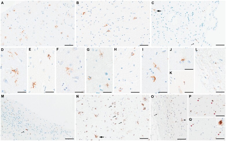
Transactive response DNA-binding protein 43 (TDP-43) pathology is categorized as type A-E in frontotemporal lobar degeneration and as type α-β in Alzheimer disease (AD) based on inclusion type. We screened amygdala slides of 131 cases with varying ages at death, clinical/neuroimaging findings, and AD neuropathologic changes for TDP-43 pathology using anti-phospho-TDP-43 antibodies. Seven cases (5%) only showed atypical TDP-43 inclusions that could not be typed. Immunohistochemistry and immunofluorescence assessed the atypical star-shaped TDP-43 pathology including its distribution, species, cellular localization, and colocalization with tau. All 7 had died at an extremely old age (median: 100 years [IQR: 94-101]) from nonneurological causes and none had dementia (4 cognitively unimpaired, 3 with amnestic mild cognitive impairment). Neuroimaging showed mild medial temporal involvement. Pathologically, the star-shaped TDP-43-positive inclusions were found in medial (subpial) amygdala and, occasionally, in basolateral regions. Hippocampus only showed TDP-43-positive neurites in the fimbria and subiculum while the frontal lobe was free of TDP-43 inclusions. The star-shaped inclusions were better detected with antibodies against N-terminal than C-terminal TDP-43. Double-labeling studies confirmed deposition of TDP-43 within astrocytes and colocalization with tau. We have identified a novel TDP-43 pathology with star-shaped morphology associated with superaging, with a homogeneous clinicopathologic picture, possibly representing a novel, true aging-related TDP-43 pathology.

Keywords: Astrocytes; Dementia; Superaging; TDP-43 proteinopathy; Tau-associated TDP-43.


Conflict of interest statement

The authors have no duality or conflicts of interest to declare.

Figures

Figure 1.
Figure 1.
Longitudinal changes in clinical and neuropsychologic scores. The spaghetti plots show the progression of clinical or neuropsychological scores across the time from baseline to death. For each patient and each test, 2 values are shown representing scores at baseline and at the last follow-up visit closest to death. The black line represents the mean score for the cohort and the shaded gray area represents confidence intervals. Noteworthy changes are seen with the drop in AVLT delayed scores, suggesting impaired episodic memory, and the lengthening of time to complete the TMT-B test, indicating worsening cognitive flexibility. Abbreviations: AVLT, auditory verbal learning test; BNT, Boston naming test; CDR-SOB, Clinical Dementia Rating Scale—Sum of Boxes; MMSE, Mini Mental State Examination; NPI, Neuropsychiatric Inventory; TMT-A, Trail Making Test-A; TMT-B, Trail Making Test-B; UPDRS, Unified Parkinson’s Disease Rating Scale; WAIS-BD, Weschler Adult Intelligence Scale-Revised Block Design Test.
Figure 2.
Figure 2.
Volume loss and hypometabolism on neuroimaging. The effect map shows differences in gray matter volumes between the star-shaped TDP-43 group (n = 5) and the healthy control group (n = 12) (A). In the top lateral views, involvement of the anterior temporal pole and the posterosuperior temporal region is seen. In the bottom medial views, involvement of the medial temporal pole, precuneus and posterior cingulate are evident. The color bar represents height threshold T-values ranging from 0 (blue) to 5.5 (red). Regions that showed significantly less gray matter volumes in the star-shaped TDP-43 group at p < .001 are those with T > 3.78 (dark yellow to red). (BD) Results from longitudinal FDG-PET scans (taken 2 years apart) from 1 case (case 4) showing progressive left medial temporal lobe hypometabolism. Abbreviations: FDG-PET, 18F-fluorodeoxyglucose-positron emission tomography; F, front; I, inferior; L, left; R, right.
Figure 3.
Figure 3.
Distribution of TDP-43-immunoreactive inclusions. Whole slide imaging of digitalized sections from the amygdala (A) and hippocampus (B) are shown. To easily identify anatomical regions, sections stained with hematoxylin and eosin from each patient are selected. The amygdaloid subregions are labeled. Areas where the star-shaped TDP-43 inclusions are observed in the amygdala are delineated by the yellow boxes (A). These mainly involve the corticomedial regions in all cases and additionally the basolateral white matter and subependymal regions in case 4 and 5. Areas where the thread-like neurites are seen in the hippocampal regions are similarly delineated by the yellow boxes (B). These include the fibers of the fimbria and the subiculum in all cases, with the addition of the entorhinal cortex in case 7. Abbreviations: Ahi, amygdala hippocampal transition area; BL, basolateral nucleus; BM, basomedial nucleus; Ce, central nucleus; Chor, choroid plexus; Co, cortical nucleus; DG, dentate gyrus; Ent cx, entorhinal cortex; Fi, fimbria; GA, gyrus accumbens; HP, hippocampal proper; LA, lateral nucleus; Me, medial nucleus; Para, parahippocampal gyrus; Sub, subiculum; Tent cx, transentorhinal cortex.
Figure 4.
Figure 4.
Representative figures of microscopic TDP-43 findings. Amygdala slides stained with antibodies against phospho-TDP-43 showed star-shaped TDP-43-immunoreactive inclusions in medial subpial regions (A) and basolateral white matter (B) and subependymal regions (thick arrow in C). Additionally, dot-like neuropil (A) and a long thread-like neurite (thin arrow in C) are seen. Different morphologies of star-shaped inclusions are shown: star-shaped with long, branching processes (D, E), with lace-like processes (F), with granular appearance (G, H), or with crown-like (I) or thorn-like (J) appearance. An occasional coiled body-like inclusion is shown (K). Superficial thin, thread-like neurites are commonly seen (L). In the hippocampus, a variety of neurites from thin to thick and short to long are seen in the fimbria (M), as well as the subiculum and entorhinal cortex (not shown). Using antibodies against N-terminal TDP-43 (N), star-shaped TDP-43 inclusions with long processes are also seen (thick arrow), although many of the inclusions are more granular in appearance (thin arrows and inset in N). Using antibodies against the C-terminal region (O), only a few granular star-shaped inclusions are seen (thick arrow) and some superficial neurites (thin arrow). However, some comma-shaped thick neurites (pink arrowheads in P) and neuronal cytoplasmic inclusions (pink arrowheads and inset in Q) are additionally seen. Scale bars: A-C, M-Q = 40 μm, D-K and all insets = 30 μm.
Figure 5.
Figure 5.
Representative images of double-labeling immunohistochemistry findings. Double-labeling immunohistochemistry showed colocalization of pTDP-43 (brown: Rb3655) and GFAP (blue: GA-5) within the star-shaped inclusions in subpial (indicated by arrowheads in (A) and magnified inclusion in (B)) and perivascular regions (indicated by black arrowheads in (C)). Double labeling immunohistochemistry for pTDP-43 (brown: Rb3655) and phospho-tau (blue: CP13) also showed colocalization of the 2 proteins in the star-shaped inclusions (arrowheads in (D) and magnified inclusion in (E)). Similar colocalizations are seen using anti-nTDP-43 antibodies (brown: MC2079) and anti-GFAP (blue: GA-5) in panels (F) and (G), as well as using anti-nTDP-43 (brown: MC2069) and anti-phospho-tau (blue: CP13) in panels (H) and (I). Scale bars: A, C, D, F, H = 40 μm, B, E, G, I = 10 μm. Abbreviation: GFAP, glial fibrillary acidic protein.
Figure 6.
Figure 6.
Representative images of double-labeling immunofluorescence findings. Double-labeling immunofluorescence (red: MC2079, green: GFAP) shows localization of TDP-43 within astrocytes (A). Similarly, double-staining immunofluorescence with MC2079 (red) and CP13 (green) showed colocalization of TDP-43 and tau within the atypical inclusions (B). No star-shaped TDP-43 pathologic inclusions localized in astrocytes (C) or colocalizing with tau (D) are seen with the negative control case. Scale bar = 10 μm. Abbreviation: GFAP, glial fibrillary acidic protein.

References

    1. Kawakami I, Arai T, Hasegawa M.. The basis of clinicopathological heterogeneity in TDP-43 proteinopathy. Acta Neuropathol 2019;138:751–70 - PMC - PubMed
    1. Buratti E, Baralle FE.. Multiple roles of TDP-43 in gene expression, splicing regulation, and human disease. Front Biosci 2008;13:867–78 - PubMed
    1. Arai T, Hasegawa M, Akiyama H, et al.TDP-43 is a component of ubiquitin-positive tau-negative inclusions in frontotemporal lobar degeneration and amyotrophic lateral sclerosis. Biochem Biophys Res Commun 2006;351:602–11 - PubMed
    1. Neumann M, Sampathu DM, Kwong LK, et al.Ubiquitinated TDP-43 in frontotemporal lobar degeneration and amyotrophic lateral sclerosis. Science 2006;314:130–3 - PubMed
    1. Arnold SJ, Dugger BN, Beach TG.. TDP-43 deposition in prospectively followed, cognitively normal elderly individuals: Correlation with argyrophilic grains but not other concomitant pathologies. Acta Neuropathol 2013;126:51–7 - PMC - PubMed

Publication types

MeSH terms