Review
. 2023 Dec 12;9(1):449.
doi: 10.1038/s41420-023-01749-8.

Programmed cell death in hepatic fibrosis: current and perspectives

Affiliations
Review

Programmed cell death in hepatic fibrosis: current and perspectives

Ju-Lu Lu et al. Cell Death Discov. .

Abstract

The initiation, development and resolution of hepatic fibrosis are influenced by various cytokines, chemokines, damage-associated molecular patterns (DAMPs) and signaling pathways. A significant number of studies in recent years have indicated that the progression of hepatic fibrosis is closely linked to programmed cell death processes such as apoptosis, autophagy, pyroptosis, necroptosis, ferroptosis, cuproptosis, and PANoptosis. Inducement of hepatic stellate cells (HSCs) death or preventing death in other liver cells can delay or even reverse hepatic fibrosis. Nevertheless, the roles of programmed cell death in hepatic fibrosis have not been reviewed. Therefore, this review summarizes the characteristics of various of hepatic fibrosis and programmed cell death, focuses on the latest progress of programmed cell death in the promotion and regression of hepatic fibrosis, and highlights the different roles of the programmed cell death of HSCs and other liver cells in hepatic fibrosis. In the end, the possible therapeutic approaches targeting programmed cell death for treating hepatic fibrosis are discussed and prospected.


Conflict of interest statement

The authors declare no competing interests.

Figures
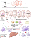
Fig. 1
Fig. 1. Overview of the role of programmed cell death in hepatic fibrosis.
In response to congenital factors, medicine, alcohol, CCl4, viral, cholestasis, parasite infection, and NASH, liver cells including hepatocyte, macrophage, and endothelial cells, undergo autophagy, apoptosis, necroptosis, pyroptosis, ferroptosis, cuproptosis, or PANoptosis. Autophagy is regulated by ATGs and is mediated by the formation of autolysosomes, which remove damaged mitochondria and excess lipids, thereby inhibiting the activation of HSCs and promoting hepatic fibrosis. In apoptotic cells, activation of caspase-3 and caspase-7 leads to the formation of apoptotic bodies, which activate HSCs directly or by activating macrophages. In necroptotic cells, activation of RIP1-RIP3-MLKL signaling causes the formation of necrosomes, resulting in cell membrane rupture. Activated caspase-1 and caspase-11 cleave GSDMD proteins to perforate cell membranes and promote the secretion of the inflammatory factors IL-1β and IL-18, with the consequent development of pyroptosis. Apoptosis, pyroptosis, and necroptosis are collectively referred as PANoptosis. Upstream of PANoptosis, the key molecules AIM2, pyrin, and ZBP1 regulate the formation of PANoptosomes, ultimately leading to cell membrane rupture. The main characteristic of ferroptosis is lipid peroxidation, which is resulted from the excessive enrichment of iron-dependent ROS in cells and the weakened clearance of GPX4. Those changes lead to decreased mitochondrial size, increased membrane density and mitochondrial membrane rupture. Cuproptosis, regulated by FDX1, is caused by the direct interaction of copper ions with fatty acylated proteins involved in the TCA cycle of mitochondrial respiration. Such interactions lead to protein toxic stress and ultimately to mitochondrial membrane rupture. Necroptosis, pyroptosis, ferroptosis, cuproptosis and PANoptosis can release DAMPs, including HMGB1, ATP, and IL-1, which lead to the aggregation of macrophages, monocytes and dendritic cells, the secretion of inflammatory factors such as TNF-α, IL-6, and IL-1, and further expand the inflammatory response. Inflammation stimulates the activation of quiescent HSCs, which then promote hepatic fibrosis. Autophagy in quiescent HSCs includes lipid degradation, also called lipophagy, which in turn leads to lipid droplet mobilization and mitochondrial β-oxidation to provide energy for HSCs activation. Small-molecule compounds such as sorafenib and metformin can induce ferroptosis and apoptosis in activated HSCs, inhibiting hepatic fibrosis. In short, programmed cell death in different cell types has different effects on hepatic fibrosis. Promotion and inhibition of hepatic fibrosis are indicated by the red line and the green line, respectively. NASH nonalcoholic steatohepatitis, ATG autophagy-related gene, HSC hepatic stellate cell, GPX4 glutathione peroxidase 4, FDX1 ferredoxin 1, ROS reactive oxygen species, TCA tricarboxylic acid, TNF tumor necrosis factor, IL interleukin, DAMP damage-associated molecular pattern, ZBP1 Z-DNA binding protein 1, AIM2 absent in melanoma 2, RIP receptor interaction protein kinase, MLKL mixed-lineage kinase domain-like pseudokinase, GSDMD Gasdermin D, ATP adenosine triphosphate.

References

    1. Iredale JP. Models of liver fibrosis: exploring the dynamic nature of inflammation and repair in a solid organ. J Clin Invest. 2007;117:539–48. doi: 10.1172/JCI30542. - DOI - PMC - PubMed
    1. Zhang C-Y, Yuan W-G, He P, Lei J-H, Wang C-X. Liver fibrosis and hepatic stellate cells: etiology, pathological hallmarks and therapeutic targets. World J Gastroenterol. 2016;22:10512–22. doi: 10.3748/wjg.v22.i48.10512. - DOI - PMC - PubMed
    1. Wang F-D, Zhou J, Chen E-Q. Molecular mechanisms and potential new therapeutic drugs for liver fibrosis. Front Pharm. 2022;13:787748. doi: 10.3389/fphar.2022.787748. - DOI - PMC - PubMed
    1. Roehlen N, Crouchet E, Baumert TF. Liver fibrosis: mechanistic concepts and therapeutic perspectives. Cells. 2020;9:875. doi: 10.3390/cells9040875. - DOI - PMC - PubMed
    1. Bataller R, Brenner DA. Liver fibrosis. J Clin Invest. 2005;115:209–18. doi: 10.1172/JCI24282. - DOI - PMC - PubMed