. 2023 Dec 12;9(1):452.
doi: 10.1038/s41420-023-01747-w.

The development of a hiPSC-based platform to identify tissue-dependencies of IDH1 R132H

Affiliations

The development of a hiPSC-based platform to identify tissue-dependencies of IDH1 R132H

N Z Mehjardi et al. Cell Death Discov. .

Abstract

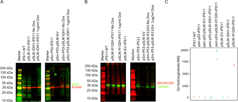
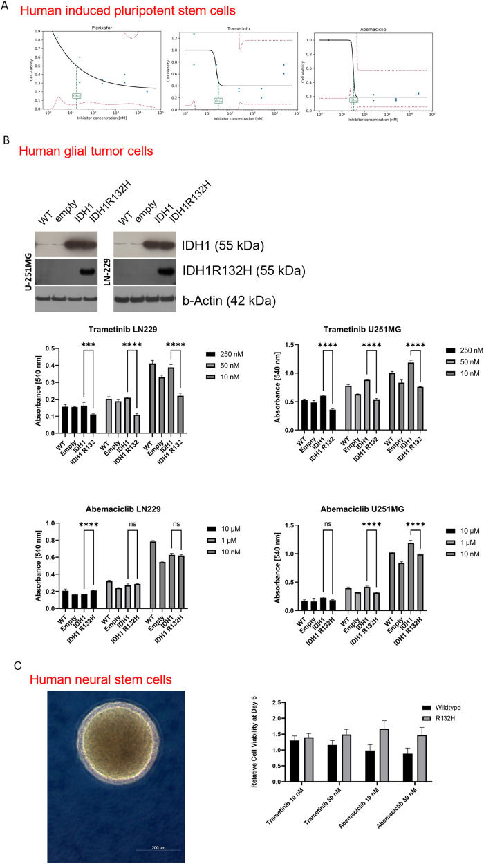
The application of patient-derived (PD) in vitro tumor models represents the classical strategy for clinical translational oncology research. Using these cellular heterogeneous cultures for the isolation of cancer stem cells (CSCs), suggested to be the main driver for disease malignancy, relies on the use of surrogate biomarkers or is based on CSC-enriching culture conditions. However, the ability of those strategies to exclusively and efficiently enrich for CSC pool has been questioned. Here we present an alternative in vitro CSC model based on the oncogenic transformation of single clone-derived human induced pluripotent stem cells (hiPSC). Hotspot mutations in the DNA encoding for the R132 codon of the enzyme isocitrate dehydrogenase 1 (IDH1) and codon R175 of p53 are commonly occurring molecular features of different tumors and were selected for our transformation strategy. By choosing p53 mutant glial tumors as our model disease, we show that in vitro therapy discovery tests on IDH1-engineered synthetic CSCs (sCSCs) can identify kinases-targeting chemotherapeutics that preferentially target tumor cells expressing corresponding genetic alteration. In contrast, neural stem cells (NSCs) derived from the IDH1R132H overexpressing hiPSCs increase their resistance to the tested interventions indicating glial-to-neural tissue-dependent differences of IDH1R132H. Taken together, we provide proof for the potential of our sCSC technology as a potent addition to biomarker-driven drug development projects or studies on tumor therapy resistance. Moreover, follow-up projects such as comparing in vitro drug sensitivity profiles of hiPSC-derived tissue progenitors of different lineages, might help to understand a variety of tissue-related functions of IDH1 mutations.


Conflict of interest statement

The authors declare no competing interests.

Figures

Fig. 1
Fig. 1. Generation of iPSC models with inducible IDH1 protein expression.
Representative Western blot membranes showing protein bands of IDH1 (A) and IDH1R132H (B), IDH1 and IDH1R132H 46 kDa, GAPDH 36 kDa, beta Actin 42 kDa. IDH1R132H protein only visible after DOX induction (12 h). Quantification of D2 Hydroxyglutarate in metabolic extracts of the corresponding cells as assessed by mass spectrometry (MS) (C).
Fig. 2
Fig. 2. All cell models represent morphological appearance typical for pluripotent cells.
A iPS11-wt; B pSLIK-IDH1-iPS11; C pSLIK-R132H-iPS11; D pSLIK-EV-iPS11; E pSin-p53-iPS11; F pSin-p53-pSLIK-IDH1-iPS11; G: pSin-p53-pSLIK-R132H-iPS11; H: pSin-p53-pSLIK-EV-iPS11.
Fig. 3
Fig. 3. Gene expression profiling of iPS11-TP53R175H model systems.
Global gene expression profiling revealed a distinct separation of transcriptome from cells with induced IDH1 and cells with induced IDH1R132H indicating the significance of this biomarker in the context of total gene expression networks in the context of human pluripotent stem cells (A). Gene Set Enrichment Analysis identified various pathways dysregulated in response to IDH1R132 induction such as increased expression of gene associated with angiogenesis or downregulation of p53 network indicating misbalanced DNA damage repair (B).
Fig. 4
Fig. 4. Testing effect of IDH1R132H protein overexpression on drug resistance in cells of different tissue differentiation status.
Cell growth–drug response curves of iPS11_IDH1R132H on top three effective drugs out of a semi-automatic executed drug screening, as defined by dose-dependent reduction of cell growth reaching lowest 50% of growth inhibition concentration (IC50) when using minimal amount of drug (Plerixafor GI50 = 18.3 nM, Trametinib GI50 = 30,7 nM, Abemaciclib GI50 = 33,0 nM (A). Protein verification of IDH1 status of p53 mutant glial tumor models used in this study and results of drug testing on those glioblastoma cells. Trametinib and Abemaciclib show increased efficacy in cells expressing IDH1R132H protein as compared to their IDH1 WT counterparts (B). Microscopic images of human neural stem cell (NSC) and results of drug sensitivity testing of NSCs under induced transgene expression, showing a trend of increased resistance of R132H cells as to Trametinib and Abemaciclib as their IDH1 WT counterparts (C). ****p ≤ 0.0001 empty empty vector control, WT wildtype.

References

    1. Reitman ZJ, Yan H. Isocitrate dehydrogenase 1 and 2 mutations in cancer: alterations at a crossroads of cellular metabolism. JNCI J Natl Cancer Inst. 2010;102:932–41. doi: 10.1093/jnci/djq187. - DOI - PMC - PubMed
    1. Liu Y, Xu W, Li M, Yang Y, Sun D, Chen L, et al. The regulatory mechanisms and inhibitors of isocitrate dehydrogenase 1 in cancer. Acta Pharm Sin B. 2023;13:1438–66. doi: 10.1016/j.apsb.2022.12.019. - DOI - PMC - PubMed
    1. Dang L, White DW, Gross S, Bennett BD, Bittinger MA, Driggers EM, et al. Cancer-associated IDH1 mutations produce 2-hydroxyglutarate. Nature. 2009;462:739. doi: 10.1038/nature08617. - DOI - PMC - PubMed
    1. Sasaki M, Knobbe CB, Itsumi M, Elia AJ, Harris IS, Chio IIC, et al. D-2-hydroxyglutarate produced by mutant IDH1 perturbs collagen maturation and basement membrane function. Genes Dev. 2012;26:2038–49. doi: 10.1101/gad.198200.112. - DOI - PMC - PubMed
    1. Figueroa ME, Abdel-Wahab O, Lu C, Ward PS, Patel J, Shih A, et al. Leukemic IDH1 and IDH2 mutations result in a hypermethylation phenotype, disrupt TET2 function, and impair hematopoietic differentiation. Cancer Cell. 2010;18:553–67. doi: 10.1016/j.ccr.2010.11.015. - DOI - PMC - PubMed