. 2024 Apr;45(4):728-737.
doi: 10.1038/s41401-023-01198-0. Epub 2023 Dec 12.

Chemokine CCL2 promotes cardiac regeneration and repair in myocardial infarction mice via activation of the JNK/STAT3 axis

Affiliations

Chemokine CCL2 promotes cardiac regeneration and repair in myocardial infarction mice via activation of the JNK/STAT3 axis

Wei Wang et al. Acta Pharmacol Sin. 2024 Apr.

Erratum in

Abstract

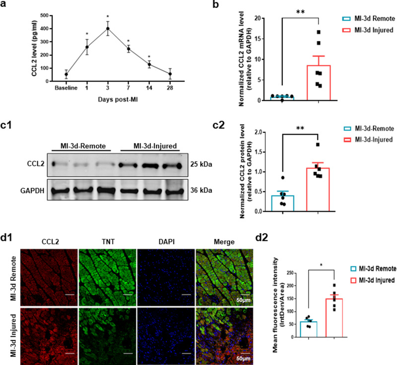
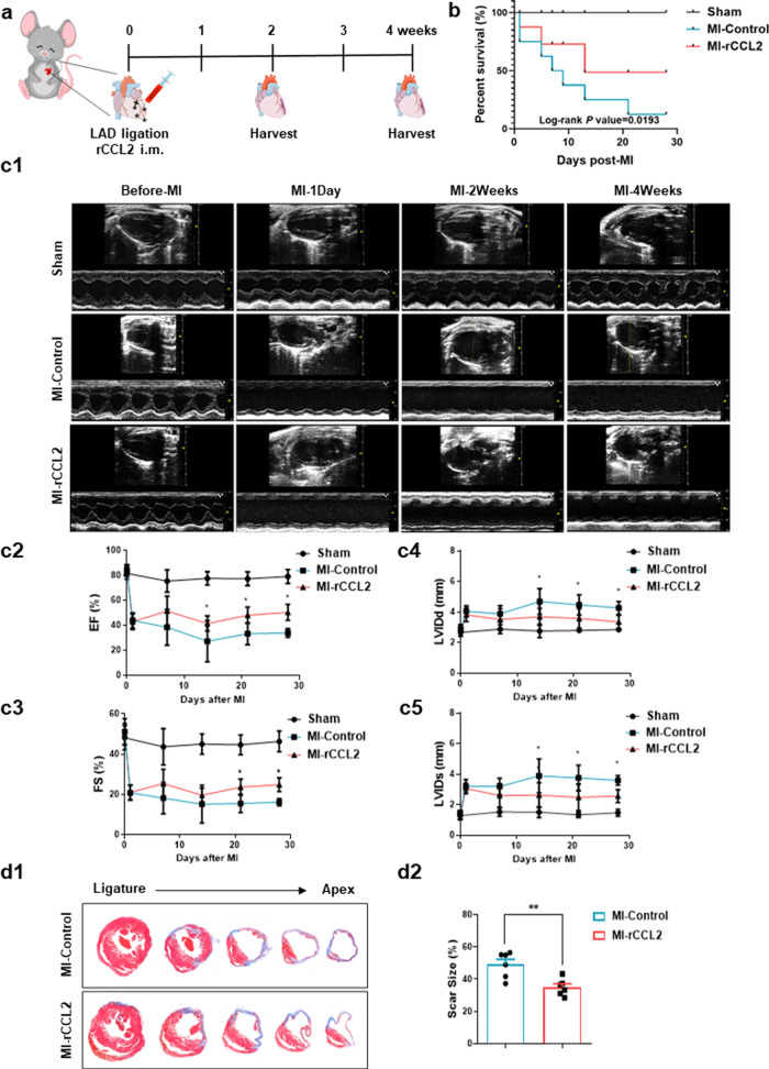
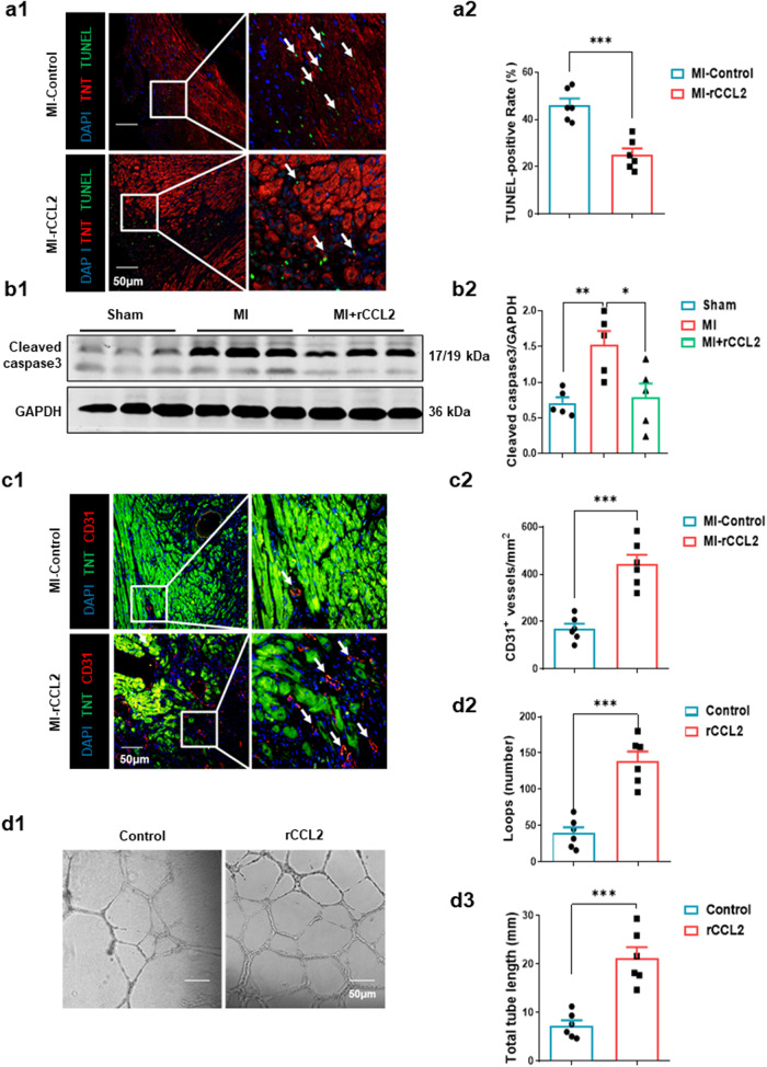
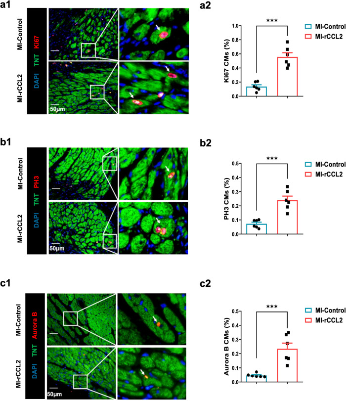
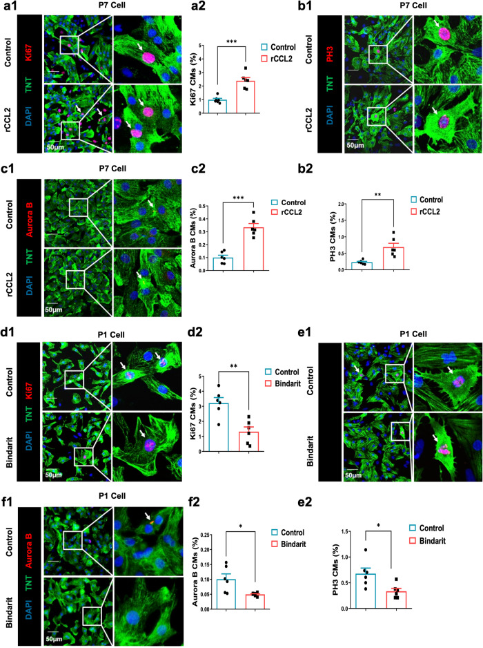
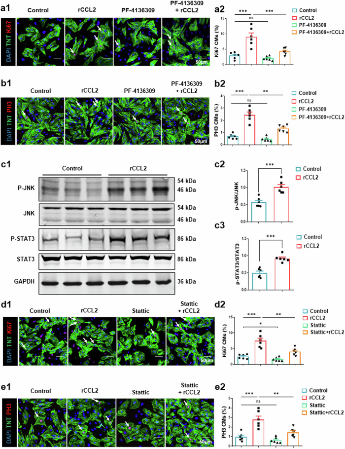
Stimulation of adult cardiomyocyte proliferation is a promising strategy for treating myocardial infarction (MI). Earlier studies have shown increased CCL2 levels in plasma and cardiac tissue both in MI patients and mouse models. In present study we investigated the role of CCL2 in cardiac regeneration and the underlying mechanisms. MI was induced in adult mice by permanent ligation of the left anterior descending artery, we showed that the serum and cardiac CCL2 levels were significantly increased in MI mice. Intramyocardial injection of recombinant CCL2 (rCCL2, 1 μg) immediately after the surgery significantly promoted cardiomyocyte proliferation, improved survival rate and cardiac function, and diminished scar sizes in post-MI mice. Alongside these beneficial effects, we observed an increased angiogenesis and decreased cardiomyocyte apoptosis in post-MI mice. Conversely, treatment with a selective CCL2 synthesis inhibitor Bindarit (30 μM) suppressed both CCL2 expression and cardiomyocyte proliferation in P1 neonatal rat ventricle myocytes (NRVMs). We demonstrated in NRVMs that the CCL2 stimulated cardiomyocyte proliferation through STAT3 signaling: treatment with rCCL2 (100 ng/mL) significantly increased the phosphorylation levels of STAT3, whereas a STAT3 phosphorylation inhibitor Stattic (30 μM) suppressed rCCL2-induced cardiomyocyte proliferation. In conclusion, this study suggests that CCL2 promotes cardiac regeneration via activation of STAT3 signaling, underscoring its potential as a therapeutic agent for managing MI and associated heart failure.

Keywords: Bindarit; CCL2; PF-4136309; STAT3; cardiomyocyte proliferation; myocardial infarction; neonatal rat ventricle myocytes; stattic.


Conflict of interest statement

The authors declare no competing interests.

Figures

Fig. 1
Fig. 1. CCL2 expression is increased in the mouse heart after MI.
a ELISA measurement of plasma CCL2 after MI (n = 6, *P < 0.05). Baseline indicates the level 1 day before MI. b Ccl2 mRNA level in injured heart and in an area remote from injury at 3 days after MI (n = 6, **P < 0.01). c Representative immunoblot image (c1) and quantification (c2) of CCL2 protein level in injured heart and in an area remote from injury at 3 days post-MI (n = 6, **P < 0.01). d Representative immunofluorescence staining (d1) and mean fluorescence intensity (d2) of CCL2 in injured heart and in an area remote from injury at 3 days post-MI (n = 6, **P < 0.01, scale bar = 50 μm). IntDen integrated density.
Fig. 2
Fig. 2. CCL2 ameliorates cardiac remodeling after MI.
a Schematic of intramyocardial injection of recombinant CCL2 and the experimental timeline to evaluate cardiac regeneration and cardiac function in the MI mouse model. b Recombinant CCL2 significantly improved the post-MI survival rate (Kaplan–Meier survival plot, n = 6). c Representative M-mode echocardiography (c1); ejection fraction (EF %), fractional shortening (FS %), left-ventricular end-diastolic internal diameter (LVIDd) and left-ventricular end-systolic internal diameter (LVIDs) (c2c5) at 1, 7, 14, and 28 days post-MI (n = 6, *P < 0.05). d Representative Masson’s trichrome staining (d1) and quantification (d2) of scar area at 28 days after MI (n = 6, **P < 0.01).
Fig. 3
Fig. 3. CCL2 promotes heart regeneration after MI.
a Representative images (a1) and quantification (a2) of TUNEL-positive cardiomyocytes in heart infarct border zone at 7 days post-MI (n = 6, ***P < 0.001, scale bar = 50 μm). b Representative immunoblot (b1) and quantification (b2) of cleaved-Caspase3 protein in heart at 3 days post-MI (n = 6, **P < 0.01, *P < 0.05). c Representative images (c1) and quantification (c2) showing CD31 immunofluorescence staining in heart sections at 14 days post-MI (n = 6, ***P < 0.001, scale bar = 50 μm). d Representative images (d1) and quantification (d2, d3) of endothelial tube formation assays (n = 6, ***P < 0.001, scale bar = 50 μm).
Fig. 4
Fig. 4. CCL2 promotes cardiomyocyte proliferation in vivo.
Representative immunofluorescence staining (a1b1c1) and quantification (a2b2c2) of Ki67, PH3, and Aurora B in the infarct area at 14 days post-MI (n = 6, ***P < 0.001, scale bar = 50 μm). CMs cardiomyocytes.
Fig. 5
Fig. 5. CCL2 induces cardiomyocyte proliferation in vitro.
a–c Representative images (a1b1c1) and quantification (a2b2c2) of Ki67, PH3 and Aurora B in P7 cardiomyocytes 24 h after recombinant CCL2 administration (100 ng/mL) (n = 6, **P < 0.01, ***P < 0.001, scale bar = 50 μm). df Representative immunofluorescence images (d1e1f1) and quantification (d2e2f2) of Ki67, PH3 and Aurora B in P1 cardiomyocytes after treatment with Bindarit (30 μM) (n = 6, scale bar = 50 μm, **P < 0.01, *P < 0.05, scale bar = 50 μm). CMs cardiomyocytes.
Fig. 6
Fig. 6. Activation of STAT3 signaling in cardiomyocytes after recombinant CCL2 stimulation.
a, b Representative immunofluorescence staining (a1, b1) and quantification (a2, b2) of Ki67 and PH3 in cardiomyocytes 24 h after recombinant CCL2 treatment (100 ng/mL) with or without PF-4136309 (30 μM), a CCR2 antagonist (n = 6, ns not significant, **P < 0.01, ***P < 0.001, scale bar = 50 μm). c Representative immunoblot image (c1) and quantification (c2, c3) of p-JNK and p-STAT3 in cardiomyocytes after recombinant CCL2 administration (n = 6, ns. not significant, ***P < 0.001, scale bar = 50 μm). d, e Representative immunofluorescence staining (d1, e1) and quantification (d2, e2) of Ki67 and PH3 in cardiomyocytes 24 h after recombinant CCL2 treatment with or without Stattic (30 μM), a STAT3 phosphorylation inhibitor (n = 6, ns. not significant, **P < 0.01, ***P < 0.001, scale bar = 50 μm). CMs cardiomyocytes.

References

    1. Reed GW, Rossi JE, Cannon CP. Acute myocardial infarction. Lancet. 2017;389:197–210. - PubMed
    1. Chablais F, Veit J, Rainer G, Jazwinska A. The zebrafish heart regenerates after cryoinjury-induced myocardial infarction. BMC Dev Biol. 2011;11:21. - PMC - PubMed
    1. Wang Z, Cui M, Shah AM, Ye W, Tan W, Min YL, et al. Mechanistic basis of neonatal heart regeneration revealed by transcriptome and histone modification profiling. Proc Natl Acad Sci USA. 2019;116:18455–65. - PMC - PubMed
    1. Uygur A, Lee RT. Mechanisms of cardiac regeneration. Dev Cell. 2016;36:362–74. - PMC - PubMed
    1. Bergmann O, Bhardwaj RD, Bernard S, Zdunek S, Barnabe-Heider F, Walsh S, et al. Evidence for cardiomyocyte renewal in humans. Science. 2009;324:98–102. - PMC - PubMed