. 2024 Jan 23;98(1):e0183023.
doi: 10.1128/jvi.01830-23. Epub 2023 Dec 13.

Skin tropism during Usutu virus and West Nile virus infection: an amplifying and immunological role

Affiliations

Skin tropism during Usutu virus and West Nile virus infection: an amplifying and immunological role

Axelle Vouillon et al. J Virol. .

Abstract

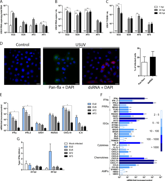
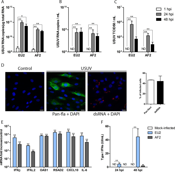
Usutu virus (USUV) and West Nile virus (WNV) are closely related emerging arboviruses belonging to the Flavivirus genus and posing global public health concerns. Although human infection by these viruses is mainly asymptomatic, both have been associated with neurological disorders such as encephalitis and meningoencephalitis. Since USUV and WNV are transmitted through the bite of an infected mosquito, the skin represents the initial site of virus inoculation and provides the first line of host defense. Although some data on the early stages of WNV skin infection are available, very little is known about USUV. Herein, USUV-skin resident cell interactions were characterized. Using primary human keratinocytes and fibroblasts, an early replication of USUV during the first 24 hours was shown in both skin cells. In human skin explants, a high viral tropism for keratinocytes was observed. USUV infection of these models induced type I and III interferon responses associated with upregulated expression of various interferon-stimulated genes as well as pro-inflammatory cytokine and chemokine genes. Among the four USUV lineages studied, the Europe 2 strain replicated more efficiently in skin cells and induced a higher innate immune response. In vivo, USUV and WNV disseminated quickly from the inoculation site to distal cutaneous tissues. In addition, viral replication and persistence in skin cells were associated with an antiviral response. Taken together, these results provide a better understanding of the pathophysiology of the early steps of USUV infection and suggest that the skin constitutes a major amplifying organ for USUV and WNV infection.IMPORTANCEUsutu virus (USUV) and West Nile virus (WNV) are closely related emerging Flaviviruses transmitted through the bite of an infected mosquito. Since they are directly inoculated within the upper skin layers, the interactions between the virus and skin cells are critical in the pathophysiology of USUV and WNV infection. Here, during the early steps of infection, we showed that USUV can efficiently infect two human resident skin cell types at the inoculation site: the epidermal keratinocytes and the dermal fibroblasts, leading to the induction of an antiviral innate immune response. Moreover, following cutaneous inoculation, we demonstrated that both viruses can rapidly spread, replicate, and persist in all distal cutaneous tissues in mice, a phenomenon associated with a generalized skin inflammatory response. These results highlight the key amplifying and immunological role of the skin during USUV and WNV infection.

Keywords: Usutu virus; West Nile virus; fibroblasts; flavivirus; innate immune response; keratinocytes; skin tropism; viral dissemination.


Conflict of interest statement

The authors declare no conflict of interest.

Figures

Fig 1
Fig 1
USUV replicates in keratinocytes and induces an innate immune response. Primary human keratinocytes were infected by USUV strains at an MOI of 1. (A) Quantification of viral RNA in cell lysates and (B) in cell supernatant. (C) Quantification of viral particle production in the supernatant using the end-point dilution assay. (D) Left panel: mock and EU2-infected cells were labeled at 24 hpi with pan-Flavivirus envelope protein antibody (green), double-stranded RNA (dsRNA) antibody (red), and DAPI (blue) (scale bar = 20 µm). Right panel: quantification of USUV-infected cells at 24 hpi using pan-Flavivirus and dsRNA antibodies. (E) Induction of inflammatory and antiviral gene expression in USUV-infected keratinocytes at 24 hpi. (F) mRNA expression of a large panel of IFNs, ISGs, cytokines, and chemokines by keratinocytes infected with EU2 strain at 24 hpi. (G) Secretion levels of type I IFNs by USUV-infected keratinocytes. Data are represented as mean ± SEM of six independent experiments. *p < 0.05, **p < 0.01, and ***p < 0.001. ND, not detected.
Fig 2
Fig 2
USUV replicates in fibroblasts and induces an innate immune response. Primary human fibroblasts were infected by USUV at a MOI of 1. (A) Quantification of viral RNA in cell lysates and (B) cell supernatant. (C) Quantification of viral particle production in the supernatant using the end-point dilution assay. (D) Left panel: mock and EU2-infected cells were labeled at 24 hpi with pan-Flavivirus envelope protein antibody (green), dsRNA antibody (red), and DAPI (blue) (scale = 20 µm). Right panel: quantification of USUV-infected cells at 24 hpi using pan-Flavivirus or dsRNA antibodies. (E) Induction of inflammatory and antiviral gene expression in USUV-infected fibroblasts at 24 hpi. (F) Secretion levels of type I IFNs by USUV-infected fibroblasts. Data are represented as mean ± SEM of six independent experiments. *p < 0.05 and **p < 0.01. ND, not detected.
Fig 3
Fig 3
USUV infection of human skin explants. Skin was infected intradermally with the USUV EU2 strain. (A) Quantification of viral RNA at the inoculation site biopsies and (B) cell culture supernatants at 1, 24, and 48 hpi. (C) Quantification of infective viral particle production in the supernatants using the end-point dilution assay. (D) Skin biopsies were labeled at 24 hpi with pan-Flavivirus envelope protein antibody (green), dsRNA antibody (red), CK10 (green), and DAPI (blue) (scale = 20 µm). (E) mRNA expression of IFNs, ISGs, cytokines, and chemokines at 24 hpi. Data are represented as mean ± SEM of five independent experiments performed in duplicate. *p < 0.05, **p < 0.01, and ***p < 0.001. ND, not detected.
Fig 4
Fig 4
Replication, dissemination, and persistence of USUV and WNV in skin tissues. Mice were infected subcutaneously with USUV or WNV in the back. (A) Viral RNA was quantified at 2 and 6 dpi for USUV and (B) at 2 and 5 dpi for WNV. (C) Transverse sections of the ears of mock-, WNV-, or USUV-infected mice showing detection of viral dsRNA and cytokeratin 14 (scale = 100 µm). mRNA fold increase of ISGs, cytokines, and chemokines from skin of the back (D and E) or in distal limbs (F and G) of infected mice as compared to mock-infected controls. *p < 0.05, **p < 0.01, and ***p < 0.001. ND, not detected.

References

    1. Pierson TC, Diamond MS. 2020. The continued threat of emerging flaviviruses. Nat Microbiol 5:796–812. doi:10.1038/s41564-020-0714-0 - DOI - PMC - PubMed
    1. Poidinger M, Hall RA, Mackenzie JS. 1996. Molecular characterization of the Japanese encephalitis serocomplex of the flavivirus genus. Virology 218:417–421. doi:10.1006/viro.1996.0213 - DOI - PubMed
    1. Cadar D, Simonin Y. 2022. Human Usutu virus infections in Europe: a new risk on horizon? Viruses 15:77. doi:10.3390/v15010077 - DOI - PMC - PubMed
    1. Muzumdar S, Rothe MJ, Grant-Kels JM. 2019. The rash with maculopapules and fever in children. Clin Dermatol 37:119–128. doi:10.1016/j.clindermatol.2018.12.005 - DOI - PubMed
    1. Garcia M, Wehbe M, Lévêque N, Bodet C. 2017. Skin innate immune response to flaviviral infection. Eur Cytokine Netw 28:41–51. doi:10.1684/ecn.2017.0394 - DOI - PubMed

Supplementary concepts