. 2023 Nov 23;26(12):108539.
doi: 10.1016/j.isci.2023.108539. eCollection 2023 Dec 15.

A novel plasmonic optical-fiber-based point-of-care test for periodontal MIP-1α detection

Affiliations

A novel plasmonic optical-fiber-based point-of-care test for periodontal MIP-1α detection

Marco Annunziata et al. iScience. .

Abstract

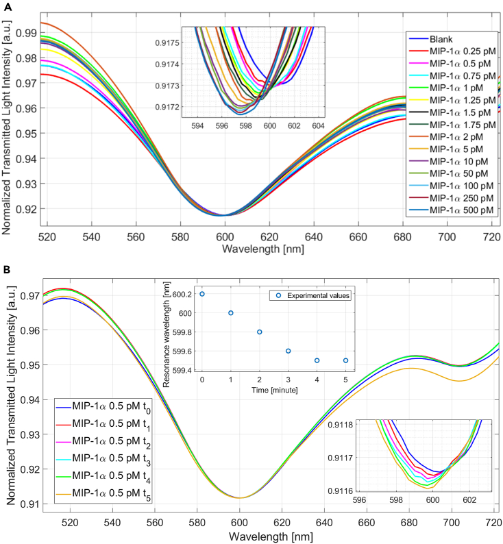
The analysis of salivary biomarkers as expression of periodontal health conditions has been proposed as a useful aid to conventional diagnostic approaches. In this study, we present a point-of-care test (POCT) exploiting a surface plasmon resonance (SPR)-based optical biosensor to detect salivary macrophage inflammatory protein (MIP)-1α, a promising marker of periodontitis. A plastic optical fiber (POF) was suitably modified and functionalized by an antibody self-assembled monolayer against MIP-1α for plasmonic detection. The proposed SPR-POF biosensor showed high selectivity and very low limit of detection for MIP-1α of 129 fM (1.0 pg/mL) in phosphate-buffered saline and 346 fM (2.7 pg/mL) in saliva. As a proof of concept, this POCT was also able to discriminate between a periodontitis patient and a healthy subject. The obtained results support the future application of this technology for an on-site detection and real-time monitoring of periodontal health conditions for diagnostic and therapeutic purposes.

Keywords: Fiber optics; Optics; Physics.


Conflict of interest statement

The authors declare no competing interests with respect to the authorship and/or publication of this article.

Figures

None
Graphical abstract
Figure 1
Figure 1
Surface plasmon resonance (SPR) plastic optical fiber (POF) biosensor system Outline and cross-section view of the SPR-POF biosensor system functionalized by an anti-MIP-1α antibody self-assembled monolayer (SAM).
Figure 2
Figure 2
Functionalization of the POF surface for MIP-1α SPR spectra obtained in PBS, before (bare surface) and after functionalization (functionalized surface). The inset highlights the resonance wavelength shift to the right for the functionalized compared with the bare surface.
Figure 3
Figure 3
Binding experiments in PBS (A) SPR spectra obtained by the developed SPR-POF biosensor at different MIP-1α concentrations in PBS. The inset highlights the resonance wavelength shift to the left, compared with the blank, at the MIP-1α concentration increase. (B) SPR spectra as a function of time at 0.5 pM MIP-1α concentration. The lower inset shows a magnification of the resonance wavelength shift. The upper inset shows the resonance wavelength versus the incubation time.
Figure 4
Figure 4
Dose-response curves in PBS and saliva Absolute values of SPR wavelength variation (Δλ), compared with the blank, versus the MIP-1α concentration, together with the Langmuir fitting and the error bars (maximum standard deviation) of the experimental values obtained in PBS (A) and saliva (B). Langmuir parameters of the fitting are also reported below the graphs. λ0, resonance value at the blank solution; Δλmax, maximum value of Δλ, calculated by the resonance saturation value minus the blank resonance value; K, dissociation constant; SE, standard error; X2, chi-square; R2, R-square.
Figure 5
Figure 5
Selectivity test Resonance wavelength variation (Δλ) obtained by different substances in PBS: MIP-3β (20 pM), BSA (20 pM), and MIP-1α (2 pM). The Δλ is calculated with respect to the blank. Means with error bars (maximum standard deviation).
Figure 6
Figure 6
MIP-1α detection in a real clinical scenario Biosensor response to a negative salivary sample (periodontally healthy subject) and a positive salivary sample (periodontitis patient). The inset highlights the shift of the resonance wavelength to the right, for the periodontally healthy subject, and to the left, for the periodontitis patient, compared with the blank.

References

    1. Pihlstrom B.L., Michalowicz B.S., Johnson N.W. Periodontal diseases. Lancet. 2005;366:1809–1820. - PubMed
    1. Deng Z.-L., Szafrański S.P., Jarek M., Bhuju S., Wagner-Döbler I. Dysbiosis in chronic periodontitis: Key microbial players and interactions with the human host. Sci. Rep. 2017;7:3703. - PMC - PubMed
    1. Hajishengallis G. Interconnection of periodontal disease and comorbidities: Evidence, mechanisms, and implications. Periodontol. 2022;89:9–18. - PMC - PubMed
    1. van der Waal S.V., Lappin D.F., Crielaard W. Does apical periodontitis have systemic consequences? The need for well-planned and carefully conducted clinical studies. Br. Dent. J. 2015;218:513–516. - PubMed
    1. Cecoro G., Annunziata M., Iuorio M.T., Nastri L., Guida L. Periodontitis, Low-Grade Inflammation and Systemic Health: A Scoping Review. Medicina. 2020;56:E272. - PMC - PubMed