Case Reports
. 2023 Nov 27:17:1284262.
doi: 10.3389/fnins.2023.1284262. eCollection 2023.

Tri-axial rubidium and helium optically pumped magnetometers for on-scalp magnetoencephalography recording of interictal epileptiform discharges: a case study

Affiliations
Case Reports

Tri-axial rubidium and helium optically pumped magnetometers for on-scalp magnetoencephalography recording of interictal epileptiform discharges: a case study

Odile Feys et al. Front Neurosci. .

Abstract

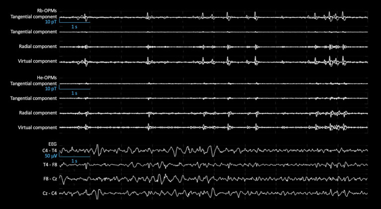
Cryogenic magnetoencephalography (MEG) enhances the presurgical assessment of refractory focal epilepsy (RFE). Optically pumped magnetometers (OPMs) are cryogen-free sensors that enable on-scalp MEG recordings. Here, we investigate the application of tri-axial OPMs [87Rb (Rb-OPM) and 4He gas (He-OPM)] for the detection of interictal epileptiform discharges (IEDs). IEDs were recorded simultaneously with 4 tri-axial Rb- and 4 tri-axial He-OPMs in a child with RFE. IEDs were identified visually, isolated from magnetic background noise using independent component analysis (ICA) and were studied following their optimal magnetic field orientation thanks to virtual sensors. Most IEDs (>1,000) were detectable by both He- and Rb-OPM recordings. IEDs were isolated by ICA and the resulting magnetic field oriented mostly tangential to the scalp in Rb-OPMs and radial in He-OPMs. Likely due to differences in sensor locations, the IED amplitude was higher with Rb-OPMs. This case study shows comparable ability of Rb-OPMs and He-OPMs to detect IEDs and the substantial benefits of triaxial OPMs to detect IEDs from different sensor locations. Tri-axial OPMs allow to maximize spatial brain sampling for IEDs detection with a limited number of sensors.

Keywords: focal epilepsy; magnetoencephalography; on-scalp magnetoencephalography; optically pumped magnetometers; refractory epilepsy.


Conflict of interest statement

NH and MB hold founding equity in Cerca Magnetics Limited, a spin-off company whose aim is to commercialize aspects of OPM-MEG technology based on QuSpin’s Rb-OPMs. EL and AP-L hold founding equity in Mag4Health SAS, a French startup company, which is developing and commercializing MEG systems based on He-OPM technology. The remaining authors declare that the research was conducted in the absence of any commercial or financial relationships that could be construed as a potential conflict of interest. The author(s) declared that EL and MB were an editorial board member of Frontiers, at the time of submission. This had no impact on the peer review process and the final decision.

Figures

Figure 1
Figure 1
Clinical EEG data and previous cryogenic MEG data. Top: 10-s EEG signal (longitudinal bipolar montage) recorded during the clinical follow-up 7 months before the multimodal OPM acquisition, band-pass filtered at 0.53–70 Hz. Middle: Non-simultaneous 10 s of cryogenic MEG signals (magnetometers) recorded 7 months before the multimodal acquisition, band-pass filtered at 3–40 Hz. Bottom: Axial brain T1-weighted MRI illustrating the resection cavity after the resection of a right temporal dysembryoplastic neuroepithelial tumor (Left). Source localization of IEDs detected with cryogenic MEG signals (for methods, see Feys et al., 2022) displayed on parasagittal (Middle left; right hemisphere), coronal (Middle right), and axial (Right) sections.
Figure 2
Figure 2
View of the multimodal EEG/He-OPM/Rb-OPM montage. Left: Map illustrating the placement of He-OPMs (blue), Rb-OPMs (green) and EEG electrodes (purple edges) with respect to a standard scalp EEG montage. The He-OPM marked with a cross was non-functional during the recording. Right: The OPMs were mounted on a dedicated helmet superimposed on scalp electrodes (not visible).
Figure 3
Figure 3
Sample of Rb-OPMs, He-OPMs, and EEG signals. Butterfly plots of 10-s signals of each tangential component, radial component, and virtual component from 4 Rb-OPMs (Top) and He-OPMs (Middle), after rejection of 11 independent components free of IEDs from the 12-channel raw data. Bipolar plots of 10-s simultaneous signals between each couple of EEG electrodes (placed according to the 10–20 montage) (Bottom). All signals were band-pass filtered at 3–38 Hz. This illustrates IEDs that can be detected simultaneously in both kinds of OPM.
Figure 4
Figure 4
IED amplitude and background activity. Top: IED amplitude across three axes, after ICA preprocessing. Tri-axial Rb-OPM with the highest IED amplitude (light gray, left). Tri-axial He-OPM with the highest IED amplitude (dark gray, right). Amplitudes correspond to the single ICA component that contained IED activity. Bar plots show mean ± SD across a sample of 102 simultaneous IEDs. The IED amplitude was significantly higher on one tangential axis for the Rb-OPM and on the radial axis for the He-OPM, with respect to the relative position of each kind of OPM to the epileptogenic zone. These differences depending on the measurement axis can be explained by a more optimal position of Rb OPM than He OPM relative to the presumed irritative zone, as supported by the previous clinical EEG data. Bottom: IED amplitude, background activity after ICA preprocessing in the optimal magnetic orientation (virtual sensor). Comparison of each IED amplitude (left), background activity from 100 ms to 50 ms before each selected IED peak (right) between Rb-OPMs and He-OPMs. All amplitudes correspond to the virtual sensor signal built from the single ICA component that isolated IED activity. The two OPM modalities show similar background noise levels. The higher IED amplitude in Rb-OPM likely reflect difference in OPM positions with respect to the presumed irritative zone localization.

References

    1. Acharya J. N., Acharya V. J. (2019). Overview of EEG montages and principles of localization. J Clin Neurophysiol Off Publ Am Electroencephalogr Soc. 36, 325–329. doi: 10.1097/WNP.0000000000000538 - DOI - PubMed
    1. Beato F., Belorizky E., Labyt E., Le Prado M., Palacios-Laloy A. (2018). Theory of a 4He parametric-resonance magnetometer based on atomic alignment. Phys. Rev. A 98:053431. doi: 10.1103/PhysRevA.98.053431 - DOI
    1. Borna A., Carter T. R., Colombo A. P., Jau Y. Y., McKay J., Weisend M., et al. . (2020). Non-invasive functional-brain-imaging with an OPM-based magnetoencephalography system. PLoS One 15:e0227684. doi: 10.1371/journal.pone.0227684, PMID: - DOI - PMC - PubMed
    1. Borna A., Iivanainen J., Carter T. R., McKay J., Taulu S., Stephen J., et al. . (2022). Cross-Axis projection error in optically pumped magnetometers and its implication for magnetoencephalography systems. Neuroimage 247:118818. doi: 10.1016/j.neuroimage.2021.118818, PMID: - DOI - PMC - PubMed
    1. Boto E., Bowtell R., Krüger P., Fromhold T. M., Morris P. G., Meyer S. S., et al. . (2016). On the potential of a new generation of magnetometers for MEG: a beamformer simulation study. PLoS One 11:e0157655. doi: 10.1371/journal.pone.0157655, PMID: - DOI - PMC - PubMed

Publication types