. 2023 Nov 25;9(12):e22784.
doi: 10.1016/j.heliyon.2023.e22784. eCollection 2023 Dec.

Kamishoyosan and Kamikihito protect against decreased KCC2 expression induced by the P. gingivalis lipopolysaccharide treatment in PC-12 cells and improve behavioral abnormalities in male mice

Affiliations

Kamishoyosan and Kamikihito protect against decreased KCC2 expression induced by the P. gingivalis lipopolysaccharide treatment in PC-12 cells and improve behavioral abnormalities in male mice

Kazuo Tomita et al. Heliyon. .

Abstract

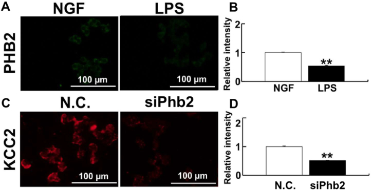
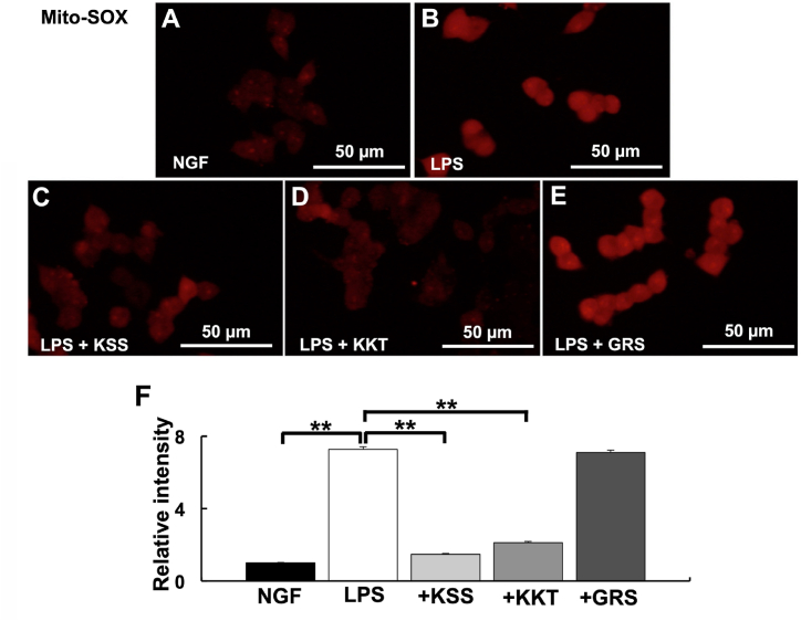
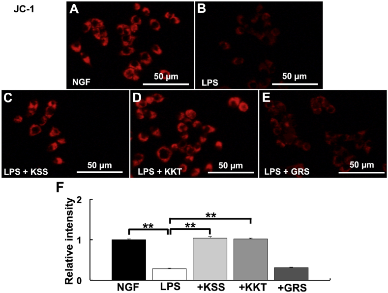
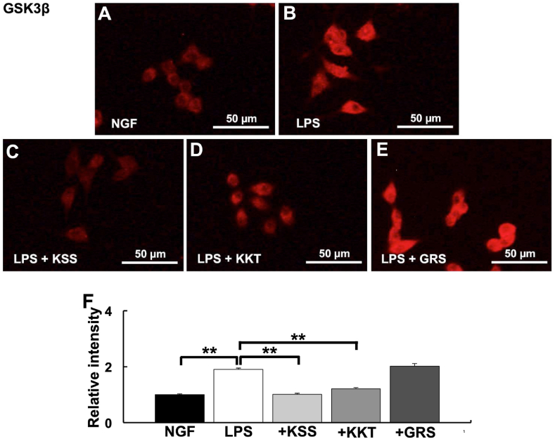
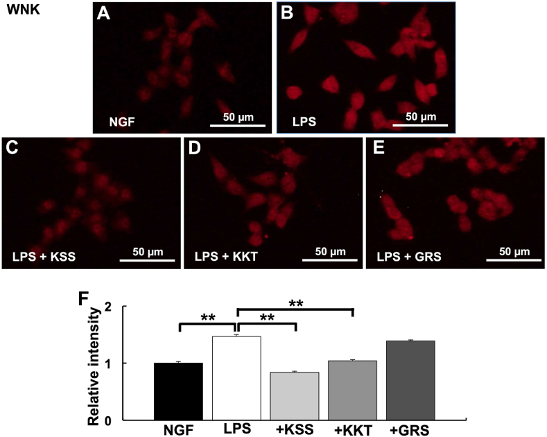
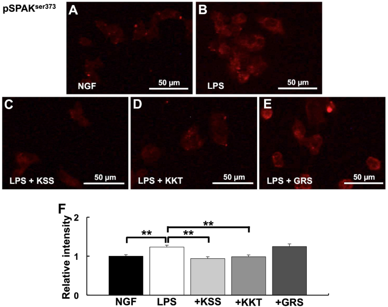
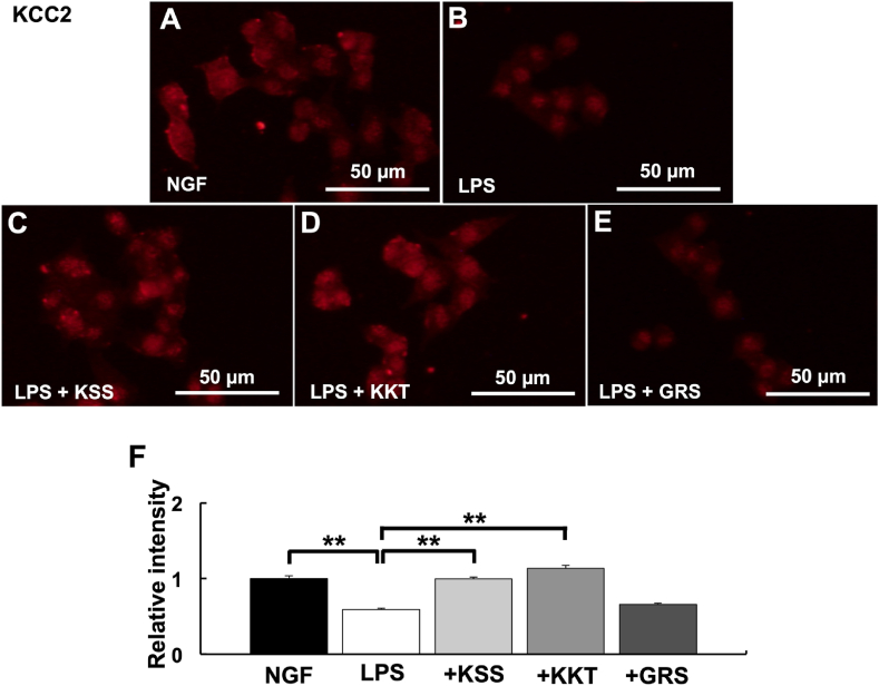
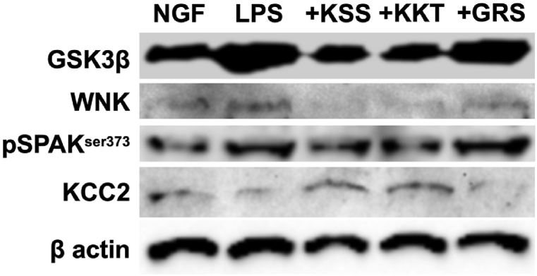
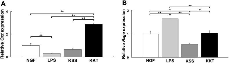
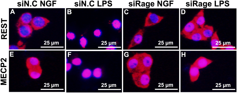
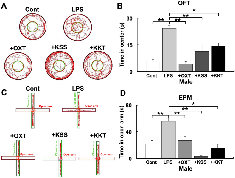
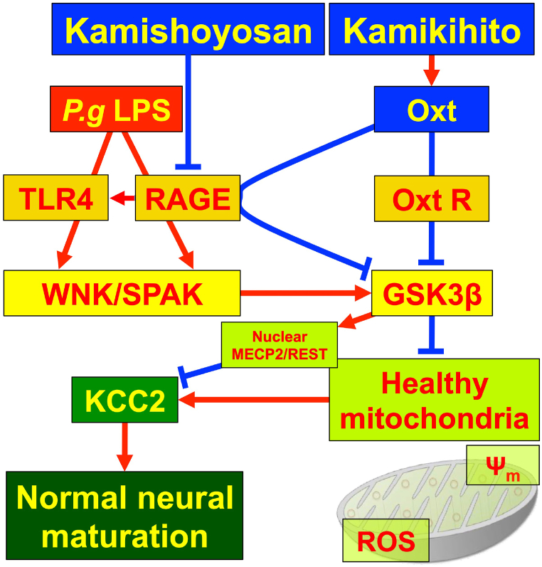
Kamishoyosan (KSS) and Kamikihito (KKT) have been traditionally prescribed for neuropsychiatric symptoms in Japan. However, the molecular mechanism of its effect is not elucidated enough. On the other hand, it has been reported that lipopolysaccharide derived from Porphyromonas gingivalis (P. g LPS) is involved not only in periodontal disease but also in the systemic diseases such as psychiatric disorders via neuroinflammation. Here, we investigated the molecular mechanism of KSS and KKT treatment by LPS-induced neuropathy using PC-12 cells. When P. g LPS was administrated during the NGF treatment, the KCC2 expression was decreased in PC-12 cells. P. g LPS treatment also decreased the WNK and phospho SPAK (pSPAK) expression and enhanced GSK-3β expression that negatively regulates WNK-SPAK signaling. Moreover, when KSS or KKT was administrated before P. g LPS treatment, the decrease of KCC2, WNK and pSPAK was rescued. KSS and KKT treatment also rescued the enhancement of GSK3β expression by P. g LPS treatment. Furthermore, KSS, KKT and/or oxytocin could rescue behavioral abnormalities caused by P. g LPS treatment by animal experiments. These effects were not shown in the Goreisan treatment, which has been reported to act on the central nervous system. These results indicate that KSS and KKT are candidates for therapeutic agents for neural dysfunction.

Keywords: GABA; KCC2; LPS; Oxytocin; PC-12 cells.


Conflict of interest statement

The authors declare that they have no known competing financial interests or personal relationships that could have appeared to influence the work reported in this paper.

Figures

Fig. 1
Fig. 1
PHB2 regulation of KCC2 expression. (A) PHB2 expression in PC-12 cells detected by immunofluorescence after LPS treatment. (B) Relative intensity of PHB2. (C) Detection of KCC2 in PC-12 cells after Phb2 knockdown. (D) Relative intensity of KCC2. LPS treatment decreases PHB2 and the knockdown of Phb2 decreases KCC2 expression in PC-12 cells. Fluorescent images were obtained from three separate dishes for each treatment. **: p < 0.01 using student's t-test.
Fig. 2
Fig. 2
Detection of mtROS after P. g LPS and Kampo treatment. (A–E) Relative mtROS intensity was detected by mitoSOX. (A) NGF treatment (control). (B) P. g LPS treatment. (C) P. g LPS + Kamishoyosan treatment. (D) P. g LPS + Kamikihito treatment. (E) P. g LPS + Goreisan treatment. (F) Relative intensities of A–E. Relative mtROS were significantly increased after P. g LPS treatment. Pretreatment with Kamishoyosan or Kamikihito prevented the P. g LPS-induced changes in mtROS. Goreisan did not prevent the mtROS increase induced by P. g LPS. Fluorescent images were obtained from three separate dishes for each treatment. **: p < 0.01 vs. LPS by Scheffe’s F test.
Fig. 3
Fig. 3
Detection of mitochondrial membrane potential (Ψm) after P. g LPS and Kampo treatment. (A–E) Relative Ψm intensity was detected by JC-1. (A) NGF treatment (control). (B) P. g LPS treatment. (C) P. g LPS + Kamishoyosan treatment. (D) P. g LPS + Kamikihito treatment. (E) P. g LPS + Goreisan treatment. (F) Relative intensities of A-E. Relative Ψm was significantly decreased after P. g LPS treatment. Pretreatment with Kamishoyosan or Kamikihito attenuated P. g LPS-induced changes in Ψm. Goreisan did not prevent the Ψm decrease induced by P. g LPS treatment. Fluorescent images were obtained from three separate dishes for each treatment. **: p < 0.01 vs. LPS by Scheffe’s F test.
Fig. 4
Fig. 4
Expression of GSK3β after P. g LPS and Kampo treatment. (A–E) Relative GSK3β intensity was detected by immunostaining. (A) NGF treatment (control). (B) P. g LPS treatment. (C) P. g LPS + Kamishoyosan treatment. (D) P. g LPS + Kamikihito treatment. (E) P. g LPS + Goreisan treatment. (F) Relative intensities of A-E. Relative GSK3β expression were significantly increased after P. g LPS treatment. Pretreatment with Kamishoyosan or Kamikihito attenuated P. g LPS-induced changes in GSK3β expression. Goreisan did not prevent the GSK3β increase induced by P. g LPS treatment. Fluorescent images were obtained from three separate dishes for each treatment. **: p < 0.01 vs. LPS by Scheffe’s F test.
Fig. 5
Fig. 5
Expression of WNK after P. g LPS and Kampo treatment. (A–E) Relative WNK intensity was detected by immunostaining. (A) NGF treatment (control). (B) P. g LPS treatment. (C) P. g LPS + Kamishoyosan treatment. (D) P. g LPS + Kamikihito treatment. (E) P. g LPS + Goreisan treatment. (F) Relative intensities of A-E. Relative WNK expression was significantly increased after P. g LPS treatment. Pretreatment with Kamishoyosan or Kamikihito attenuated the P. g LPS-induced changes in WNK expression. Goreisan did not prevent WNK increases induced by P. g LPS treatment. Fluorescent images were obtained from three separate dishes for each treatment. **: p < 0.01 vs. LPS by Scheffe’s F test.
Fig. 6
Fig. 6
Expression of pSPAKser-373 after P. g LPS and Kampo treatment. (A–E) Relative pSPAKser-373 intensity detected by immunostaining. (A) NGF treatment (control). (B) P. g LPS treatment. (C) P. g LPS + Kamishoyosan treatment. (D) P. g LPS + Kamikihito treatment. (E) P. g LPS + Goreisan treatment. (F) Relative intensities of A-E. Relative pSPAKser-373 expression was significantly increased after P. g LPS treatment. Kamishoyosan or Kamikihito pretreatment attenuated P. g LPS-induced changes in pSPAKser-373 expression. Goreisan did not prevent the pSPAKser-373 increase induced by P. g LPS treatment. Fluorescent images were obtained from three separate dishes for each treatment. **: p < 0.01 vs. LPS by Scheffe’s F test.
Fig. 7
Fig. 7
Expression of KCC2 after P. g LPS and Kampo treatment. (A–E) Relative KCC2 intensity detected by immunostaining. (A) NGF (control) treatment. (B) P. g LPS treatment. (C) P. g LPS + Kamishoyosan treatment. (D) P. g LPS + Kamikihito treatment. (E) P. g LPS + Goreisan treatment. (F) Relative intensities of A-E. Relative KCC2 expression was significantly decreased after P. g LPS treatment. Pretreatment with Kamishoyosan or Kamikihito attenuated the P. g LPS-induced changes in KCC2 expression. Goreisan did not prevent the KCC2 decrease induced by P. g LPS treatment. Fluorescent images were obtained from three separate dishes for each treatment. **: p < 0.01 vs. LPS by Scheffe’s F test.
Fig. 8
Fig. 8
Western Blotting of GSK3β, WNK, pSPAKser373, and KCC2 after P. g LPS and Kampo treatment. Twenty μg of each cell lysates were subjected to SDS-PAGE and blotted to PVDF membranes. LPS treatment up-regulated GSK3β, WNK, and pSPAKser-373. KCC2 expression was down-regulated by LPS treatment. Pretreatment with Kamishoyosan or Kamikihito attenuated the P. g LPS-induced changes in these proteins. Goreisan did not prevent these protein expression changes induced by P. g LPS treatment. The full, non-adjusted images of Western blotting in this figure were shown in Supplemental Fig. 6.
Fig. 9
Fig. 9
The relative gene expression changes of oxytocin (Oxt) and Rage after Kampo treatment by quantitative PCR (2h after treatment). NGF: control, LPS: P. g LPS, KSS; Kamishoyosan, KKT; Kamikihito. A: Kamikihito increased the expression of Oxt. P. g LPS treatment significantly decreased Oxt expression. In PC-12 cells, Kamikihito treatment significantly increased Oxt expression compared with the LPS treatment. B: Kamishoyosan decreased the expression of Rage. P. g LPS treatment significantly increased the expression of Rage. In PC-12 cells, KSS and KKT significantly decreased Rage expression compared with the LPS treatment. *: p < 0.05, **: p < 0.01 using Scheffe’s F test.
Fig. 10
Fig. 10
Nuclear translocation of REST and MECP2 after P. g LPS treatment is attenuated by siRage. The effects of Rage on the nuclear translocation of REST and MECP2 after LPS treatment were investigated. Immunostaining of REST (A–D), Immunostaining of MECP2 (E–H). LPS treatment induced the translocation of MECP2 and REST to the nucleus in PC-12 cells after transfection with the control si RNA (siN.C.) (A, B, E, F). Nuclear localization induced by LPS treatment was prevented when Rage was knocked down by siRNA treatment (C, D, G, H).
Fig. 11
Fig. 11
The influence of P. g LPS and Kampo on behavior tests in male mice. (A) Typical traces of mouse movement during the open-field test. Cont: saline, LPS: P. g LPS, +OXT; oxytocin + P. g LPS, + KSS; Kamishoyosan + P. g LPS, + KKT; Kamikihito + P. g LPS. (B) Time spent in the central compartment during the open-field test in each group. (C) Typical traces of mouse movement during the elevated plus maze test. (D) Time spent in the open arm during the elevated plus maze test. Mouse behavior was quantified and analyzed using SMART v2.5. *: p < 0.05, **: p < 0.01 using Scheffe’s F test (vs. P. g LPS).
Fig. 12
Fig. 12
Schematic diagram of the signal cascade induced by P. g LPS treatment. P. g LPS reduces KCC2 expression. In this process, P. g LPS binds to TLR4 or RAGE. P. g LPS activates WNK/SPAK and WNK/SPAK activates GSK3β. GSK3β enhances the nuclear localization of REST and MECP2, which bind to the KCC2 promoter and downregulate KCC2 expression. GSK3β decreases mitochondrial membrane potential (Ψm) and increases mitochondrial ROS (mtROS). Decreased Ψm and increased mtROS inactivate KCC2 expression. Decreased KCC2 expression inhibits normal neural maturation. Kmishoyosan decreases the expression of Rage, which decreases P. g LPS receptor Tlr4 expression (Supplemental Fig. 5). Kamikihito increases the expression of oxytocin (Oxt), which inactivates GSK3β and leads to an increase in KCC2 expression.

Similar articles

Cited by

  • Therapeutic potential for KCC2-targeted neurological diseases.
    Tomita K, Kuwahara Y, Igarashi K, Kitanaka J, Kitanaka N, Takashi Y, Tanaka KI, Roudkenar MH, Roushandeh AM, Kurimasa A, Nishitani Y, Sato T. Tomita K, et al. Jpn Dent Sci Rev. 2023 Dec;59:431-438. doi: 10.1016/j.jdsr.2023.11.001. Epub 2023 Nov 11. Jpn Dent Sci Rev. 2023. PMID: 38022385 Free PMC article. Review.

References

    1. Matsuda K. The effect of KAMISHOYOSAN on two male cases of vegetative stigmata. J. Japan Soc. Orient. Med. 1975;26:158–160. doi: 10.14868/kampomed1950.26.158. - DOI
    1. Lee J.Y., Oh H.K., Ryu H.S., Yoon S.S., Eo W., Yoon S.W. Efficacy and safety of the traditional herbal medicine, Gamiguibi-tang, in patients with cancer-related sleep disturbance: a prospective, randomized, wait-list-controlled, pilot study. Integr. Cancer Ther. 2018;17:524–530. doi: 10.1177/1534735417734914xd. - DOI - PMC - PubMed
    1. Igarashi K., Kuchiiwa T., Kuchiiwa S., Iwai H., Tomita K., Kamishoyosan T. Sato. (a Japanese traditional herbal formula), which effectively reduces the aggressive biting behavior of male and female mice, and potential regulation through increase of Tph1, Tph2, and Esr2 mRNA levels. Brain Res. 2021;1768 - PubMed
    1. Tsukada M., Ikemoto H., Lee X.P., Takaki T., Tsuchiya N., Mizuno K., Inoue T., Tsunokawa Y., Okumo T., Matsuyama T., Kamikihito M. Sunagawa. A traditional Japanese Kampo medicine, increases the secretion of oxytocin in rats with acute stress. J. Ethnopharmacol. 2021;276 doi: 10.1016/j.jep.2021.114218. - DOI - PubMed
    1. Bakos J., Srancikova A., Havranek T., Bacova Z. Molecular mechanisms of oxytocin signaling at the synaptic connection. Neural Plast. 2018;2018 doi: 10.1155/2018/4864107. - DOI - PMC - PubMed

LinkOut - more resources