Review
. 2023 Nov 27:14:1245421.
doi: 10.3389/fimmu.2023.1245421. eCollection 2023.

Cancer stem cell-immune cell crosstalk in breast tumor microenvironment: a determinant of therapeutic facet

Affiliations
Review

Cancer stem cell-immune cell crosstalk in breast tumor microenvironment: a determinant of therapeutic facet

Aishwarya Guha et al. Front Immunol. .

Abstract

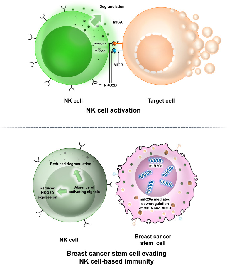
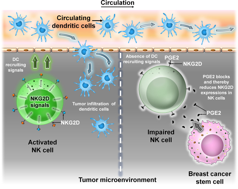
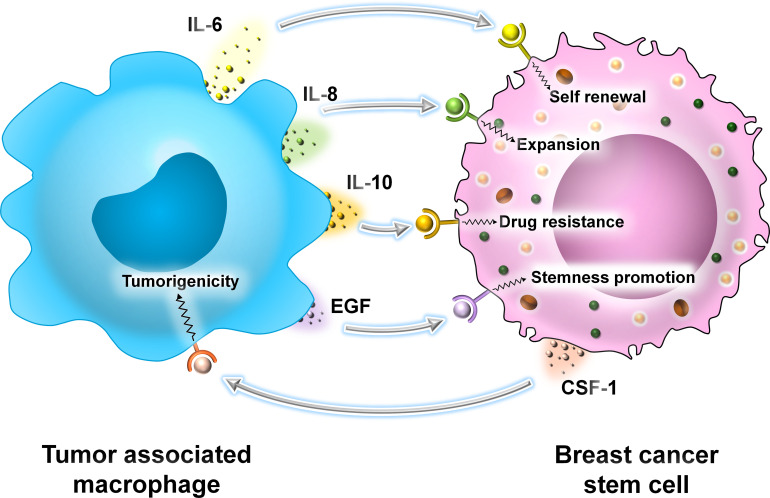
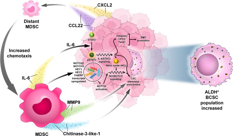
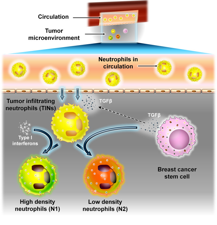
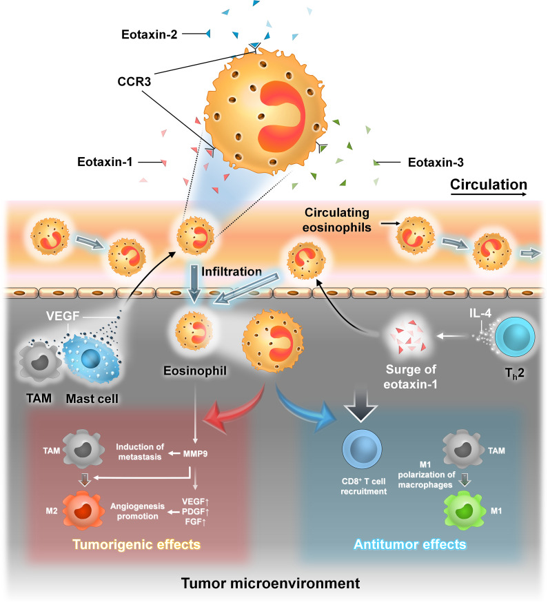
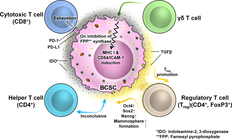
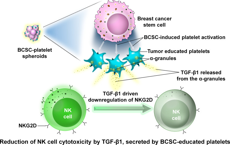
Breast cancer (BC) is globally one of the leading killers among women. Within a breast tumor, a minor population of transformed cells accountable for drug resistance, survival, and metastasis is known as breast cancer stem cells (BCSCs). Several experimental lines of evidence have indicated that BCSCs influence the functionality of immune cells. They evade immune surveillance by altering the characteristics of immune cells and modulate the tumor landscape to an immune-suppressive type. They are proficient in switching from a quiescent phase (slowly cycling) to an actively proliferating phenotype with a high degree of plasticity. This review confers the relevance and impact of crosstalk between immune cells and BCSCs as a fate determinant for BC prognosis. It also focuses on current strategies for targeting these aberrant BCSCs that could open avenues for the treatment of breast carcinoma.

Keywords: adaptive immune cells; breast cancer (BC); breast cancer stem cells (BCSCs); innate immune cells; tumor microenvironment (TME).


Conflict of interest statement

The authors declare that the research was conducted in the absence of any commercial or financial relationships that could be construed as a potential conflict of interest.

Figures

Figure 1
Figure 1
Molecular classification of breast cancer (BC). Schematic representation of the molecular classification of breast cancer: based on the presence or absence of estrogen receptor (ER), progesterone receptor (PR), and human epidermal growth factor receptor 2 (HER2), human breast carcinoma has been categorized into four different types: luminal A (ER+, PR+, HER2), luminal B (ER+, PR+/−, HER2+/−), HER2+, and triple negative breast cancer (TNBC) (ER, PR, HER2). TNBCs are further subdivided into transcriptome-based subtypes: basal cell-like type 1 (BL-1), basal cell-like type 2 (BL-2), immune-modulatory (IM), mesenchymal-like (M), mesenchymal stem cell-like (MSL), luminal androgen receptor (LAR), and claudin low.
Figure 2
Figure 2
Properties of breast cancer stem cells (BCSCs). BCSCs show characteristics resembling those of typical stem cells. They are dormant cells that divide slowly. They have the capacity for self-renewal, thereby maintaining their population of undifferentiated cells. They divide asymmetrically to create daughter cells that undergo differentiation. This kind of cell division allows them to maintain their own pool while also producing the bulk of the tumor. They are immortal cells because they can withstand chemotherapy or radiation treatment. Following that, these therapy-resistant cells display all of EMT’s characteristics and a heightened ability for metastasis. They can grow in poor adherence cell culture plates under in-vitro conditions to form tumorspheres.
Figure 3
Figure 3
The origin of BCSCs. Numerous theories are prevalent regarding the origin of BCSCs. According to one theory, genetic and epigenetic alternations of non-stem cells within the TME cause the dedifferentiation of these cells into CSCs. A second theory suggests the presence of unipotent progenitor cells which accumulate mutations over time to give rise to CSCs. A third theory predicts that CSCs arise from multipotent mammary stem cells that have undergone mutational changes.
Figure 4
Figure 4
Relative content of BCSCs among different subtypes of BC. The proportion of BCSCs varies among different subtypes of BC and this correlates with their prognosis. Luminal A has the lowest proportion followed by luminal B, HER2+, and TNBC subtypes of BC. The landscape of developing TME involves infiltration, adaptation, and/or alteration as well as crosstalk-dependent cellular evolution involving cancer cells, immune cells, and the extracellular matrix (ECM), which altogether determines the fate of the tumor. A significant body of evidence suggests that a bidirectional crosstalk is involved in developing TME. On the one hand, immune cells of the TME modulate stemness in BC cells, and on the other hand, cancer cells escape immune surveillance by exercising their effects on cells like tumor-associated macrophages (TAMs), dendritic cells (DCs), myeloid-derived suppressor cells (MDSCs), T regulatory (Treg) cells, natural killer (NK) cells, and tumor-infiltrating lymphocytes (TILs). The co-evolution of the TME and BCSCs determines the fate of BC.
Figure 5
Figure 5
The interaction between NK cells and BCSCs. Representative image of the interaction between NK cells and BCSCs. Upper panel: The NK cell interacts with the target cell via NKG2D. NKG2D interacts with its two ligands MICA and MICB on the surface of the target cell thereby mediating NK cell cytotoxicity. Lower panel: Interaction of NK cells with BCSCs causes reduction of MICA and MICB on the surface of BCSCs via miR20a. This prevents the functioning of NKG2D thereby preventing degranulation of NK cells and allowing BCSCs to escape NK cell-mediated cytotoxicity.
Figure 6
Figure 6
The interaction between DCs and BCSCs. Representative image of the interaction between DCs and BCSCs. Left panel: Recruitment of circulating DC into the TME is mediated by NKG2D signals from NK cells. Right panel: BCSCs block the activity of NKG2D by secreting prostaglandin E2 (PGE2), thereby preventing NK cell-mediated recruitment of DC into the TME.
Figure 7
Figure 7
The interaction between TAMs and BCSCs. Representative image of the interaction between TAMs and BCSCs. TAMs secrete cytokines like IL-6, IL-8, and IL-10 and growth factor EGF, promoting self-renewal, expansion, drug resistance, and overall stemness in BCSCs, respectively. BCSCs, on the other hand, secrete CSF-1 that promotes the tumorigenicity of TAMs.
Figure 8
Figure 8
The interaction between MDSCs and BCSCs. Representative image of the interaction between MDSCs and BCSCs. MDSCs are recruited to the tumor site by ΔNp63-dependent activation of chemokines CXCL2 and CCL22. They secrete pro-metastatic factors such as MMP9 and chitinase 3-like 1 to induce BCSC enrichment. MDSCs cause IL-6-dependent phosphorylation of STAT3 that promotes NOTCH signaling (NOTCH2, NOTCH3, HEY1, HEY2, and CHERP transcripts and the intracellular domain of NOTCH) expression through nitric oxide (NO), leading to persistent STAT3 activation that results in the induction of EMT with high expression genes like Vimentin, CK14, and TWIST.
Figure 9
Figure 9
The interaction between neutrophils and BCSCs. Representative image of the interaction between neutrophils and BCSCs. Recruitment of neutrophils into the TME from circulation is brought about by TGF-β secreted from BCSCs. TINs can polarize into N1 (antitumor) or high-density neutrophils or N2 (protumor) or low-density neutrophils depending on the signal. Type 1 interferons convert TINs to the N1 type, whereas TGF-β from BCSCs polarizes them to the N2 type.
Figure 10
Figure 10
The interaction between eosinophil cells and BCSCs. Representative image of the interaction between eosinophils and BCSCs. Left panel: Recruitment of eosinophils into the TME from circulation is brought about by eotaxin-1, eotaxin-2, and eotaxin-3 that activate CCR3 on the surface of eosinophils. Also, VEGF secreted by TAM and mast cells recruits eosinophils into the TME. In the TME, eosinophils produce MMP-9 that promotes metastasis, polarizes TAM to the M2 phenotype, and also favors angiogenesis by secretion of VEGF, PDGF, and FGF, thereby exerting protumorigenic effects. Right panel: Indirect recruitment of eosinophils into the TME is mediated by IL-4 from Th-2 cells by the local production of eotaxin-1. In the TME, they promote the recruitment of CD8+ T cells and induce M1 macrophage polarization, thereby exerting antitumor effects.
Figure 11
Figure 11
The interaction between different T cells and BCSCs. Representative image of the interaction between T cells and BCSCs. BCSCs express programmed death-ligand 1 (PD-L1) on their surface which interacts with programmed cell death protein 1 (PD-1) on the surface of CD8+ T cells to cause T-cell exhaustion. The interaction of BCSCs with CD4+ T cells is inconclusive. BCSCs have elevated levels of indoleamine-2,3-dioxygenase (IDO), which promotes the generation and activation of Tregs. Tregs regulate the stemness of BCSCs through the TGF-β signaling pathway that increased mammosphere formation with enhanced expression of OCT4, SOX2, and NANOG. BCSCs are resistant to γδ T-cell-mediated killing due to the presence of farnesyl pyrophosphate synthase. Inhibition of farnesyl pyrophosphate synthase results in MHC-I and CD54/ICAM-1 upregulation resulting in susceptibility to γδ T-cell- and CD8+ T-cell-mediated lysis.
Figure 12
Figure 12
The interaction between platelets and BCSCs. Representative image of the interaction between platelets and BCSCs. The interaction between platelets and BCSCs causes the release of transforming growth factor β1 (TGF-β1) from the α-granules of platelets. TGF-β1 inhibits NK cell activity by downregulating the expression of NKG2D which prevents the antitumor activity of NK cells.

References

    1. Yin L, Duan JJ, Bian XW, Yu SC. Triple-negative breast cancer molecular subtyping and treatment progress. Breast Cancer Res (2020) 22(1):1–3. doi: 10.1186/s13058-020-01296-5 - DOI - PMC - PubMed
    1. Lehmann BD, Bauer JA, Chen X, Sanders ME, Chakravarthy AB, Shyr Y, et al. . Identification of human triple-negative breast cancer subtypes and preclinical models for selection of targeted therapies. J Clin Invest (2011) 121(7):2750–67. doi: 10.1172/JCI45014 - DOI - PMC - PubMed
    1. Crabtree JS, Miele L. Breast cancer stem cells. Biomedicines. (2018) 6(3):77. doi: 10.3390/biomedicines6030077 - DOI - PMC - PubMed
    1. Lagadec C, Vlashi E, Della Donna L, Dekmezian C, Pajonk F. Radiation-induced reprogramming of breast cancer cells. Stem Cells (2012) 30(5):833–44. doi: 10.1002/stem.1058 - DOI - PMC - PubMed
    1. Rios AC, Fu NY, Lindeman GJ, Visvader JE. In situ identification of bipotent stem cells in the mammary gland. Nature. (2014) 506(7488):322–7. doi: 10.1038/nature12948 - DOI - PubMed

Publication types