

Quantitative iron-neuromelanin MRI associates with motor severity in Parkinson's disease and matches radiological disease classification
- PMID: 38090717
- PMCID: PMC10711072
- DOI: 10.3389/fnagi.2023.1287917
Quantitative iron-neuromelanin MRI associates with motor severity in Parkinson's disease and matches radiological disease classification
Abstract
Background: Neuromelanin- and iron-sensitive MRI studies in Parkinson's disease (PD) are limited by small sample sizes and lack detailed clinical correlation. In a large case-control PD cohort, we evaluated the diagnostic accuracy of quantitative iron-neuromelanin MRI parameters from the substantia nigra (SN), their radiological utility, and clinical association.
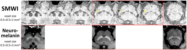
Methods: PD patients and age-matched controls were prospectively recruited for motor assessment and midbrain neuromelanin- and iron-sensitive [quantitative susceptibility mapping (QSM) and susceptibility map-weighted imaging (SMWI)] MRI. Quantitative neuromelanin-iron parameters from the SN were assessed for their discriminatory performance in PD classification using ROC analysis compared to those of qualitative visual classification by radiological readers of differential experience and used to predict motor severity.
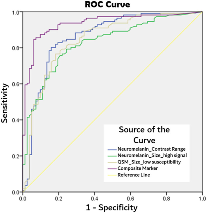
Results: In total, 191 subjects (80 PD, mean age 65.0 years; 111 controls, 65.6) were included. SN masks showed (a) higher mean susceptibility (p < 0.0001) and smaller sizes after thresholding for low susceptibility (p < 0.0001) on QSM and (b) lower contrast range (p < 0.0001) and smaller sizes after thresholding for high-signal voxels (p < 0.0001) on neuromelanin-sensitive MRI in patients than in controls. Quantitative iron and neuromelanin parameters showed a moderate correlation with motor dysfunction (87.5%: 0.4< | r | <0.6, p < 0.0001), respectively. A composite quantitative neuromelanin-iron marker differentiated the groups with excellent performance (AUC 0.94), matching the diagnostic accuracy of the best-performing reader (accuracy 97%) using SMWI.
Conclusion: Quantitative neuromelanin-iron MRI is associated with PD motor severity and matched best-performing radiological PD classification using SMWI, with the potential to improve diagnostic confidence in the clinics and track disease progression and response to neuroprotective therapies.
Keywords: MRI; Parkinson's disease; classification; comparative study; correlation analysis; iron; neuromelanin; substantia nigra.
Copyright © 2023 Hartono, Chen, Welton, Tan, Lee, Teh, Chen, Hou, Tham, Lim, Prakash, Shih, Lee, Tan, Tan and Chan.
Conflict of interest statement
The authors declare that the research was conducted in the absence of any commercial or financial relationships that could be construed as a potential conflict of interest.
Figures
Similar articles
-
Classification of Parkinson's disease by deep learning on midbrain MRI.Front Aging Neurosci. 2024 Aug 20;16:1425095. doi: 10.3389/fnagi.2024.1425095. eCollection 2024. Front Aging Neurosci. 2024. PMID: 39228827 Free PMC article.
-
Quantifying iron deposition within the substantia nigra of Parkinson's disease by quantitative susceptibility mapping.J Neurol Sci. 2018 Mar 15;386:46-52. doi: 10.1016/j.jns.2018.01.008. Epub 2018 Jan 12. J Neurol Sci. 2018. PMID: 29406966
-
Magnetic resonance correlation of iron content with neuromelanin in the substantia nigra of early-stage Parkinson's disease.Eur J Neurol. 2016 Feb;23(2):368-74. doi: 10.1111/ene.12838. Epub 2015 Oct 31. Eur J Neurol. 2016. PMID: 26518135
-
Nigrosome Imaging and Neuromelanin Sensitive MRI in Diagnostic Evaluation of Parkinsonism.Mov Disord Clin Pract. 2018 Feb 22;5(2):131-140. doi: 10.1002/mdc3.12590. eCollection 2018 Mar-Apr. Mov Disord Clin Pract. 2018. PMID: 30363419 Free PMC article. Review.
-
Iron Imaging as a Diagnostic Tool for Parkinson's Disease: A Systematic Review and Meta-Analysis.Front Neurol. 2020 May 28;11:366. doi: 10.3389/fneur.2020.00366. eCollection 2020. Front Neurol. 2020. PMID: 32547468 Free PMC article.
Cited by
-
Radiomics score derived from T1-w/T2-w ratio image can predict motor symptom progression in Parkinson's disease.Eur Radiol. 2024 Dec;34(12):7921-7933. doi: 10.1007/s00330-024-10886-2. Epub 2024 Jul 3. Eur Radiol. 2024. PMID: 38958697
-
Classification of Parkinson's disease by deep learning on midbrain MRI.Front Aging Neurosci. 2024 Aug 20;16:1425095. doi: 10.3389/fnagi.2024.1425095. eCollection 2024. Front Aging Neurosci. 2024. PMID: 39228827 Free PMC article.
-
Integrated evaluation of Nigrosome 1 sign, neuromelanin-sensitive MR and iron deposition.Jpn J Radiol. 2025 Aug 18. doi: 10.1007/s11604-025-01858-7. Online ahead of print. Jpn J Radiol. 2025. PMID: 40824349
-
Neuroimaging and fluid biomarkers in Parkinson's disease in an era of targeted interventions.Nat Commun. 2024 Jul 5;15(1):5661. doi: 10.1038/s41467-024-49949-9. Nat Commun. 2024. PMID: 38969680 Free PMC article. Review.
References
LinkOut - more resources
Full Text Sources