. 2023 Dec 19;120(51):e2303713120.
doi: 10.1073/pnas.2303713120. Epub 2023 Dec 13.

The mitochondrial ATP synthase is a negative regulator of the mitochondrial permeability transition pore

Affiliations

The mitochondrial ATP synthase is a negative regulator of the mitochondrial permeability transition pore

Ryan Pekson et al. Proc Natl Acad Sci U S A. .

Abstract

The mitochondrial permeability transition pore (mPTP) is a channel in the inner mitochondrial membrane whose sustained opening in response to elevated mitochondrial matrix Ca2+ concentrations triggers necrotic cell death. The molecular identity of mPTP is unknown. One proposed candidate is the mitochondrial ATP synthase, whose canonical function is to generate most ATP in multicellular organisms. Here, we present mitochondrial, cellular, and in vivo evidence that, rather than serving as mPTP, the mitochondrial ATP synthase inhibits this pore. Our studies confirm previous work showing persistence of mPTP in HAP1 cell lines lacking an assembled mitochondrial ATP synthase. Unexpectedly, however, we observe that Ca2+-induced pore opening is markedly sensitized by loss of the mitochondrial ATP synthase. Further, mPTP opening in cells lacking the mitochondrial ATP synthase is desensitized by pharmacological inhibition and genetic depletion of the mitochondrial cis-trans prolyl isomerase cyclophilin D as in wild-type cells, indicating that cyclophilin D can modulate mPTP through substrates other than subunits in the assembled mitochondrial ATP synthase. Mitoplast patch clamping studies showed that mPTP channel conductance was unaffected by loss of the mitochondrial ATP synthase but still blocked by cyclophilin D inhibition. Cardiac mitochondria from mice whose heart muscle cells we engineered deficient in the mitochondrial ATP synthase also demonstrate sensitization of Ca2+-induced mPTP opening and desensitization by cyclophilin D inhibition. Further, these mice exhibit strikingly larger myocardial infarctions when challenged with ischemia/reperfusion in vivo. We conclude that the mitochondrial ATP synthase does not function as mPTP and instead negatively regulates this pore.

Keywords: mitochondrial ATP synthase; mitochondrial permeability transition pore; necrosis.


Conflict of interest statement

Competing interests statement:The authors declare no competing interest.

Figures
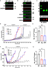
Fig. 1.
Fig. 1.
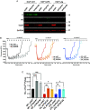
Loss of ATP synthase potentiates mPTP in HAP1 cells. Western blot of SDS-PAGE of (A) total cell and (B) mitochondrial fraction lysates from HAP1 cells. (C) Western blot of blue native polyacrylamide gel electrophoresis (BN-PAGE) for ATP synthase complexes extracted from mitochondrial lysates of HAP1 cells. ATP synthase complexes indicated on the left. O, oligomers; D, dimers; M, monomers; AI, assembly intermediates. Representative calcium retention capacity (CRC) assay for Ca2+-induced mPTP opening in (D) isolated mitochondria from HAP1 cells and (F) permeabilized HAP1 cells. Quantification of number of Ca2+ pulses required to induce mPTP opening in (E) isolated mitochondria from HAP1 cells and (G) permeabilized HAP1 cells. All data represent mean ± SEM, n = 3 to 4 independent experiments. Statistical analyses were performed using one-way ANOVA. *P < 0.05; **P < 0.01. CsA - cyclosporine A.
Fig. 2.
Fig. 2.
Knockdown of Ppif inhibits mPTP opening in the absence of ATP synthase. (A) Western blot of SDS-PAGE for shRNA-mediated knockdown of Ppif in HAP1 cells. (B) CRC assay for Ca2+-induced mPTP opening in permeabilized CypD KD HAP1 cells. (C) Quantification of number of Ca2+ pulses required to induce mPTP opening in permeabilized Ppif KD HAP1 cells. All data represent mean ± SEM, n = 3 to 4 independent experiments. Statistical analyses were performed using one-way ANOVA. *P < 0.05; **P < 0.01; ***P < 0.001; ****P < 0.0001.
Fig. 3.
Fig. 3.
HAP1 mitoplasts depleted of ATP synthase retain mPTP conductance. (A) Representative patch clamp current traces of mitoplasts isolated from HAP1-WT, HAP1-Δg, and HAP1-ΔF6. The patching medium included 250 mM CaCl2, 5 mM succinate, and 5 mM rotenone to induce permeability transition. The high conductance openings in the WT and mutant lines were sensitive to addition of 5 to 20 µM cyclosporine A. (B) Quantification of mPTP conductances in HAP1 mitoplasts. Data represent mean ± SEM of 11 to 16 mitoplast recordings per group. Statistical analyses were performed using one-way ANOVA. ns, nonsignificance. CsA - cyclosporine A.
Fig. 4.
Fig. 4.
Deletion of Atp5l potentiates mPTP opening in cardiac mitochondria. (A) Western blot of SDS-PAGE for subunit g in cardiac mitochondria from WT and Atp5l KO mice at the indicated weeks post-TMX. (B) Western blot of BN-PAGE for blue native gel assessing ATP synthase complexes in cardiac mitochondria from WT and Atp5l KO mice indicated weeks post-TMX. ATP synthase complexes indicated on the Left. O, oligomers; D, dimers; M, monomers; AI, assembly intermediates. (C) Representative CRC assay for Ca2+-induced mPTP opening and (D) quantification of number of Ca2+ pulses required to induce mPTP opening in cardiac mitochondria isolated from mice at 8 wk post-TMX. (E) Representative CRC assay for Ca2+-induced mPTP opening and (F) quantification of number of Ca2+ pulses required to induce mPTP opening in cardiac mitochondria isolated from mice at 12 wk post-TMX. All data represent mean ± SEM, n = 6 WT mice, 6 Atp5l KO mice. Statistical analyses were performed using one-way ANOVA *P < 0.05; **P < 0.01; ***P < 0.001; ****P < 0.0001. CsA - cyclosporine A.
Fig. 5.
Fig. 5.
Deletion of Atp5l augments myocardial damage during ischemia-reperfusion injury. (A) Schematic for myocardial ischemia-reperfusion (I/R) protocol in WT and Atp5l KO mice at weeks 8 to 11 post-TMX. (B) Representative images of TTC staining to assess infarct size in WT and Atp5l KO mice subjected to myocardial I/R. (C) Quantification of AAR/LV, INF/AAR, and INF/LV following myocardial I/R in WT and Atp5l KO mice. (D) Troponin I release following myocardial I/R in WT and Atp5l KO mice. All data represent mean ± SEM, n = 13 male WT mice, 12 male Atp5l KO mice. Statistical analyses were performed using two-tailed Student’s t test. ***P < 0.001; ****P < 0.0001.

References

    1. Raaflaub J., Die schwellung isolierter leberzell mitochondrien und ihre physikalisch beeinflußarkeit. Helv. Physiol. Pharmacol. Acta. 11, 142–156 (1953). - PubMed
    1. Hunter D. R., Haworth R. A., Southard J. H., Relationship between configuration, function, and permeability in calcium-treated mitochondria. J. Biol. Chem. 251, 5069–5077 (1976). - PubMed
    1. Massari S., Azzone G. F., The equivalent pore radius of intact and damaged mitochondria and the mechanism of active shrinkage. Biochim. Biophys. Acta 283, 23–29 (1972), 10.1016/0005-2728(72)90094-1. - DOI - PubMed
    1. Crompton M., Ellinger H., Costi A., Inhibition by cyclosporin A of a Ca2+-dependent pore in heart mitochondria activated by inorganic phosphate and oxidative stress Biochem. J. 255, 357–360 (1988). - PMC - PubMed
    1. Kinnally K. W., Campo M. L., Tedeschi H., Mitochondrial channel activity studied by patch-clamping mitoplasts. J. Bioenerg. Biomembr. 21, 497–506 (1989), 10.1007/BF00762521. - DOI - PubMed

MeSH terms

Substances