. 2023 Dec 15;9(50):eadj4407.
doi: 10.1126/sciadv.adj4407. Epub 2023 Dec 13.

Myeloid/natural killer (NK) cell precursor acute leukemia as a distinct leukemia type

Affiliations

Myeloid/natural killer (NK) cell precursor acute leukemia as a distinct leukemia type

Akira Nishimura et al. Sci Adv. .

Abstract

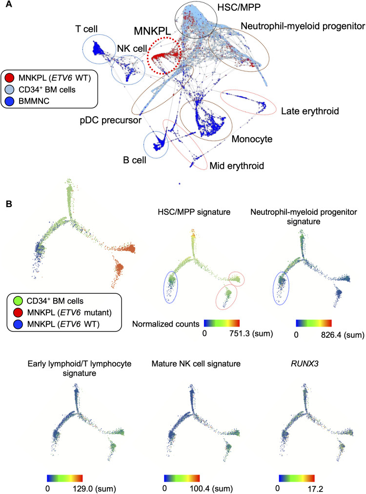
Myeloid/natural killer (NK) cell precursor acute leukemia (MNKPL) has been described on the basis of its unique immunophenotype and clinical phenotype. However, there is no consensus on the characteristics for identifying this disease type because of its rarity and lack of defined distinctive molecular characteristics. In this study, multiomics analysis revealed that MNKPL is distinct from acute myeloid leukemia, T cell acute lymphoblastic leukemia, and mixed-phenotype acute leukemia (MPAL), and NOTCH1 and RUNX3 activation and BCL11B down-regulation are hallmarks of MNKPL. Although NK cells have been classically considered to be lymphoid lineage-derived, the results of our single-cell analysis using MNKPL cells suggest that NK cells and myeloid cells share common progenitor cells. Treatment outcomes for MNKPL are unsatisfactory, even when hematopoietic cell transplantation is performed. Multiomics analysis and in vitro drug sensitivity assays revealed increased sensitivity to l-asparaginase and reduced levels of asparagine synthetase (ASNS), supporting the clinically observed effectiveness of l-asparaginase.


Figures

Fig. 1.
Fig. 1.. Distinct genomic landscape and profile of MNKPL.
(A) Genomic landscape of MNKPL. Frequencies of recurrent mutations, copy number alterations, or translocations identified by WES or WGS. Mutation categories are indicated by color. EM, extramedullary involvement; HCT, hematopoietic cell transplantation; R/R, relapse/refractory. The sidebar graph indicates the percentage of gene alterations. The genes involved in the NOTCH, RAS-MAPK, and PI3K-AKT signaling pathways are highlighted in light blue, light orange, and light green, respectively. (B) Three structural alterations of the ETV6 gene. ETV6::C3orf62 and ETV6::MAML3 were identified as in-frame mutations, respectively. (C) Five ETV6 mutations in MNKPL. Each color represents a domain or mutation type. (D) PCA of MNKPL (n = 6), AML (n = 37), T-ALL (n = 25), and MPAL (T-MPAL, n = 16, and B-MPAL, n = 8) based on RNA expression of the 1000 most differentially expressed genes. A dot represents one sample. PC1, principle component 1. (E) MNKPL exhibited a distinct genomic profile in the SNF analysis, which is an integrated multiomics (transcriptome and methylome) analysis. Consensus matrix heatmaps for k = 2 are shown. The most robust clustering was obtained with the 100 most differentially expressed genes and 1000 most differentially methylated probes (k = 2). The evaluation of the clustering performance is shown in fig. S4. TMM, trimmed mean of M.
Fig. 2.
Fig. 2.. Genomic hallmarks characterized by high expression of RUNX3 and NOTCH1 and low expression of BCL11B in MNKPL.
(A) Gene expression of TFs involved in hematopoietic lineage commitment in MNKPL, AML, T-ALL, and MPAL is visualized by a heatmap. (B) MNKPL showed higher expression of NOTCH1 and RUNX3 and lower expression of BCL11B than other leukemias. (C to F) Gene expression, β values indicating DNA methylation status, and chromatin immunoprecipitation (ChIP) sequencing (ChIP-seq) peaks showing histone H3 lysine 27 acetylation (H3K27ac) in representative cases of each disease and normal NK cells and double-positive T cells. (C) RUNX3 mRNA expression (RNA-seq) and β values (DNA methylation) of the RUNX3 promotor are indicated (P1, cg12459932, and P2, cg21406271). The P2 promoter plays a critical role in NK cell development (17). (D) BCL11B mRNA expression (RNA-seq) and β value (DNA methylation) of the BCL11B promoter probe (cg23324086). (E) Expression of enhancer RNA from the RUNX3 superenhancer region. ChIP-seq peaks of H3K27ac in normal NK cells in the lower column (GSE112813). (F) Expression of enhancer RNA at ThymoD, a superenhancer of BCL11B. ChIP-seq peaks of H3K27ac in double-positive T cells in the lower column (GSE151078). DP, double positive. Bars indicate the means ± range. * denotes statistical significance.
Fig. 3.
Fig. 3.. NK cell differentiation assay with KG-1 cells.
(A) FLAG-tagged RUNX3 expression in KG-1 cells. SMC1 expression was blotted as the loading control. (B) Cell surface CD56 expression in KG-1 cells on day 14 under condition 1. All cytokine conditions are shown in fig. S7C. Bars indicate the means ± range. * denotes statistical significance. (C) Representative data are shown in a contour plot. SSC, side scatter.
Fig. 4.
Fig. 4.. Features of the ETV6-mutant subgroup.
(A) GSEA using a gene set of HSC differentiation in ETV6-mutant and ETV6 wild-type (WT) subgroups. (B) Volcano plot showing differentially expressed genes in the ETV6-mutant subgroup compared to those in the ETV6 wild-type subgroup (bulk RNA-seq data) and mapping of 292 up-regulated genes (red plots) and 915 down-regulated genes (blue plots). (C to F) UMAP visualization of the single-cell transcriptome analysis of ETV6-mutant and ETV6 wild-type cases. (C) The subsets of each cluster were identified using PhenoGraph. (D to F) Gene expression of target genes or gene sets (51) is shown as a heatmap. (D) Expression of ETV6. (E) Expression of the HSC/MPP signature. (F) Expression of the neutrophil-myeloid progenitor signature. (G) GSEA using gene sets of regulation of myeloid cell and lymphocyte differentiation and NCAM1 interaction in ETV6 wild-type and ETV6-mutant subgroups.
Fig. 5.
Fig. 5.. MNKPL mapped to the hematopoietic cell trajectory.
(A) Trajectory analysis based on SPRING using ETV6 wild-type MNKPL cells, BM-derived CD34+ cells, and mononuclear cells (MNCs). Cell types are categorized by gene sets in each cell signature (51). (B) Pseudotime analysis was performed with Monocle based on differentially expressed genes (table S9). The expression of genes or gene sets is shown as a heatmap.
Fig. 6.
Fig. 6.. Clinical characteristics, outcomes, and utility of L-Asp for MNKPL.
(A) Clinical characteristics and outcomes of patients with MNKPL. CR, complete remission. (B) OS and EFS of patients with MNKPL (n = 15). (C) OS of patients who were treated with AML-type chemotherapy combined with L-Asp (n = 2) or other types of chemotherapy in this cohort (n = 13) (5-year OS, 100% versus 29.7%). (D) DES of L-Asp in ETV6 wild type (n = 1) and ETV6 mutant (n = 1) MNKPL, AML (n = 2), and T-ALL (n = 2). (E) Gene expression of ASNS using RNA-seq data in MNKPL (ETV6 wild type, n = 3, and ETV6 mutant, n = 3), AML (n = 37), T-ALL (n = 25), and MPAL (n = 24). Bars indicate the means ± range. (F) DNA methylation status of the ASNS promotor (cg14785449) indicated by the β value in MNKPL (ETV6 wild type, n = 3, and ETV6 mutant, n = 4), AML (n = 140), T-ALL (n = 101), and MPAL (n = 31). Bars indicate the mean. * denotes statistical significance.
Fig. 7.
Fig. 7.. High-throughput drug sensitivity screening and the PI3K-AKT signaling pathway as a therapeutic target in MNKPL.
(A) DES shown as a heatmap. Drugs categorized as good response or very good response in two MNKPL cell types are shown. (B) DES of the representative drugs PI-103 and rapamycin in MNKPL, AML, and T-ALL cells are shown. (C) GSEA of the PI3K-AKT signaling pathway in MNKPL compared to AML and T-ALL. (D) Gene expression of PIK3R2 and AKT1 in MNKPL (n = 6), AML (n = 37), T-ALL (n = 25), and MPAL (n = 24). Bars indicate the mean ± range. * denotes statistical significance.

References

    1. R. Suzuki, K. Yamamoto, M. Seto, Y. Kagami, M. Ogura, Y. Yatabe, T. Suchi, Y. Kodera, Y. Morishima, T. Takahashi, H. Saito, R. Ueda, S. Nakamura, CD7+ and CD56+ myeloid/natural killer cell precursor acute leukemia: A distinct hematolymphoid disease entity. Blood 90, 2417–2428 (1997). - PubMed
    1. Y. Noguchi, D. Tomizawa, H. Hiroki, S. Miyamoto, M. Tezuka, R. Miyawaki, M. Tanaka-Kubota, T. Okano, C. Kobayashi, N. Mitsuiki, Y. Aoki, K. Imai, M. Kajiwara, H. Kanegane, T. Morio, M. Takagi, Hematopoietic cell transplantation for myeloid/NK cell precursor acute leukemia in second remission. Clin. Case Rep. 6, 1023–1028 (2018). - PMC - PubMed
    1. Y. Ma, B. Chen, X. Xu, G. Lin, Myeloid/natural killer cell precursor acute leukemia with multiple subcutaneous nodules as the initial presentation: A case report and literature review. Int. J. Hematol. 90, 243–247 (2009). - PubMed
    1. X. Liang, D. K. Graham, Natural killer cell neoplasms. Cancer 112, 1425–1436 (2008). - PubMed
    1. A. A. Scott, D. R. Head, K. J. Kopecky, F. R. Appelbaum, K. S. Theil, M. R. Grever, I. M. Chen, M. H. Whittaker, B. B. Griffith, J. D. Licht, S. Waxman, M. M. Whalen, A. D. Bankhurst, L. C. Richter, T. M. Grogan, C. L. Willman, HLA-DR, CD33+, CD56+, CD16- myeloid/natural killer cell acute leukemia: A previously unrecognized form of acute leukemia potentially misdiagnosed as French-American-British acute myeloid leukemia-M3. Blood 84, 244–255 (1994). - PubMed

Publication types