. 2023 Dec 12;56(12):2790-2802.e6.
doi: 10.1016/j.immuni.2023.11.011.

Circulating senescent myeloid cells infiltrate the brain and cause neurodegeneration in histiocytic disorders

Affiliations

Circulating senescent myeloid cells infiltrate the brain and cause neurodegeneration in histiocytic disorders

C Matthias Wilk et al. Immunity. .

Abstract

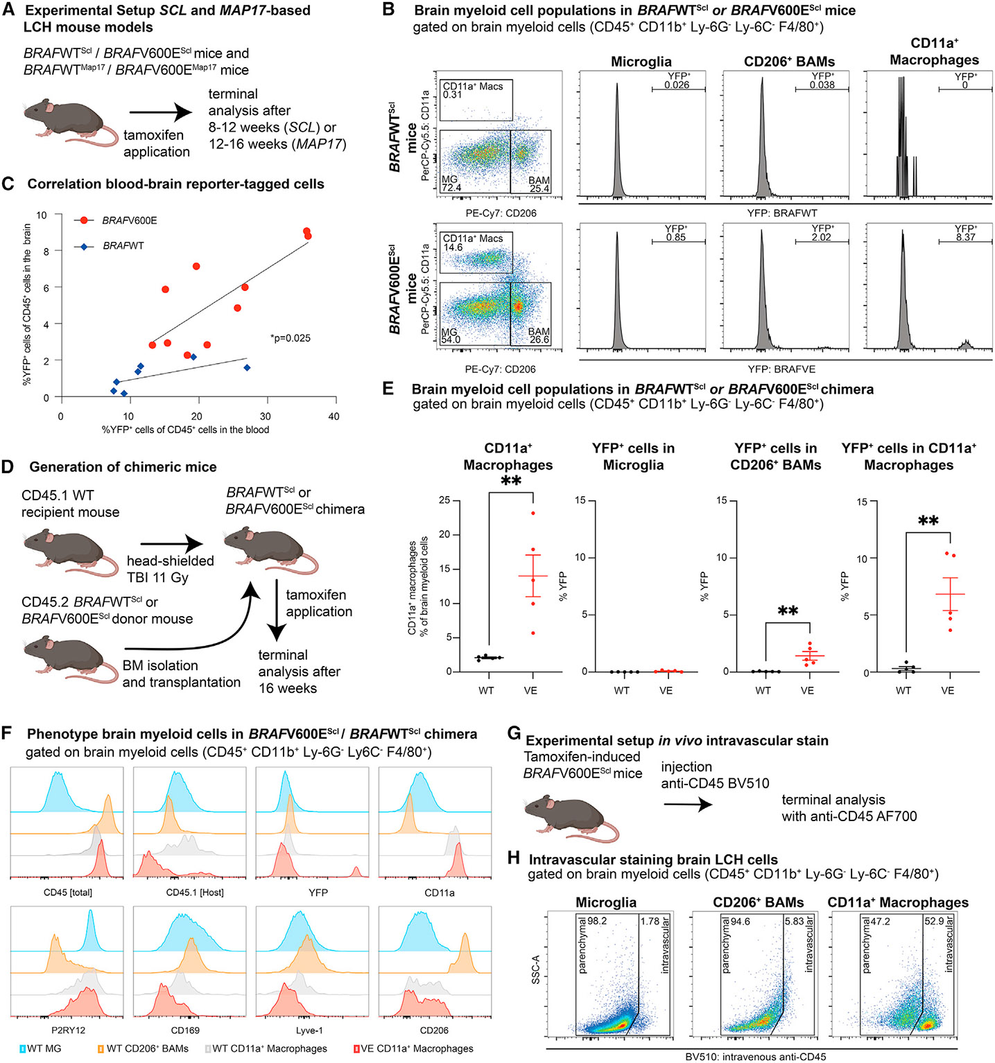
Neurodegenerative diseases (ND) are characterized by progressive loss of neuronal function. Mechanisms of ND pathogenesis are incompletely understood, hampering the development of effective therapies. Langerhans cell histiocytosis (LCH) is an inflammatory neoplastic disorder caused by hematopoietic progenitors expressing mitogen-activated protein kinase (MAPK)-activating mutations that differentiate into senescent myeloid cells that drive lesion formation. Some individuals with LCH subsequently develop progressive and incurable neurodegeneration (LCH-ND). Here, we showed that LCH-ND was caused by myeloid cells that were clonal with peripheral LCH cells. Circulating BRAFV600E+ myeloid cells caused the breakdown of the blood-brain barrier (BBB), enhancing migration into the brain parenchyma where they differentiated into senescent, inflammatory CD11a+ macrophages that accumulated in the brainstem and cerebellum. Blocking MAPK activity and senescence programs reduced peripheral inflammation, brain parenchymal infiltration, neuroinflammation, neuronal damage and improved neurological outcome in preclinical LCH-ND. MAPK activation and senescence programs in circulating myeloid cells represent targetable mechanisms of LCH-ND.

Keywords: LCH-ND; Langerhans cell histiocytosis; blood-brain barrier; histiocytic disorders; neurodegeneration; neuroinflammation.


Conflict of interest statement

Declaration of interests The authors declare no competing interests.

Figures

Figure 1.
Figure 1.. Genetic fate-mapping reveals that circulating BRAFV600E+ myeloid cells accumulate in the brains of mice with LCH-like disease
(A) Experimental setup of HSC-Scl and Map17-based LCH mouse models: tamoxifen was applied for Cre recombination, and mice were then terminally analyzed after 8–16 weeks as indicated. (B) Representative spectral cytometry pseudocolor and histogram plots of brain myeloid cells enriched using CD45+ beads with microglia (MG), CD206+ border-associated macrophages (BAMs) and CD11a+ macrophages from BRAFV600EScl mice and BRAFWTScl control mice. The abundance of reporter-tagged cells in these populations is displayed in the respective histogram plots. (C) Correlation between percentage of YFP-tagged cells in the peripheral blood and percentage of YFP-tagged cells in the brains of BRAFV600EScl mice and BRAFWTScl control mice (n = 7–10 mice per group, one experiment). (D) Experimental setup to generate BRAFV600EScl chimera and BRAFWTScl control chimera. (E) Statistical analysis of CD11a+ macrophages and percentage of YFP-expressing cells in Microglia, CD206+ BAMs, and CD11a+ macrophages of BRAFV600EScl chimera and BRAFWTScl control chimera (n = 5 mice per group, two independent experiments, Student’s t test). (F) Expression of key lineage markers in the different brain myeloid cell populations of BRAFV600EScl chimera and BRAFWTScl control chimera (n = 3 per group, two independent experiments). (G) Experimental setup of intravascular cell staining to determine vascular versus parenchymal localization of cells: BRAFV600EScl mice were injected with a BV510-conjugated anti-CD45 antibody and were shortly after terminally analyzed. (H) Representative spectral cytometry pseudocolor plots of intravenous CD45 staining for each myeloid cell population (n = 4 mice per group, two independent experiments). See also Figure S1.
Figure 2.
Figure 2.. LCH mice with brain-infiltrating BRAFV600E-mutated cells share phenotypic and transcriptional characteristics with human LCH-ND
(A) Manifestation of LCH-ND in a schematic human brain (created with biorender.com) and (B) Quantification of BRAFV600E transcripts from this specimen. (C) Visualization of the extent to which mouse brains are affected by infiltration with BRAFV600E-mutated cells (created with biorender.com) based on the quantification from different brain regions depicted in (D) (n = 4 mice, two independent experiments). (E) Representative IHC images from the brain of a BRAFWTScl chimera on the left and BRAFV600EScl chimera on the right stained for YFP-tagged, BM-derived cells marked with yellow arrows. In BRAFWTScl chimera, these cells are lineage-traced unmutated cells (left), and in BRAFV600EScl chimera, these stained cells are BRAFV600E+ cells (right). Scale bar, 150 μm in overview images and 25 μm in enlarged sections. (F–K) CD11a+ macrophages (F), peripheral blood monocytes (G), and microglia (H) from BRAFWTScl and BRAFV600EScl mice were subjected to bulk RNA sequencing from each 10 mice per group (pooled, one experiment). Gene expression profiles of CD11a+ macrophages (F), monocytes (G), and microglia (H) from BRAFWTScl mice were compared with those from BRAFV600EScl mice in (I) by analysis of the deviance of transcriptomes showing that the transcriptomic remodeling was strongest in CD11a+ macrophages. While monocytes and CD11a+ macrophages in BRAFV600EScl mice are BRAFV600E mutated, microglia are unmutated in BRAFV600EScl mice. (J) The transcriptional remodeling in CD11a+ macrophages was driven by senescence-associated genes, matrix metalloproteinases, and integrins. (K) Comparative analysis of gene expression of key lineage markers in mouse CD11a+ macrophages, peripheral blood monocytes, and microglia. (L) Comparison of these lineage markers between a human bulk RNA sequencing dataset from an LCH-ND case and human myeloid cells from a healthy brain demonstrating an enrichment for CD11a+ macrophage-defining Itgal (encoding integrin alpha L chain, CD11a) as well as Itga4 (encoding integrin alpha 4 chain, CD49d). (M) Cross-species comparison of mouse BRAFV600E+ CD11a+ macrophages and human LCH-ND material showing that 57.5% of the genes were enriched in mouse and human LCH material. Data in (D) are shown as means ± s.e.m, ****p < 0.0001. LCH-ND, neurodegenerative LCH; WT, wild type; VE, BRAFV600E. See also Figure S2.
Figure 3.
Figure 3.. BRAFV600EScl chimera have a compromised BBB and exhibit behavioral and neurologic abnormalities
(A) Experimental setup to quantify inflammatory cytokines from brain lysates from BRAFV600EScl and BRAFWTScl mice. (B) Multiplexed cytokine detection in the brains of BRAFV600EScl and BRAFWTScl control mice (n = 3–4 mice per group, one experiment). (C) From the multiplex protein dataset, a comparison between the fold change of cytokine concentration between WT and VE in the blood (x axis) and the brain (y axis). (D) Experimental setup of an Evans Blue-based assay to quantify the permeability of the BBB for small molecules. (E) Evans Blue experiment demonstrates increased permeability of BBB in BRAFV600Escl mice (n = 4–5 mice per group, two independent experiments). (F) Experimental setup to assess and quantify the transition of biotinylated IL-1β into the brain of BRAFV600EScl mice and BRAFWTScl control mice. (G) Representative images of IL-1β (cyan) and Collagen IV (red) showing a scattered perivascular signal derived from biotinylated IL-1β that can be detected in BRAFV600EScl mice but not BRAFWTScl control mice (n = 3–4 mice per group, two independent experiments). Scale bar, 50 μm. (H) Quantification of IL-1β-derived fluorescent signal shows a significant increase in extravasation of biotinylated IL-1β into the brain parenchyma in BRAFV600EScl mice compared to BRAFWTScl mice (n = 3–4 mice per group, two independent experiments, Student’s t test). (I) Experimental setup to assess the pericyte coverage of brain vessels from BRAFV600EScl and BRAFWTScl chimera. (J) BRAFV600EScl chimera have a decreased blood vessel coverage with pericytes compared to BRAFWTScl chimera (n = 3–4 mice per group, two independent experiments, Student’s t test). (K) Experimental setup for behavioral assays and histological studies for (L)–(Z). (L–P) Performance of BRAFV600EScl chimera (VE) and BRAFWTScl chimera (WT) in an open field setup showing a deteriorated performance with reduced resting time (L), distance traveled (M), time in center (N), and median velocity (O) summarized in the activity plots (P) (n = 7 mice per group, two independent experiments, Student’s t test). (Q and R) Grip strength (Q) and latency to fall (R) quantification of BRAFV600EScl and BRAFWTScl chimera in a Rotarod assay (n = 7 mice per group, two independent experiments, Student’s t test). (S–W) Footprint analysis of hindlimb and forelimb stride (S and T, respectively) as well as hindlimb and forelimb base as signs of a motor deficit in BRAFV600EScl chimera (U and V, respectively; n = 6–10 mice per group, pooled from two independent experiments). (W) Representative case of unilateral paralysis in a BRAFV600EScl chimeric mouse compared to a BRAFWTScl mouse; hind paws painted with blue ink and front paws painted with red ink. (X) Quantification of Purkinje cells in BRAFV600EScl chimera and BRAFWTScl chimera (n = 4 mice per group, two independent experiments, Student’s t test) and representative images of Calbindin-1 staining of mouse cerebella, scale bar, 50 μm in overview images and in enlarged sections. (Y) Quantification of GFAP+ brain regions of multiplex immunohistochemistry of the brains of BRAFV600EScl chimera (n = 4 mice per group, two independent experiments, Student’s t test). (Z) Representative images of multiplex immunohistochemistry of brains of BRAFV600EScl chimera staining for activated astrocytes (anti-GFAP, left), BRAFV600E-mutated cells (anti-YFP, middle), and showing co-localization (overlay, right). Student’s t test was used with *p < 0.05, **p < 0.01, ***p < 0.001, ****p < 0.0001, n.s., not significant. Data are shown as means ± s.e.m. r.o., retro orbital; GFAP, glial fibrillary acidic protein. See also Figure S3.
Figure 4.
Figure 4.. Accumulation of circulating, BRAFV600E+ cells is a driver of LCH-like disease and combined preventive senolytic/MAPK inhibitor therapy alleviates the disease burden
(A) Experimental setup for preventive in vivo drug treatment of BRAFWTScl mice and BRAFV600EScl mice with navitoclax/trametinib (NT) or vehicle control. (B) Percentage of YFP+ cells in the lungs of BRAFWTScl (WT) and BRAFV600EScl (VE) mice receiving vehicle or NT treatment analyzed by spectral cytometry (n = 5–6 per group, two independent experiments, ANOVA). (C) Percentage of CD11a+ macrophages in the brains of BRAFWTScl (WT) and BRAFV600EScl (VE) mice receiving vehicle or NT combination treatment (n = 5–6 per group, two independent experiments, ANOVA). (D) Experimental setup for in vivo drug treatment of BRAFWTScl chimera and BRAFV600EScl chimera with NT or vehicle control. (E and F) Quantification of the weight of the spleens (E) and the livers (F) of BRAFWTScl (WT) and BRAFV600EScl (VE) chimera receiving vehicle or NT treatment (n = 3–4 mice per group, two independent experiments, ANOVA). (G–J) Open Field assessment of vehicle- or NT-treated BRAFWTScl (WT) and BRAFV600EScl (VE) chimera with quantification of the resting time (G), distance traveled (H), time in the center (I) and mouse median velocity (J). (K) Movement heatmap (top row) and path traveled (middle row) in the Open Field assessment with paired anti-YFP IHC from cerebellar regions (bottom row, scale bar, 100 μm) of the respective mice. ANOVA with post-hoc analysis was used with *p < 0.05, **p < 0.01, ***p < 0.001, ****p < 0.0001, n.s., not significant. Data are shown as means ± s.e.m. NT, navitoclax/trametinib; IHC, immunohistochemistry. See also Figure S3.

Update of

Comment in

References

    1. Allen CE, Flores R, Rauch R, Dauser R, Murray JC, Puccetti D, Hsu DA, Sondel P, Hetherington M, Goldman S, and McClain KL (2010). Neurodegenerative central nervous system Langerhans cell histiocytosis and coincident hydrocephalus treated with vincristine/cytosine arabinoside. Pediatr. Blood Cancer 54, 416–423. - PMC - PubMed
    1. Héritier S, Barkaoui MA, Miron J, Thomas C, Moshous D, Lambilliotte A, Mazingue F, Kebaili K, Jeziorski E, Plat G, et al. (2018). Incidence and risk factors for clinical neurodegenerative Langerhans cell histiocytosis: a longitudinal cohort study. Br. J. Haematol 183, 608–617. - PubMed
    1. Bigenwald C, Berichel JL, Wilk CM, Chakraborty R, Chen ST, Tabachnikova A, Mancusi R, Abhyankar H, Casanova-Acebes M, Laface I, et al. (2021). BRAFV600E-induced senescence drives Langerhans cell histiocytosis pathophysiology. Nat Med 27, 851–861. - PMC - PubMed
    1. Allen CE, Merad M, and McClain KL (2018). Langerhans-Cell Histiocytosis. N. Engl. J. Med 379, 856–868. - PMC - PubMed
    1. Yeh EA, Greenberg J, Abla O, Longoni G, Diamond E, Hermiston M, Tran B, Rodriguez-Galindo C, Allen CE, and McClain KL; North American Consortium for Histiocytosis (2018). Evaluation and treatment of Langerhans cell histiocytosis patients with central nervous system abnormalities: Current views and new vistas. Pediatr. Blood Cancer 65, e26784. - PubMed

Publication types

MeSH terms

Substances