. 2024 Mar 30;120(4):345-359.
doi: 10.1093/cvr/cvad175.

An inflammation resolution-promoting intervention prevents atrial fibrillation caused by left ventricular dysfunction

Affiliations

An inflammation resolution-promoting intervention prevents atrial fibrillation caused by left ventricular dysfunction

Roddy Hiram et al. Cardiovasc Res. .

Abstract

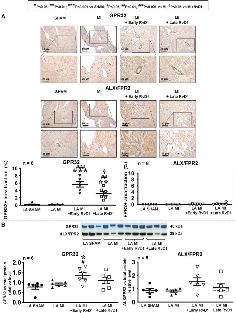
Aims: Recent studies suggest that bioactive mediators called resolvins promote an active resolution of inflammation. Inflammatory signalling is involved in the development of the substrate for atrial fibrillation (AF). The aim of this study is to evaluate the effects of resolvin-D1 on atrial arrhythmogenic remodelling resulting from left ventricular (LV) dysfunction induced by myocardial infarction (MI) in rats.

Methods and results: MI was produced by left anterior descending coronary artery ligation. Intervention groups received daily intraperitoneal resolvin-D1, beginning before MI surgery (early-RvD1) or Day 7 post-MI (late-RvD1) and continued until Day 21 post-MI. AF vulnerability was evaluated by performing an electrophysiological study. Atrial conduction was analysed by using optical mapping. Fibrosis was quantified by Masson's trichrome staining and gene expression by quantitative polymerase chain reaction and RNA sequencing. Investigators were blinded to group identity. Early-RvD1 significantly reduced MI size (17 ± 6%, vs. 39 ± 6% in vehicle-MI) and preserved LV ejection fraction; these were unaffected by late-RvD1. Transoesophageal pacing induced atrial tachyarrhythmia in 2/18 (11%) sham-operated rats, vs. 18/18 (100%) MI-only rats, in 5/18 (28%, P < 0.001 vs. MI) early-RvD1 MI rats, and in 7/12 (58%, P < 0.01) late-RvD1 MI rats. Atrial conduction velocity significantly decreased post-MI, an effect suppressed by RvD1 treatment. Both early-RvD1 and late-RvD1 limited MI-induced atrial fibrosis and prevented MI-induced increases in the atrial expression of inflammation-related and fibrosis-related biomarkers and pathways.

Conclusions: RvD1 suppressed MI-related atrial arrhythmogenic remodelling. Early-RvD1 had MI sparing and atrial remodelling suppressant effects, whereas late-RvD1 attenuated atrial remodelling and AF promotion without ventricular protection, revealing atrial-protective actions unrelated to ventricular function changes. These results point to inflammation resolution-promoting compounds as novel cardio-protective interventions with a particular interest in attenuating AF substrate development.

Keywords: Atrial fibrillation; Electrophysiology; Fibrosis; Myocardial infarction; Resolvin.


Conflict of interest statement

Conflict of interest: None declared.

Figures

Graphical Abstract
Graphical Abstract
Effects of RvD1 on an atrial fibrillation (AF) substrate resulting from myocardial infarction (MI)–induced left ventricular (LV) dysfunction. MI is characterized by a non-contractile scar that produces LV dysfunction. Early treatment with RvD1 (pre-MI) reduces the scar area and prevents LV dysfunction, whereas later RvD1 therapy (starting 7 days post-MI) does not affect MI scar or LV dysfunction. MI and associated LV dysfunction cause increased atrial inflammatory signalling and recruitment of pro-inflammatory M1 macrophages. RvD1 therapy reduces atrial inflammatory signalling and M1 macrophage recruitment, while enhancing the presence of anti-inflammatory M2 macrophages and increasing pro-resolution signalling. MI-induced inflammatory signalling causes fibrosis and atrial conduction abnormalities that lead to an AF-maintaining substrate; these changes are prevented by RvD1 treatment.
Figure 1
Figure 1
AF vulnerability, cardiac function, and atrial fibrosis. (A) Inducibility of AF and AFl during transoesophageal EPS in vivo. (B) Scar area on transverse sections 5 mm from the apex. (C) LVEF and (D) WMSI. (E–G). Fibrosis and atrial diameters (by echocardiography) for right (E and G) and left (F and H) atria. (Statistical analysis: one-way ANOVA followed by Bonferroni correction. Each point results from an individual animal.) The horizontal lines are mean ± SEM. n = 6 rats/group.
Figure 2
Figure 2
Atrial optical mapping and conduction-related genes. (A) AF induction by in situ EPS of the LA. (B) Representative LA activation maps at BCL 60 ms. (C) LA CV at BCL 60 ms. (D) LA ERP in MI, Sham, and RvD1-treated rats. (Statistical analysis: (A) Fisher’s exact test. (C) and (D) One-way ANOVA followed by Bonferroni correction.) Each point results from an individual animal. The horizontal lines are mean ± SEM. n = 6 rats/group.
Figure 3
Figure 3
Macrophage expression. Immunohistochemical staining for pro-inflammatory (M1) macrophage marker CD68 and anti-inflammatory (M2) macrophage marker CD206 (A). The arrows indicate immunochemically identified macrophages. (B–D) The quantification of immunohistochemical staining for LA (B), RA (C), and LV (D). The representative histological images are shown in Supplementary material online, Figures S17 and S18. (Statistical analysis: one-way ANOVA followed by Bonferroni correction.) Each point represents results from an individual animal. The horizontal lines are mean ± SEM. n = 6 rats/group.
Figure 4
Figure 4
Inflammasome-related and fibrosis-related genes. Gene expression level evaluated by RT–qPCR analysis for inflammation-related genes Il6 (A), Il1b (B), Cxcl1 (C), and Cxcl2 (D) and for NLRP3 inflammasome components Nlrp3 (E), Asc (F), Casp1 (G), and Casp8 (H) in RA and LA from Sham, MI, and RvD1-treated rats. (Each point represents the level of expression from an individual animal. The results are expressed as mean ± SEM. Statistically significant differences were defined as two-tailed P-values <0.05; n = 6 rats/group.)
Figure 5
Figure 5
Inflammation-related and fibrosis-related protein expression. A western blot analysis of the LA protein expression of NLRP3-related compounds: NLRP3 (A and B), ASC (A and C), PRO-CASP1 (A and D); proinflammatory interleukin IL6 (A and E); and fibrosis-related protein TGFβ3 (A and F). The images correspond to protein bands from gels on which they were run compared with total proteins on the blot (A). Uncropped membrane images are available in Supplementary material online, Figure S22. (Each point represents the level of expression from an individual animal. The results are expressed as mean ± SEM. Statistically significant differences were defined as two-tailed P-values <0.05; n = 6 rats/group.)
Figure 6
Figure 6
The LA expression of RvD1 receptors. (A) Immunohistochemical staining of RvD1 receptors GPR32 and ALX/FPR2 in LA from Sham, MI, and RvD1-treated rats, 21st day post-MI. The graphs show the area fraction (cross-sectional area occupied by immunostaining relative to the total cross-sectional area as a percentage, by colorimetric analysis) of the atrial GPR32-positive and ALX/FPR2-positive zones in LA. (B) A western blot analysis of the LA protein expression of RvD1 receptors GPR32 (left panel) and ALX/FPR2 (right panel). The images correspond to protein bands from gels on which they were run. Uncropped membrane images are available in Supplementary material online, Figure S22. Total protein blot images are available in Supplementary material online, Figure S23. (Statistical analysis: one-way ANOVA followed by Bonferroni correction.) Each point represents results from an individual animal. The horizontal lines are mean ± SEM. n = 6 rats/group.
Figure 7
Figure 7
The expression of RvD1 receptors in AF patients. A western blot analysis of the atrial protein expression of GPR32 (A and B) and ALX/FPR2 (A and C). The images correspond to protein bands from gels on which they were run compared with GAPDH. Uncropped-membrane images are available in Supplementary material online, Figure S24. The gene expressions of GPR32 (D) and ALX/FPR2 (E) were also analysed with dPCR. (Statistical analysis: Student’s t-test.) Each point represents results from an individual patient. The horizontal lines are mean ± SEM. n = 11 patients/group for immunoblot, n = 6 patients/group for dPCR.

Comment in

References

    1. Thihalolipavan S, Morin DP. Atrial fibrillation and congestive heart failure. Heart Fail Clin 2014;10:305–318. - PubMed
    1. Hiram R, Naud P, Xiong F, Al-U’Datt D, Algalarrondo V, Sirois MG, Tanguay JF, Tardif JC, Nattel S. Right atrial mechanisms of atrial fibrillation in a rat model of right heart disease. J Am Coll Cardiol 2019;74:1332–1347. - PubMed
    1. Harada M, Nattel S. Implications of inflammation and fibrosis in atrial fibrillation pathophysiology. Card Electrophysiol Clin 2021;13:25–35. - PubMed
    1. Nattel S, Heijman J, Zhou L, Dobrev D. Molecular basis of atrial fibrillation pathophysiology and therapy: a translational perspective. Circ Res 2020;127:51–72. - PMC - PubMed
    1. Serhan CN. Pro-resolving lipid mediators are leads for resolution physiology. Nature 2014;510:92–101. - PMC - PubMed

Publication types

MeSH terms

Grants and funding