. 2024 Jan 9;57(1):52-67.e10.
doi: 10.1016/j.immuni.2023.11.007. Epub 2023 Dec 12.

trans-Endothelial neutrophil migration activates bactericidal function via Piezo1 mechanosensing

Affiliations

trans-Endothelial neutrophil migration activates bactericidal function via Piezo1 mechanosensing

Amitabha Mukhopadhyay et al. Immunity. .

Abstract

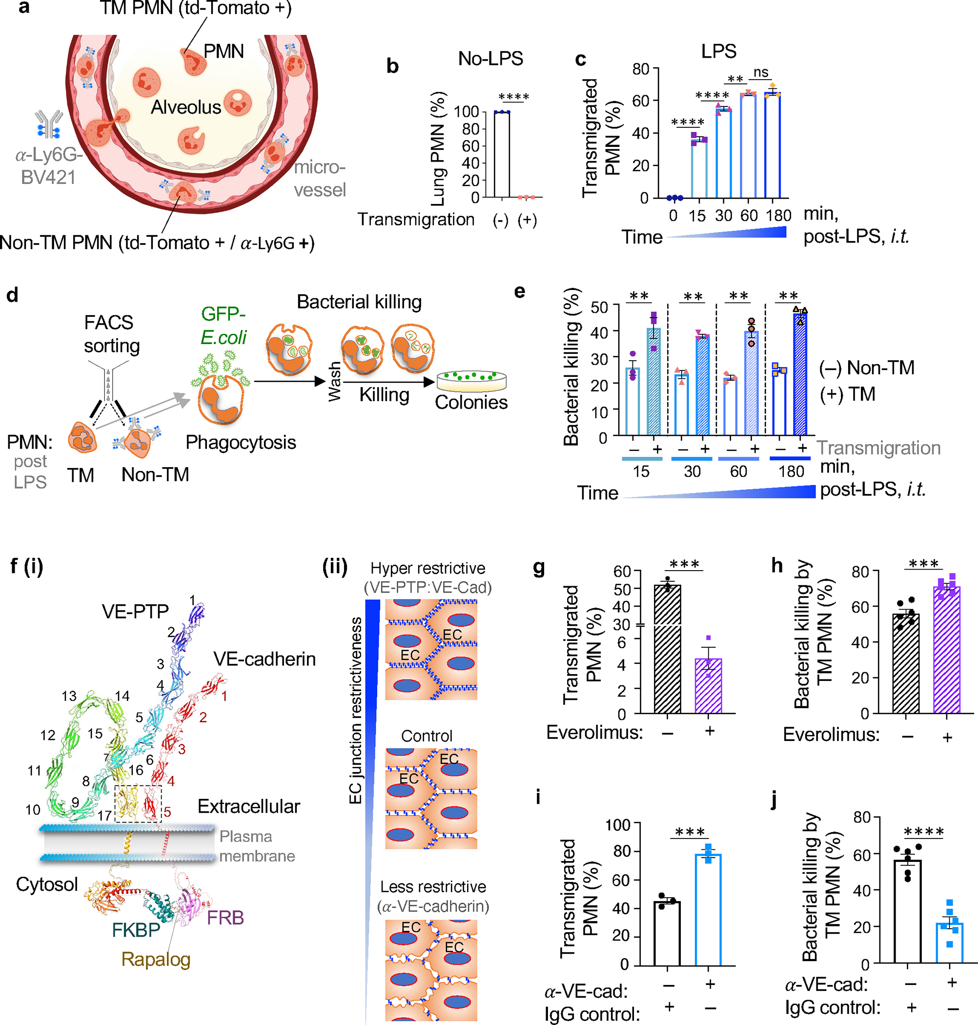
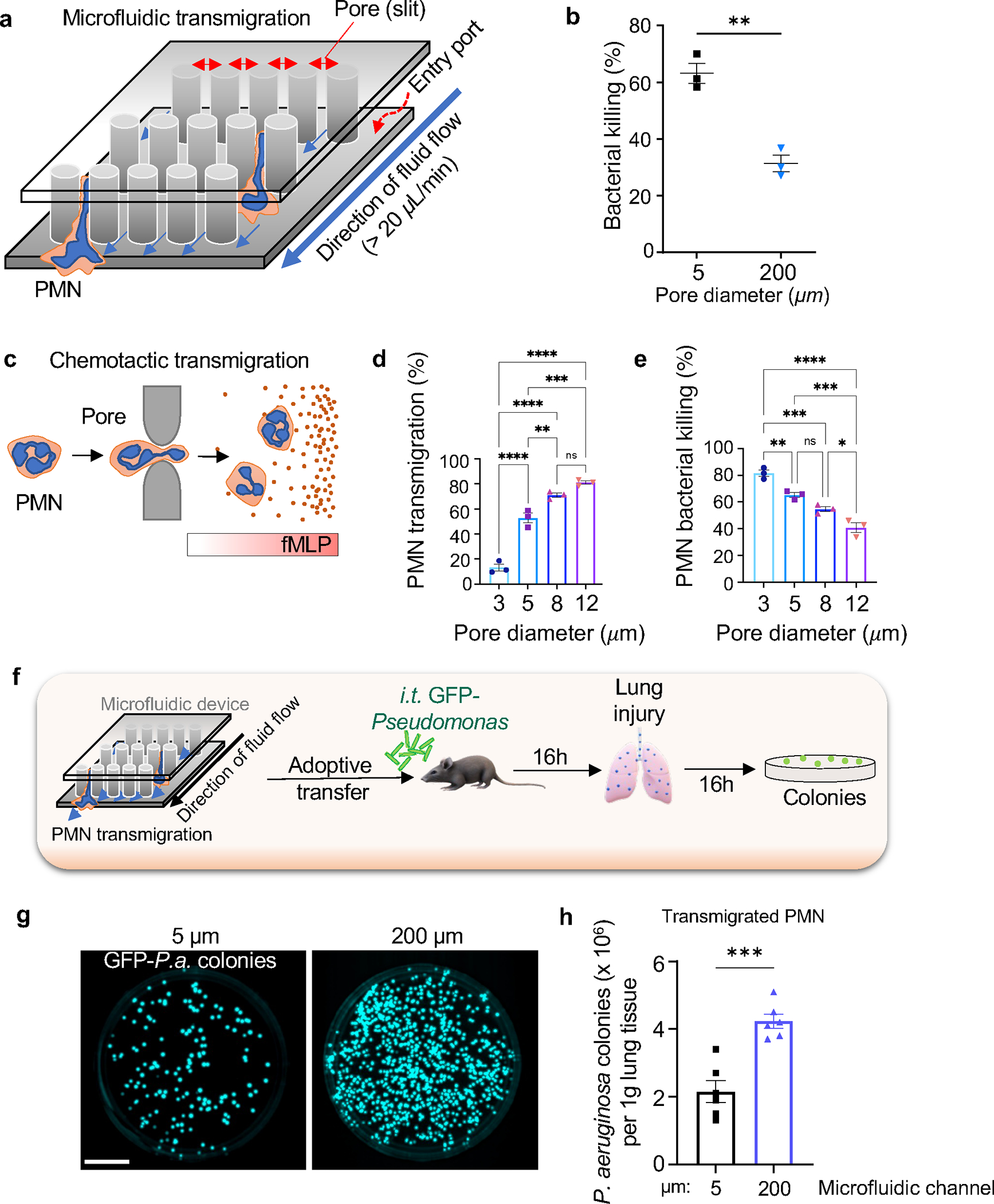
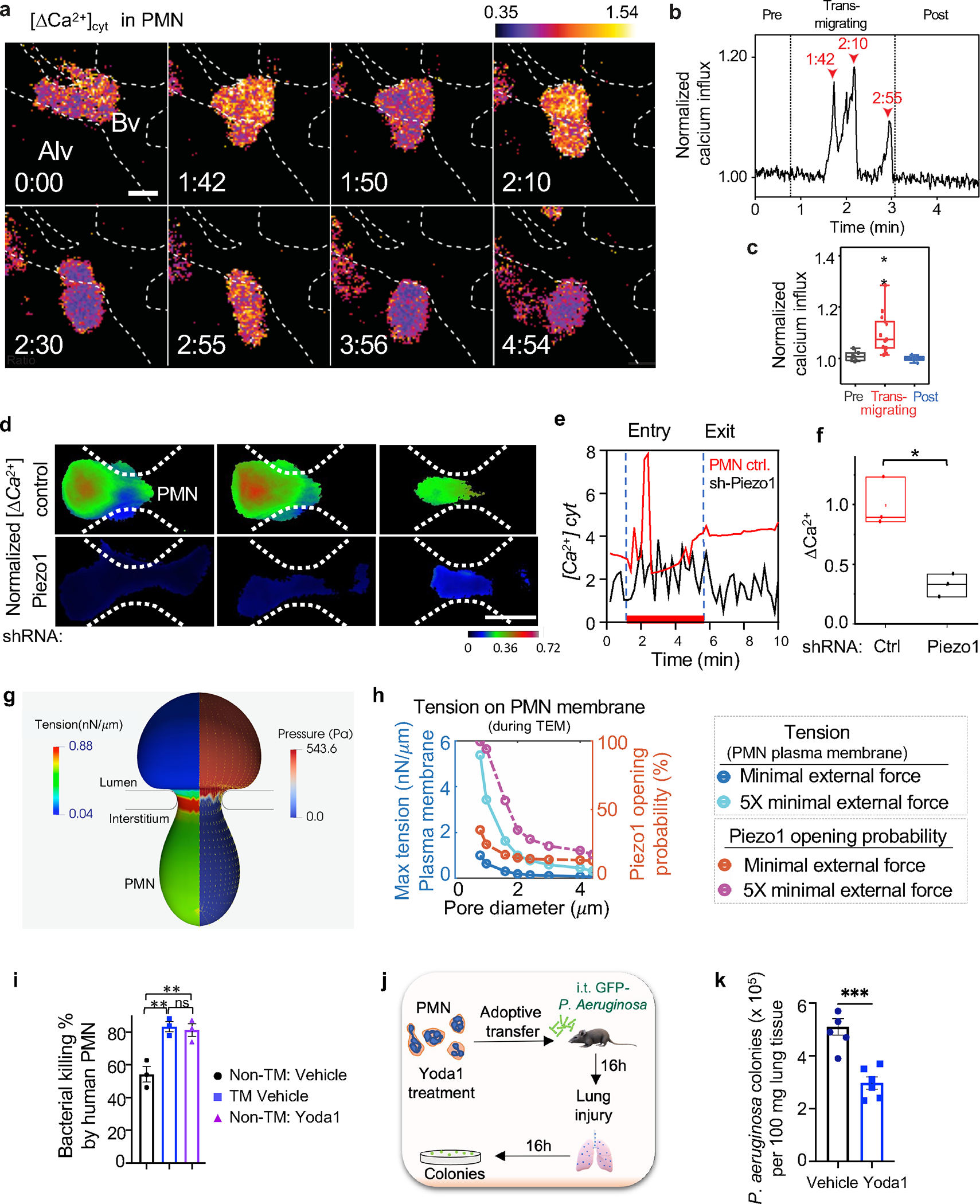
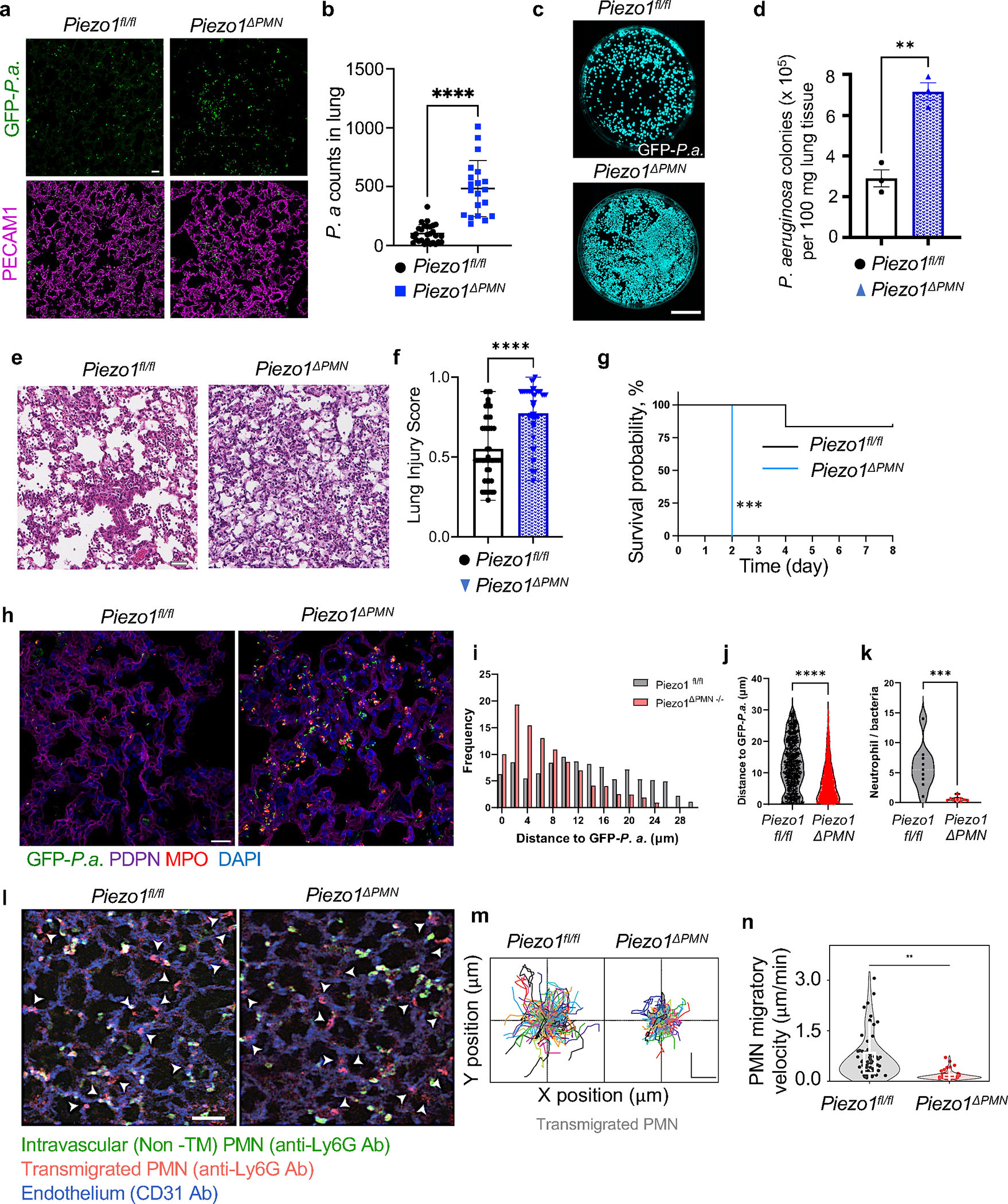
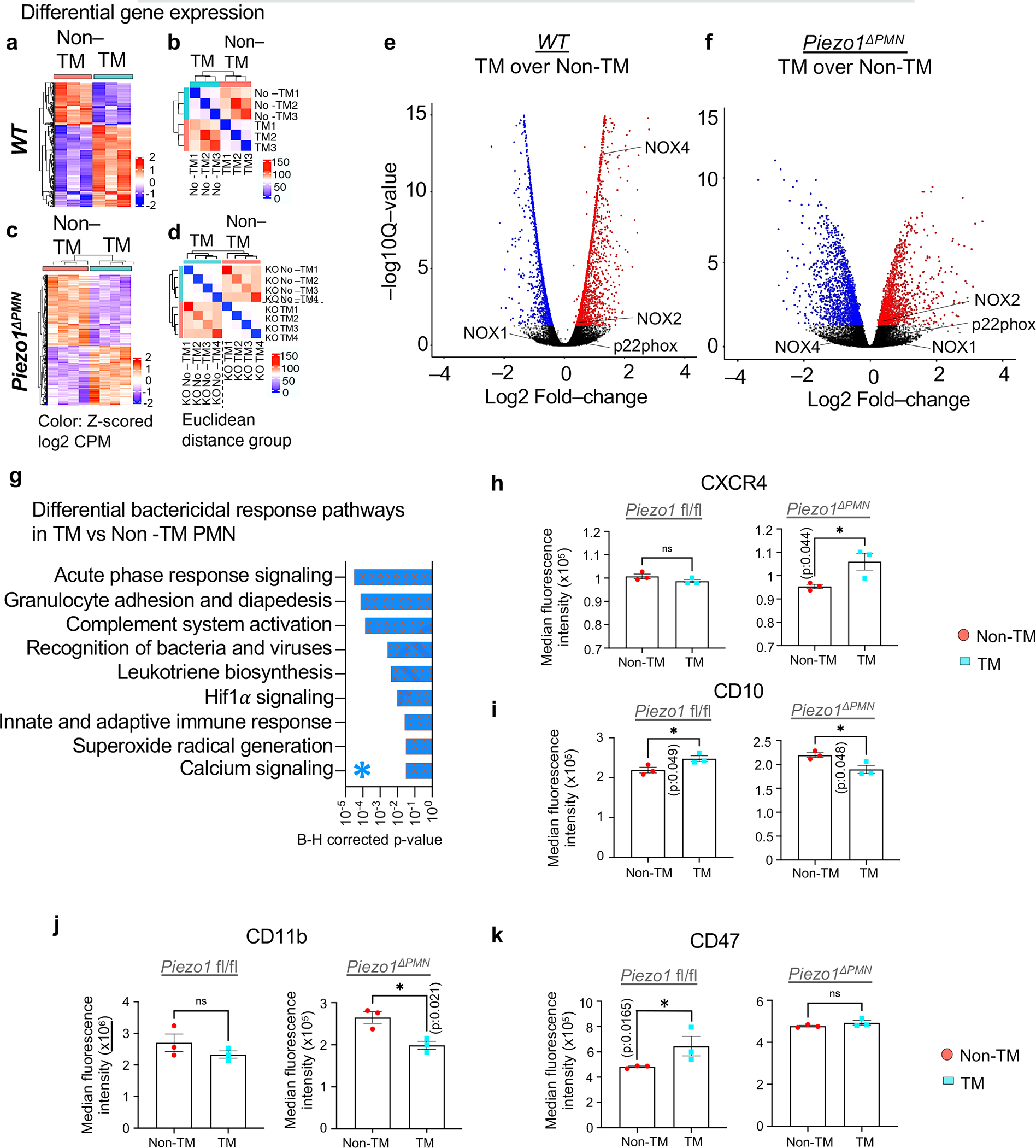
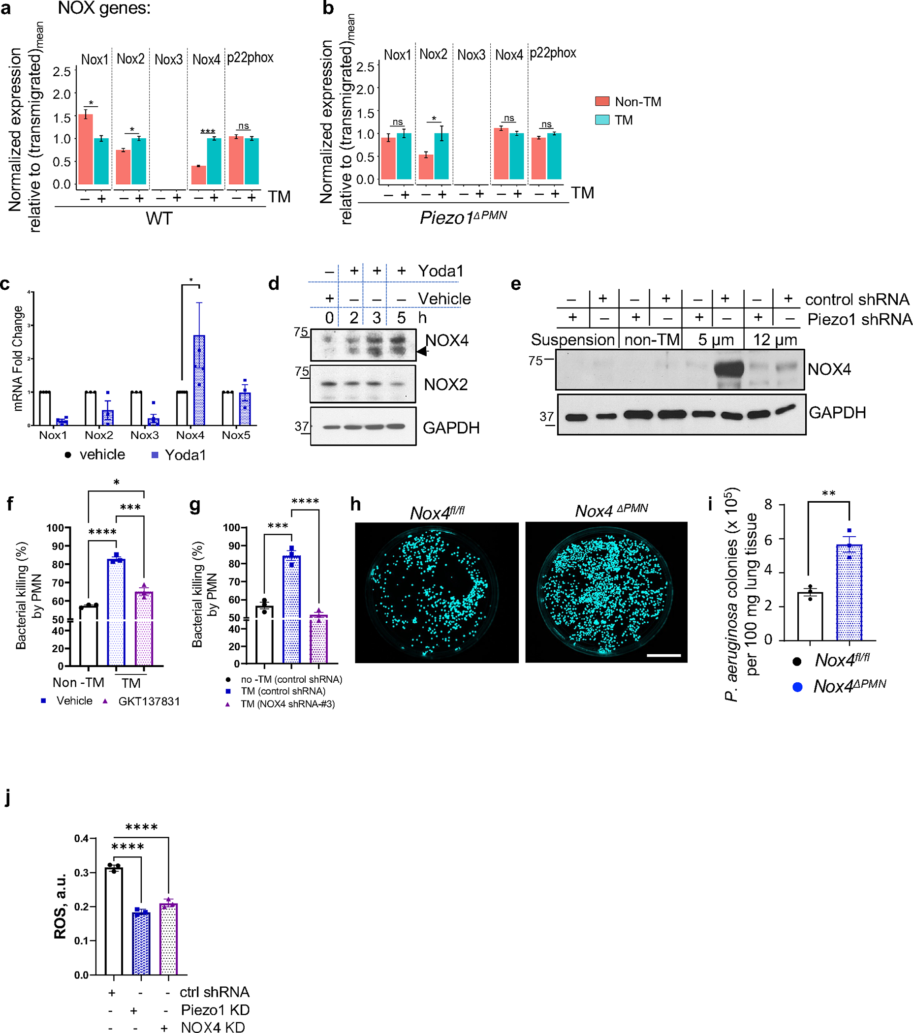
The regulation of polymorphonuclear leukocyte (PMN) function by mechanical forces encountered during their migration across restrictive endothelial cell junctions is not well understood. Using genetic, imaging, microfluidic, and in vivo approaches, we demonstrated that the mechanosensor Piezo1 in PMN plasmalemma induced spike-like Ca2+ signals during trans-endothelial migration. Mechanosensing increased the bactericidal function of PMN entering tissue. Mice in which Piezo1 in PMNs was genetically deleted were defective in clearing bacteria, and their lungs were predisposed to severe infection. Adoptive transfer of Piezo1-activated PMNs into the lungs of Pseudomonas aeruginosa-infected mice or exposing PMNs to defined mechanical forces in microfluidic systems improved bacterial clearance phenotype of PMNs. Piezo1 transduced the mechanical signals activated during transmigration to upregulate nicotinamide adenine dinucleotide phosphate (NADPH) oxidase 4, crucial for the increased PMN bactericidal activity. Thus, Piezo1 mechanosensing of increased PMN tension, while traversing the narrow endothelial adherens junctions, is a central mechanism activating the host-defense function of transmigrating PMNs.

Keywords: Nox4; Piezo1; calcium signaling; mechanical signaling; neutrophil; phagocytosis; pneumonia.


Conflict of interest statement

Declaration of interests The authors declare no competing interests.

Figures

Figure 1.
Figure 1.. PMN transmigration in endothelial AJs promotes PMN bactericidal activity.
a. Schematic of the method used to separate transmigrated and non-transmigrated PMN across lung micro-vessels. All PMN from Ly6GCre td-Tomato +/− mice expressed td-Tomato as shown in Suppl Fig 1a. Intravascular PMN were additionally labelled by α-Ly6G-BV421 antibody injected i.v. 2m prior to euthanasia. The number of extravasated td-Tomato PMN relative to intravascular PMN stained with α-Ly6G-BV421 was calculated to quantify trans-vascular PMN migration. b-c. Percentage transmigrated (TM) and non-transmigrated (non-TM) PMN assessed by FACS analysis in naïve mice and mice exposed to 5mg/ml insufflated (i.t.) LPS for different times (as shown in Suppl. Figure 1c). d. Workflow to assess bactericidal activity of transmigrated vs. non-transmigrated PMN. Flow sorted transmigrated and non-transmigrated lung PMN were incubated with GFP-E.coli for 1.5h determine bacteria phagocytosis and number of live bacteria was determined at 3.5h after bacteria washout. e. Bactericidal activity represented as % of killed GFP-E.coli normalized to phagocytosed GFP-E.coli (Suppl Figure 1f) of transmigrated vs. non-transmigrated lung PMN obtained at different times post-LPS challenge as in c. At each time point, transmigrated PMN induced 2-fold increase in bacterial killing. Data are obtained from 3 mice per group from 3 independent experiments. f(i-ii). (i) Model demonstrating the method of increasing the restrictiveness of endothelial AJs through expression of engineered VE-cadherin-FK506 and VE-PTP-FRB fusion proteins in mice as in ; this method stabilized VE-cadherin-VE-PTP interaction, and increased junction barrier restrictiveness on treating with the drug everolimus. (ii) Schema of the approaches used to either increase or decrease restrictiveness of AJs. g-h. Percent of transmigrated PMN into lungs (g) and bactericidal activity of transmigrated PMN (h) in transgenic mice treated with everolimus (“+”) or vehicle control (“−“). Bactericidal activity of PMN transmigrating through restrictive AJs of mice treated with everolimus was greater than control. i-j. Percent of transmigrated PMN into lungs (i) and bactericidal activity of transmigrated lung PMN (j) of C57B6 mice treated with isotype-matched antibody as well as functional blocking α-VE-cadherin antibody used to disassemble AJs. PMN bactericidal activity was reduced in lungs after α-VE-cadherin antibody-induced opening of AJs. Data are obtained from 3–6 mice per group from 3 independent experiments. Additional information is provided as Suppl. Figure 1.
Figure 2.
Figure 2.. PMN transmigration via restrictive pores in a microfluidic system enhances bactericidal activity of PMN.
a. Design of the microfluidic system containing pores through which PMN transmigrated. b. Bone-marrow murine PMN transmigrated through 5μm diameter pores showed 2-fold greater E. coli killing ex vivo as compared to PMN transmigrating through 200μm diameter pores. Data are obtained from 3 independent experiments. c. Bone-marrow murine PMN transmigration through the Transwell system with pore diameters ranging from 3 to 12μm in response to a gradient of the chemoattractant fMLP. d. Percentage of transmigrated PMN through different pore sizes as shown in c. PMN transmigration is a direct function of pore diameter. e. Bactericidal activity of PMN transmigrated through different pore diameters as in c. PMN transmigrating through smaller pore augmented bacterial killing. Data are from 3 independent experiments. f. Schematic of the adoptive transfer experiment in which mice challenged with GFP-Pseudomonas aeruginosa (GFP-P.a.) received 1×10⁶ PMN through i.t. route. g-h. Infected mice adoptively transferred with bone-marrow-derived murine PMN subjected to transmigration through 5μm diameter pores (as in a) showed increased clearance of Pseudomonas aeruginosa as compared to control PMN passed through 200 μm pores. Data are obtained from 6 mice per group from 6 independent experiments. Scale bar, 25 mm. Additional information is provided as Suppl. Figure 2a–b.
Figure 3.
Figure 3.. PMN migration through endothelial junctions activates calcium signaling via Piezo1 in PMN.
a. Ca2+ transients in PMN determined by motion-corrected 2-photon lung intra-vital imaging of the Ca2+ indicator GCaMP6f (green) before, during, and after PMN transmigration in endothelial AJs of lung micro-vessels. PMN reporter was td-Tomato expressed under Ly6G promoter (red); endothelial junction labeled by anti-PECAM1antibody (blue). Realtime data is supplied as supplementary Video S1. Scale, 0.3 mm/pixel; image capture rate, 2 seconds/frame. The color scheme shows blue as the lowest and yellow as highest increase in cytosolic [Ca2+]. The dotted line shows the lung capillary wall with a monolayer of endothelial cells. Bv, blood vessel; Alv, alveolus. Time is in min and sec. Scale bar, 5μm. b. Changes in cytosolic [Ca2+] during PMN transmigration in a. Arrowheads indicate Ca2+ spikes and corresponding times during PMN transmigration. c. Quantification of Ca2+ peaks normalized to baseline fluorescence prior to transmigration. Data are presented for 5 transmigration events observed in 5 mice. The boundaries of the box-plot indicate the 25th and 75th percentiles; whiskers show 1.5x interquartile range; median is shown by the straight line. d. Time-lapse images of Fluo4 in control and shRNA silenced Piezo1 PMN (HL-60 derived) migrating through 5μm pores of the microfluidic system. Scale bar, 8 μm. Individual Fluo4 tracings (e) and quantifications (f) demonstrating changes in cytosolic [Ca2+] in control and Piezo1 deleted PMN in d. The boundaries of the boxplot indicate the 25th and 75th percentiles. The median is shown by the straight line. Data are obtained from 3 independent experiments. g-h. Finite element simulations of PMN (8.5μm in diameter) transmigrating through the restrictive AJs. g. A cross-sectional view of coupled finite element and boundary element simulations of predicted tension and pressure on PMN plasma membrane during passage through a pore with diameter of 2.4μm under external force of 1.8nN, which is about 5 times the minimal external force required to push the PMN through the pore. The color maps show color-coded ranges for PMN membrane tension and pressure. h. Graph demonstrating maximum tension values at the PMN membrane (dark blue and cyan) and activation probability of the tension sensor Piezo1 (brown and pink) during PMN migration as a function of pore diameter under two different external forces, the minimal critical external force (dark blue and brown) and five times (5x) the minimal external force (cyan and pink) needed for PMN to pass through pores. i. Percent of E. coli killed by human PMN cultured under static condition and treated with vehicle control or Yoda1 to induce Piezo1 activation or post-transmigration through 5μm pores of the microfluidic system. Data are obtained from 3 independent experiments. The response was increased by Yoda1 treatment to the same degree as passing PMN through 5μm pores. j-k. Mice received i.t. adoptive transfer of 106 bone marrow derived murine PMN pretreated with Yoda1 showed marked improvement of Pseudomonas aeruginosa clearance at 16h post-i.t. instillation of 106 cfu bacteria per mouse as compared to vehicle control. Data were obtained from 5–6 mice per group from 3 independent experiments. Additional information is provided as Suppl. Figure 2c–j & 3a–e.
Figure 4.
Figure 4.. Activation of Piezo1 in transmigrated PMN increases bactericidal activity.
a-k. Genetic deletion of Piezo1 in PMN augmented lung infection due to diminished antimicrobial activity of PMN. a. Representative images of lung sections stained for GFP-Pseudomonas aeruginosa (green) and PECAM-1 (magenta) and b. quantification of GFP-Pseudomonas aeruginosa counts in lung tissue of control (Piezo1fl/fl) and PMN-specific Piezo1 deleted (Piezo1ΔPMN) mice challenged with 106 cfu bacteria per mouse for 12h. Scale bar, 20μm. c. Images of GFP-expressing Pseudomonas aeruginosa colonies grown on agar plates as in a-b. Scale bar, 25 mm. d. Quantification of data in c. Data were obtained from 3 mice per group from 3 independent experiments. e-f. Histopathological assessment of lung injury induced by Pseudomonas aeruginosa in Piezo1ΔPMN vs. control mice. Representative images of H&E-stained lung tissue from 3 independent experiments (e) and lung injury scores (f) of mice as in a-b. Scale bar, 20μm. g. Survival rates of mice challenged with i.t. 106 cfu Pseudomonas aeruginosa per mouse. Genetic deletion of Piezo1 in PMN markedly enhanced mortality. Data were obtained from 9 mice per group from 3 independent experiments. h-k. Genetic deletion of Piezo1 in PMN reduced PMN-to-bacteria ratio. h. Representative images of lung sections stained for GFP-Pseudomonas aeruginosa (green), podoplanin (PDPN, magenta) from 3 independent experiments were used for tissue architecture assessment, PMN marker myeloperoxidase (MPO, red), and 4′,6-diamidino-2-phenylindole (DAPI, blue). Scale bar, 20μm. i-k. histogram distribution (i) and mean distance (j) between PMN and GFP-Pseudomonas aeruginosa in lung tissue; and PMN-to-bacteria ratio (k) in lungs of control (Piezo1fl/fl) and PMN-specific Piezo1-deleted (Piezo1ΔPMN) mice as in a-b. l-n. Genetic deletion of Piezo1 in PMN reduced migratory velocity of PMN in lung tissue. l. 2 photon microscopic images of lung tissue PMN of control (Piezo1fl/fl) and PMN-specific Piezo1-deleted (Piezo1ΔPMN) mice 4h after challenge with insufflated LPS. Total lung intravascular and tissue PMN (red) were detected by i.v. staining of PMN with Alexa594-labeled α-Ly6G antibody before LPS insufflation. Intravascular PMN (green) were stained with BV421-labeled α-Ly6G antibody prior to intravital imaging. Endothelium (blue) was stained with α-CD31 antibody. Arrow heads show transmigrated PMN in extravascular tissue. Scale bar, 50μm. m-n. PMN migratory trajectory (m) and PMN migratory velocity (n) of transmigrated PMN. Piezo1ΔPMN PMN showed reduced migration velocity which was coupled to reduced bacteria killing as shown in a-d and k. Scale bar, 20μm. Data are obtained from 3 mice per group from 6 independent experiments. Additional information is provided as Suppl. Figure 3.
Figure 5.
Figure 5.. Genetic analysis of transmigrated and non-transmigrated PMN in lungs.
a-d. RNA-seq analysis of transmigrated vs non-transmigrated lung PMN of control (a-b) and PMN-specific Piezo1 mutant (Piezo1ΔPMN) mice (c-d). See supplementary Table S1 for details. a, c. Heat maps of gene expression profiles of non-transmigrated vs. transmigrated PMN in lungs of control mice and LPS insufflated mice as in Fig 1a–b, b, d Sample-to-sample distance matrix with color intensity representing Euclidean distance in the gene expression space. Data were obtained from 4 mice per group from 3 independent experiments. e-f. Volcano plots showing the overall changes in gene expression in wild-type and Piezo1ΔPMN PMN as a function of transmigration. NOX4 was upregulated in transmigrated wildtype PMN as compared to Piezo1ΔPMN PMN. g. The most significant bactericidal pathways derived from IPA analysis are shown (see supplementary Table S1 for further details). HIf1α and Ca2+ signaling pathways are among most altered pathways in Piezo1ΔPMN PMN. h-k. Changes in protein-level expression of cell surface markers CXCR4 (h), CD10 (i), CD11b (j), and CD47 (k) in control and Piezo1ΔPMN PMN upon transmigration as assessed by spectral flow-cytometry. Compared to control, the surface expression of CD47 and CD10 were reduced in transmigrated Piezo1ΔPMN PMN. Results are shown as mean fluorescence intensity. Data are obtained from 3 mice per group. Additional information is provided as Suppl. Figure 4 & 5.
Figure 6.
Figure 6.. Piezo1 upregulates Nox4 expression to activate bactericidal activity of transmigrated PMN.
a-b. Changes in expression of NOX isoforms in transmigrated vs. non-transmigrated lung PMN from control (a) and Piezo1ΔPMN PMN (b), as in Fig. 5a, as assessed by RNA-seq analysis (details in supplementary Table S1). Increase in NOX4 expression was greater relative to NOX2 in the transmigrated PMN of control but not Piezo1ΔPMN mice. c. Yoda1 treatment of freshly isolated human PMN showed increased NOX4 mRNA expression as early as 30 min. Data were obtained from 5 healthy donors from 5 independent experiments. d. Western blot showing time-course of NOX2 and NOX4 expression in human PMN treated with Yoda1. Yoda1 increased only NOX4 protein expression. e. NOX4 protein expression is upregulated in control but not in Piezo1 depleted HL-60 derived PMN at 2 hr post-transmigration through the Transwell system with of 5μm pore diameter. NOX4 was not upregulated in fMLP-exposed PMN grown in suspension or in non-transmigrated PMN. Data are obtained of 3 independent experiments. f-g. Pharmacological inhibition (f) or genetic ablation (g) of NOX4 function in bone marrow and HL-60 derived PMN, respectively, reversed the augmented bacterial killing responses induced by PMN transmigration. Data are obtained from 3 independent experiments. h-i. Bacterial clearance was abrogated in lungs of Nox4ΔPMN mice. Results show Pseudomonas aeruginosa colonies in lungs of Nox4fl/fl and Nox4ΔPMN mice challenged with 106 cfu Pseudomonas aeruginosa per mouse for 16 hr. Lung tissue was lysed and plated on agar plates, and images of Pseudomonas aeruginosa colonies (h) and quantification (i) are shown. Data were obtained from 3 mice from 3 independent experiments. Scale bar, 25 mm. j. Piezo1 and downstream expression of NOX4 are required for ROS generation in transmigrated PMN. ROS production measured in HL-60 derived PMN stably transduced with control, Piezo1, or NOX4 shRNA lentiviral particles. ROS concentration was assessed at 1.5 h post-PMN transmigration through the microfluidic system. Data are obtained from 3 independent experiments. Additional information is provided as Suppl. Figure 6.

Comment in

References

    1. Degrossoli A, Muller A, Xie K, Schneider JF, Bader V, Winklhofer KF, Meyer AJ, and Leichert LI (2018). Neutrophil-generated HOCl leads to non-specific thiol oxidation in phagocytized bacteria. Elife 7. 10.7554/eLife.32288. - DOI - PMC - PubMed
    1. Dziarski R, Platt KA, Gelius E, Steiner H, and Gupta D (2003). Defect in neutrophil killing and increased susceptibility to infection with nonpathogenic gram-positive bacteria in peptidoglycan recognition protein-S (PGRP-S)-deficient mice. Blood 102, 689–697. 10.1182/blood-2002-12-3853. - DOI - PubMed
    1. Balamayooran G, Batra S, Fessler MB, Happel KI, and Jeyaseelan S (2010). Mechanisms of neutrophil accumulation in the lungs against bacteria. Am J Respir Cell Mol Biol 43, 5–16. 10.1165/rcmb.2009-0047TR. - DOI - PMC - PubMed
    1. Yipp BG, Kim JH, Lima R, Zbytnuik LD, Petri B, Swanlund N, Ho M, Szeto VG, Tak T, Koenderman L, et al. (2017). The Lung is a Host Defense Niche for Immediate Neutrophil-Mediated Vascular Protection. Sci Immunol 2. 10.1126/sciimmunol.aam8929. - DOI - PMC - PubMed
    1. Sekheri M, El Kebir D, Edner N, and Filep JG (2020). 15-Epi-LXA(4) and 17-epi-RvD1 restore TLR9-mediated impaired neutrophil phagocytosis and accelerate resolution of lung inflammation. Proc Natl Acad Sci U S A 117, 7971–7980. 10.1073/pnas.1920193117. - DOI - PMC - PubMed

Publication types