Review
. 2024 Jan:98:101231.
doi: 10.1016/j.preteyeres.2023.101231. Epub 2023 Dec 12.

The endoplasmic reticulum: Homeostasis and crosstalk in retinal health and disease

Affiliations
Review

The endoplasmic reticulum: Homeostasis and crosstalk in retinal health and disease

Sarah X Zhang et al. Prog Retin Eye Res. 2024 Jan.

Abstract

The endoplasmic reticulum (ER) is the largest intracellular organelle carrying out a broad range of important cellular functions including protein biosynthesis, folding, and trafficking, lipid and sterol biosynthesis, carbohydrate metabolism, and calcium storage and gated release. In addition, the ER makes close contact with multiple intracellular organelles such as mitochondria and the plasma membrane to actively regulate the biogenesis, remodeling, and function of these organelles. Therefore, maintaining a homeostatic and functional ER is critical for the survival and function of cells. This vital process is implemented through well-orchestrated signaling pathways of the unfolded protein response (UPR). The UPR is activated when misfolded or unfolded proteins accumulate in the ER, a condition known as ER stress, and functions to restore ER homeostasis thus promoting cell survival. However, prolonged activation or dysregulation of the UPR can lead to cell death and other detrimental events such as inflammation and oxidative stress; these processes are implicated in the pathogenesis of many human diseases including retinal disorders. In this review manuscript, we discuss the unique features of the ER and ER stress signaling in the retina and retinal neurons and describe recent advances in the research to uncover the role of ER stress signaling in neurodegenerative retinal diseases including age-related macular degeneration, inherited retinal degeneration, achromatopsia and cone diseases, and diabetic retinopathy. In some chapters, we highlight the complex interactions between the ER and other intracellular organelles focusing on mitochondria and illustrate how ER stress signaling regulates common cellular stress pathways such as autophagy. We also touch upon the integrated stress response in retinal degeneration and diabetic retinopathy. Finally, we provide an update on the current development of pharmacological agents targeting the UPR response and discuss some unresolved questions and knowledge gaps to be addressed by future research.

Keywords: Autophagy; Diabetic retinopathy; Endoplasmic reticulum; Integrated stress response; Mitochondria; Protein homeostasis; Retina; Retinal degeneration; Unfolded protein response.


Conflict of interest statement

Declaration of competing interest None.

Figures

Fig. 1.
Fig. 1.
ER localization in retinal neurons. A) Morphology and distribution of ER in the soma, axon, presynaptic terminals of a neuron. 1) Rough ER is distributed around the nuclear envelope in the soma and in somato-dendritic regions. 2) Smooth ER is localized predominantly to distal dendritic regions and axons. 3) The ER forms physical contacts with mitochondria, microtubules, endosomes, and lysosomes to support axon growth and organelle transportation. 4) In presynaptic terminals, the ER contributes to neurotransmitter production and regulation of calcium signaling. B) Distribution of ER in rod and cone photoreceptors.
Fig. 2.
Fig. 2.
The IRE1/XBP1 signaling pathway. In resting cells, ER stress sensors including IRE1 bind to Bip/GRP78, which keeps them in inactive state. 1) Upon ER stress, Bip dissociates from IRE1 and binds to accumulated unfolded or misfolded proteins. 2) IRE1 is activated by dimerization and autophosphorylation. Increased kinase activity of IRE1 promotes JNK and IKK activation resulting in inflammation and apoptosis. 3–4) The endoribonucease domain of IRE1 is activated resulting in an unconventional splicing of XBP1 mRNA (3) and regulated IRE1-dependent decay of mRNA (RIDD) (4). The resulting spliced XBP1 (XBP1s) encodes an active transcription factor that upregulates ER chaperones and genes encoding ER-associated degradation (ERAD) proteins.
Fig. 3.
Fig. 3.
The PERK signaling pathway. 1) PERK is held inactive by the binding of its luminal domain by Grp78/BiP. BiP dissociates from PERK’s luminal domain upon misfolded protein accumulation, leaving PERK unbound. 2) PERK dimerizes and becomes activated by autophosphorylation. 3) PERK phosphorylates eIF2α at S51, resulting in the eIF2 complex’s inhibition of eIF2B’s guanine nucleotide exchange function, halting general protein synthesis. 4) Inhibition of general protein synthesis allows key stress related mRNAs (such as ATF4) to be selectively translated. ATF4 is a transcription factor that travels to the nucleus to promote transcription of pro-apoptotic genes such as CHOP, GADD34 and TRB3.
Fig. 4.
Fig. 4.
The ATF6 signaling pathway. Full length ATF6 can be present as a monomer, dimer, or oligomer via disulfide bond. Under ER stress, reduced ATF6 monomers traffics from the ER to the Golgi compartment. S1P and S2P proteases cleave ATF6 in the Golgi apparatus to release the cytosolic bZIP transcriptional activator ATF6 domain. Liberated ATF6 moves to nucleus to transcribe target genes. Class 1 ATF6 mutants Y567N, D564G, G512Lfs*39, and L479Vfs*11 show impaired ER-to-Golgi trafficking (blue box). Class 2 ATF6 mutants R376*, V371Sfs*3, N366Hfs*12, and R324C have fully intact ATF6 cytosolic domain and show constitutive transcriptional activator function (green box). The ATF6 mutant I304_R573del has defect in the in-frame bZip, transmembrane, and luminal domains of ATF6. Class 3 ATF6 mutants N267*, E119Gfs*8, P118Lfs*31, M67V, D28Gfs*36, and D28_T82del do not have a functional bZIP domain (purple box) and fail to up-regulate ATF6 target genes. Activation of the ATF6 pathway of the UPR.
Fig. 5.
Fig. 5.
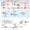
The mitochondria-associated ER membrane (MAM). A). Schematic diagram of MAM structure and function. MAM is formed through several pairs of tethering proteins localized to the outer mitochondrial membrane and ER membrane, including Mfn1/2-Mfn2, FPTPIP51-VABP, VDAC-GRP75-IP3R, PTPIP51-MOSPD2, and others. The VDAC-GRP75-IP3R is responsible for Ca2+ trafficking from the ER to the mitochondria. Sigma-1R can bind to IP3R and regulate calcium transfer to the mitochondria. B). Electron microscopic image shows a close contact of mitochondria with the ER in the RPE of a wild-type mouse. Double-headed arrow denotes the distance between the ER and the mitochondria.
Fig. 6.
Fig. 6.
The UPR and autophagy. A) All three UPR pathways alter autophagy. IRE1 and ATF6 can regulate autophagy by acting on regulators of Beclin1 while the PERK pathway results in the upregulation of transcription of autophagy related genes. These pathways alter the transcriptional landscape to promote autophagy. B) The UPR alters autophagy through TRB3’s interaction with P62 resulting in its subsequent impairment of autophagy.
Fig. 7.
Fig. 7.
Small molecules targeting ATF6, PERK, and IRE1 pathways. Ceapin-A7 inhibits ATF6 signaling by trapping the ATF6 molecule in the ER, thereby preventing the generation of ATF6 transcriptional activator. AA147 selectively activate the ATF6 signaling by inhibiting the activity of protein disulfide isomerases to increase reduced ATF6 monomers in the ER. IXA4 selectively upregulates IRE1/XBP1s target genes. 4u8c, covalently modifies and inactivates that RNase domain of IRE1. The PERK inhibitor, GSK2606414 and GSK2656157 bind to the PERK kinase and thereby inhibit the PERK pathway. The ISRIB binds and stabilizes the active form of eIF2B which is normally rendered inactive by PERK signaling. Salubrinal inhibits p-eIF2α dephosphorylation.

References

    1. Adachi Y, Yamamoto K, Okada T, Yoshida H, Harada A, Mori K, 2008. ATF6 is a transcription factor specializing in the regulation of quality control proteins in the endoplasmic reticulum. Cell Struct. Funct 33, 75–89. - PubMed
    1. Adekeye A, Haeri M, Solessio E, Knox BE, 2014. Ablation of the proapoptotic genes CHOP or Ask1 does not prevent or delay loss of visual function in a P23H transgenic mouse model of retinitis pigmentosa. PLoS One 9, e83871. - PMC - PubMed
    1. Agosto MA, Anastassov IA, Robichaux MA, Wensel TG, 2018. A large endoplasmic reticulum-resident pool of TRPM1 in retinal ON-bipolar cells. eNeuro 5. ENEURO.0143-18.2018. - PMC - PubMed
    1. Alavi MV, Chiang WC, Kroeger H, Yasumura D, Matthes MT, Iwawaki T, LaVail MM, Gould DB, Lin JH, 2015. In vivo visualization of endoplasmic reticulum stress in the retina using the ERAI reporter mouse. Invest. Ophthalmol. Vis. Sci 56, 6961–6970. - PMC - PubMed
    1. Almanza A, Mnich K, Blomme A, Robinson CM, Rodriguez-Blanco G, Kierszniowska S, McGrath EP, Le Gallo M, Pilalis E, Swinnen JV, Chatziioannou A, Chevet E, Gorman AM, Samali A, 2022. Regulated IRE1α-dependent decay (RIDD)-mediated reprograming of lipid metabolism in cancer. Nat. Commun 13, 2493. - PMC - PubMed

MeSH terms