Review
. 2023 Dec 28;66(24):16515-16545.
doi: 10.1021/acs.jmedchem.3c01130. Epub 2023 Dec 13.

Small Molecule Inhibitors Targeting the "Undruggable" Survivin: The Past, Present, and Future from a Medicinal Chemist's Perspective

Affiliations
Review

Small Molecule Inhibitors Targeting the "Undruggable" Survivin: The Past, Present, and Future from a Medicinal Chemist's Perspective

Qingbin Cui et al. J Med Chem. .

Abstract

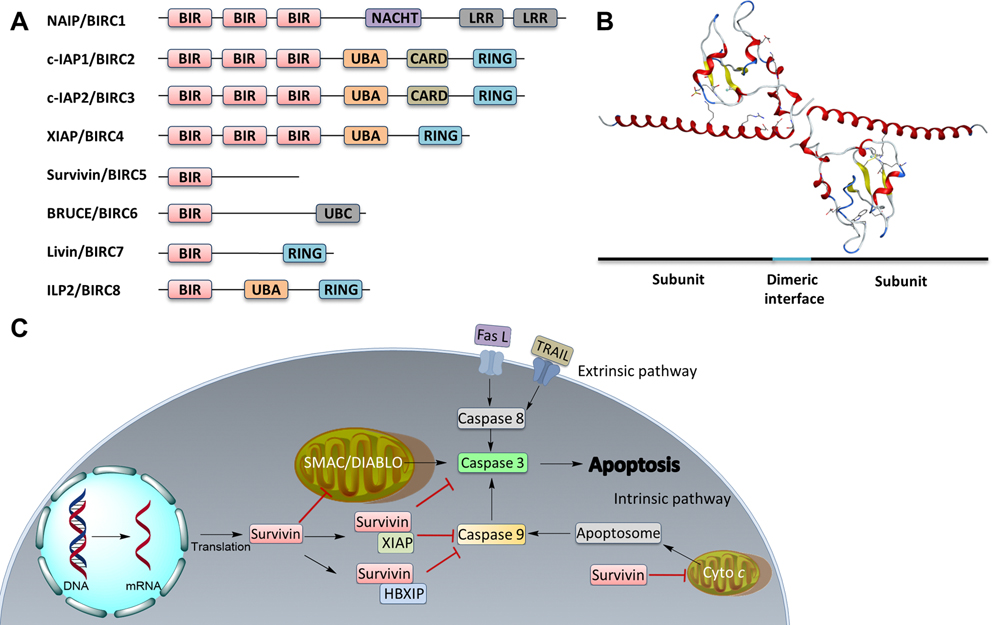
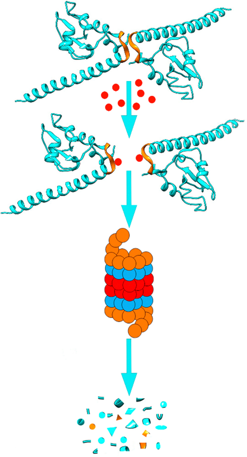
Survivin, a homodimeric protein and a member of the IAP family, plays a vital function in cell survival and cycle progression by interacting with various proteins and complexes. Its expression is upregulated in cancers but not detectable in normal tissues. Thus, it has been regarded and validated as an ideal cancer target. However, survivin is "undruggable" due to its lack of enzymatic activities or active sites for small molecules to bind/inhibit. Academic and industrial laboratories have explored different strategies to overcome this hurdle over the past two decades, with some compounds advanced into clinical testing. These strategies include inhibiting survivin expression, its interaction with binding partners and homodimerization. Here, we provide comprehensive analyses of these strategies and perspective on different small molecule survivin inhibitors to help drug discovery targeting "undruggable" proteins in general and survivin specifically with a true survivin inhibitor that will prevail in the foreseeable future.


Conflict of interest statement

The authors declare no competing financial interest.

Figures

Figure 1.
Figure 1.
IAP family members and survivin. (A) Linear domain structures of all eight IAP family members. (B) Crystal structure of survivin as a homodimer (PDB code: 1F3H). (C) Survivin function in extrinsic and intrinsic apoptosis. Fas L, Fas ligand; TRAIL, TNF-related apoptosis-inducing ligand; Cyto c, cytochrome c.
Figure 2.
Figure 2.
Structures of small molecule inhibitors that are included (A) in or excluded (B) from discussion in this work.
Figure 3.
Figure 3.
Medicinal chemistry of compound 1 and its derivatives. (A) Chemical structure of compound 1. (B–C) Analogs of compound 1 with substitutions on N1 (B) and N3 (C) as highlighted in cyan and pink, respectively. (D) Proposed lead derivatives 20–23.
Figure 4.
Figure 4.
Perspectives of compound 1. (A) Radar Chart analysis of 1. The scale of cLog P (calculated by ChemDraw 20.0) and HBD is equivalent to the original value multiplied by 100 and that of HBA multiplied by 50. (B) Proposed modifications of 1. (C) The structures of compounds 32 (G-555) and 33 (doxofylline). (D) Ketal moiety is used in the synthesis of 34 (lactonamycin).
Figure 5.
Figure 5.
Medicinal chemistry of 2. (A) Chemical structures of 35 (NDGA) and 2. (B–E) Ether bond (B), ester bond (C), end-ring (D), and linear or cycled carbon chain bridge (E) modifications on compound 2. (F) Chemical structure of 36 (NDGA-Cl2). (G) Compound 2 analogs with different length of the carbon-bridge or number of hydroxyl groups on benzene rings.
Figure 6.
Figure 6.
Perspective of 2. (A) Radar Chart analysis of 2. The scale of cLog P and HBDs is equivalent to the original value multiplied by 100 and that of HBAs multiplied by 50. (B) Chemical structure of proposed derivatives 42–45.
Figure 7.
Figure 7.
Medicinal chemistry of 3. (A) Chemical structures of 3 and 46 (camptothecin). (B) Chemical structures of camptothecin-based topoisomerase 1 inhibitors 47 (irinotecan), 48 (topotecan), and irinotecan metabolite 49 (SN-38). (C) Derivatives 50–58 with modified R7. (D) Derivatives 59 (9-Q6) and 60 (9-Q20) with modified R9. (E) Derivative 61 (Val-FL118) with addition of valine and 62–65 with other modifications at the hydroxyl group. The positions that are tolerable to modification are highlighted as R7 or R9 in claret.
Figure 8.
Figure 8.
Perspective of 3. (A-B) Radar Chart analysis of compounds 3 (A) and 52 (B). The scale of cLog P and HBDs is equivalent to the original value multiplied by 100 and that of HBAs multiplied by 50. (C) Proposed modifications/optimizations of 3. Compound 67 is derived from topotecan; 68 and 69 are inspired by compound 2, and the discovery of 74 (icotinib), respectively. (D) 74 (icotinib) and 75 (erlotinib).
Figure 9.
Figure 9.
Medicinal chemistry of 4. (A–B) Chemical structures of 7-(pyrrolidin-1-ylmethyl)quinolin-8-ol motif (A) and 4 (B). Replacement of the linker and benzene ring IV in 4 with different groups led to 76 and 77 (C and D). (E) Using methyl as a linker and replacing benzene with indole motif led to the discovery of 78 and its analogs 79 and 80. Further study revealed another lead 5 (F) and 6 (G).
Figure 10.
Figure 10.
Perspective of 4 and its analogs. (A) Radar Chart analysis of 4. The scale of cLog P and HBDs is equivalent to the original value multiplied by 100 and that of HBAs multiplied by 50. (B) Proposed modification/optimization of 4. Quinoxaline (UC-P1 series) or quinazoline (UC-P2 series) are used to replace quinolone and −NH2, −SH, or CN are used to replace −OH in R2. Residues in R1 (a–f) are adopted from 5, 6, 76, 77, 79, and 80.
Figure 11.
Figure 11.
Scheme of survivin dissociation and degradation in the proteasome. The two identical survivin subunits in a homodimer are shown in blue with the hydrophobic patch in the dimerization interface indicated in gold. The red dot represents inhibitors that bind to the hydrophobic dimerization interface, inhibiting dimerization, leading to destruction of survivin in proteasome.
Figure 12.
Figure 12.
Medicinal chemistry associated with 81 (LQZ-7). (A, B) Chemical structures of the hit compound 81 and its six analogs including 7 and its hydrolysis product 8. (C) Chemical structures of 87 (LQZ-7G), 88 (LQZ-7H), 9 (LQZ-7I), 89 (LQZ-7J), and 90 (LQZ-7K) originated from 81. Labile hydrazone linker and other linkers are highlighted in cyan. 7 has a fused tetracyclic ring. 9 differs from other analogs 87–90 mainly by the substitution patterns on the two benzene rings.
Figure 13.
Figure 13.
Perspectives of compounds 7–9. (A–C) The radar charts of 7 (A), 8 (B) and 9 (C). (D–E) Proposed further structural modifications of 8.
Figure 14.
Figure 14.
Medicinal chemistry associated with 97 (Abbott 8). (A-B) Chemical structures of the hit compound 96 (Abbott 1), (A) and its analog 97 (B). (C) Functional groups linked to the two benzene rings on 96. (D) Hydrophilic moieties that are used to modify ring I. (E) The leading compound 10 (LLP3) and its analog 98 (LLP9). (F) A series of novel fused tricyclic ring analogs. (G–H) Six series of 97 analogs with different substitutions. Bn, benzyl. Key structural differences were highlighted.
Figure 15.
Figure 15.
Perspective of 97 and its analogs. (A–C) The Radar Charts for 97 (A), 10 (B), and 11 (C). (D) The proposed future structural optimization based on leading compounds 10, 11, and 108.

References

    1. Fulda S Molecular pathways: targeting inhibitor of apoptosis proteins in cancer-from molecular mechanism to therapeutic application. Clin. Cancer Res. 2014, 20, 289–295. - PubMed
    1. Hay BA; Wassarman DA; Rubin GM Drosophila homologs of baculovirus inhibitor of apoptosis proteins function to block cell death. Cell 1995, 83, 1253–1262. - PubMed
    1. Salvesen GS; Duckett CS IAP proteins: blocking the road to death’s door. Nat. Rev. Mol. Cell Bio. 2002, 3, 401–410. - PubMed
    1. Mace PD; Shirley S; Day CL Assembling the building blocks: structure and function of inhibitor of apoptosis proteins. Cell Death Differ. 2010, 17, 46–53. - PubMed
    1. Morizane Y; Honda R; Fukami K; Yasuda H X-linked inhibitor of apoptosis functions as ubiquitin ligase toward mature caspase-9 and cytosolic Smac/DIABLO. J. Biochem. 2005, 137, 125–132. - PubMed

Publication types