. 2023 Dec 13;14(1):8273.
doi: 10.1038/s41467-023-43579-3.

DPPIV+ fibro-adipogenic progenitors form the niche of adult skeletal muscle self-renewing resident macrophages

Affiliations

DPPIV+ fibro-adipogenic progenitors form the niche of adult skeletal muscle self-renewing resident macrophages

Farshad Babaeijandaghi et al. Nat Commun. .

Abstract

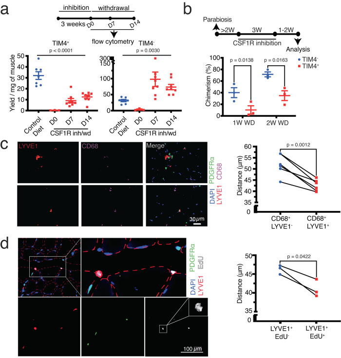
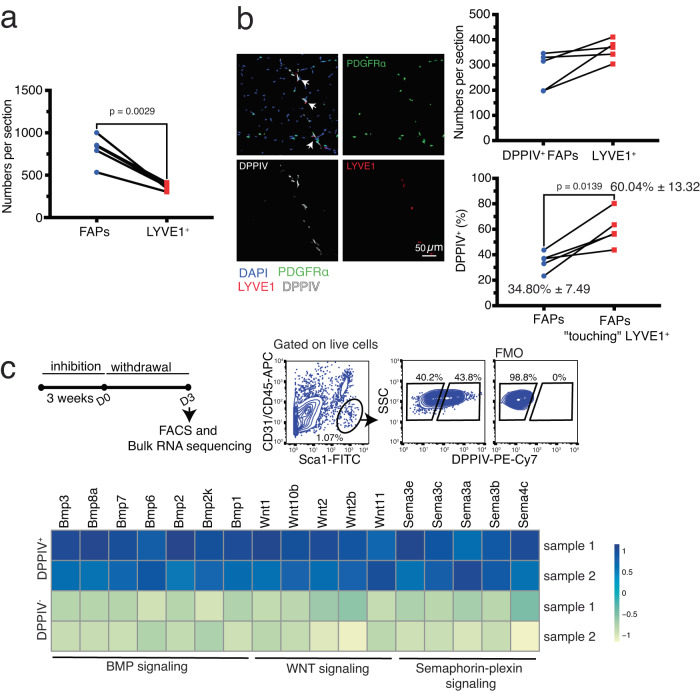
Adult tissue-resident macrophages (RMs) are either maintained by blood monocytes or through self-renewal. While the presence of a nurturing niche is likely crucial to support the survival and function of self-renewing RMs, evidence regarding its nature is limited. Here, we identify fibro-adipogenic progenitors (FAPs) as the main source of colony-stimulating factor 1 (CSF1) in resting skeletal muscle. Using parabiosis in combination with FAP-deficient transgenic mice (PdgfrαCreERT2 × DTA) or mice lacking FAP-derived CSF1 (PdgfrαCreERT2 × Csf1flox/null), we show that local CSF1 from FAPs is required for the survival of both TIM4- monocyte-derived and TIM4+ self-renewing RMs in adult skeletal muscle. The spatial distribution and number of TIM4+ RMs coincide with those of dipeptidyl peptidase IV (DPPIV)+ FAPs, suggesting their role as CSF1-producing niche cells for self-renewing RMs. This finding identifies opportunities to precisely manipulate the function of self-renewing RMs in situ to further unravel their role in health and disease.


Conflict of interest statement

The authors declare no competing interests.

Figures

Fig. 1
Fig. 1. Local CSF1 from FAPs is required for the survival of muscle RMs.
a Gating strategy to identify different muscle resident cell populations. b Expression of Csf1 in different muscle resident cell populations at steady state assessed by droplet digital PCR (n = 5 mice pooled from 2 experiments, Brown-Forsythe and Welch ANOVA tests). c The number of FAPs detected by flow cytometry in skeletal muscle from tamoxifen (TAM)-induced PdgfrαCreERT2 × DTA mice compared to TAM-induced transgene (Tg) controls (each dot represents one mouse, data were pooled from 2 experiments, unpaired t-test). Because there is a minor decrease in muscle mass after depleting FAPs, their numbers are presented per muscle (gastrocnemius) and not per milligram of tissue. d The amount of CSF1 detected by ELISA in quadriceps muscle of TAM-induced PdgfrαCreERT2 × DTA mice compared to Tg control (each dot represents one mouse, data were pooled from ≥3 experiments, unpaired t-test). e The number of TIM4- and TIM4+ RMs detected by flow cytometry in skeletal muscle from TAM-induced PdgfrαCreERT2 × DTA mice compared to Tg control (each dot represents one mouse, data were pooled from 2 experiments, unpaired t-test). f The number of TIM4- and TIM4+ RMs detected by flow cytometry in skeletal muscle from TAM-induced PdgfrαCreERT2 × Csf1flox/null mice and Tg controls (each dot represents one mouse, data were pooled from ≥3 experiments, unpaired t-test for TIM4- and unpaired t test with Welch’s correction for TIM4+). g Replacement of depleted muscle RMs in TAM-induced PdgfrαCreERT2 × Csf1flox/null mice by bone marrow blood progenitors from the wild type (WT) parabiotic partner (n = 4 parabiotic pairs from ≥3 experiments, paired t-test).
Fig. 2
Fig. 2. Local CSF1 restores depleted RMs.
a The amount of CSF1 detected by ELISA in skeletal muscle at different time points after CSF1R inh/wd (each dot represents one mouse, data were pooled from ≥3 experiments). b The amount of CSF1 detected by ELISA in the blood within three days after withdrawal of CSF1R inhibition or the control diet (each dot represents one mouse, data were pooled from 2 experiments, unpaired t-test). c Expression of Csf1 in different muscle resident cell populations at different time points after CSF1R inh/wd assessed by droplet digital PCR (ddPCR, n = 8 mice for FAPs, n = 5 mice for the other populations, data were pooled from ≥3 experiments, Brown-Forsythe and Welch ANOVA tests). d The number of TIM4- and TIM4+ RMs detected by flow cytometry in skeletal muscle from tamoxifen (TAM)-induced PdgfrαCreERT2 × Csf1flox/null or transgene (Tg) controls following CSF1R inh/wd (n = 11 for control and n = 10 for CSF1R inh/wd, data were pooled from ≥3 experiments, unpaired t test with Welch’s correction).
Fig. 3
Fig. 3. SRRMs are located close to FAPs.
a The number of TIM4- and TIM4+ RMs detected by flow cytometry in skeletal muscle following CSF1R inh/wd (n = 7 mice for the control group, n = 3, 8, and 8 for the CSF1R inh/wd group on D0, D7, and D14, respectively; data were pooled from ≥3 experiments, ordinary one-way ANOVA). b Replacement rate of muscle RMs by blood progenitors following pharmacological depletion: The replacement rate of RMs was normalized to the percentage of chimerism in mononuclear myelomonocytic cells in the blood (n = 3 female parabiotic pairs per time point, data were pooled from ≥3 experiments, paired t test). c Immunofluorescent staining of Tibialis Anterior (TA) muscle sections and quantification of the distance between LYVE1+ SRRMs or LYVE1- RMs and PDGFRα-EGFP+ FAPs (n = 6 mice, pooled from 2 experiments; paired t-test). On average, we manually measured distances of approximately 575 CD68 + LYVE1- and 450 CD68 + LYVE1+ RMs from their closest FAPs in each mouse. d Immunofluorescent staining of TA sections and quantification of the distance between LYVE1+EdU+ or LYVE1+EdU- cells and PDGFRα-EGFP+ FAPs (n = 3 mice pooled from 2 experiments; paired t-test). On average, we manually measured distances of approximately 175 LYVE1+EdU- and 25 LYVE1+ EdU+ cells from their closest FAPs in each mouse. The figure displays two LYVE1+ EdU+ cells situated in proximity to FAPs. The cell on the left exhibits a dividing nucleus, as illustrated in greater detail in the inset.
Fig. 4
Fig. 4. DPPIV+ FAPs form the niche of skeletal muscle SRRMs.
a The number of LYVE1+ SRRMs and FAPs (PDGFRα-EGFP+) in TA sections (n = 4 mice pooled from two experiments, paired t-test). b Immunofluorescent staining of TA sections and quantification of the number of LYVE1+ SRRMs and DPPIV + PDGFRα-EGFP+ FAPs (top) and the percentage of DPPIV+ cells among all PDGFRα-EGFP+ FAPs or among PDGFRα-EGFP+ FAPs with a SRRM in their close vicinity (n = 5 mice pooled from 2 experiments, paired t-test, bottom). On average, approximately 800 FAPs per mouse were included for quantification. Among them, an average of about 100 FAPs had SRRMs in close vicinity. c Experimental design and gating strategy to sort DPPIV- and DPPIV+ FAPs for bulk RNA sequencing (top) and heatmap showing selected differentially upregulated genes in DPPIV+ FAPs compared to DPPIV- FAPs categorized by different signaling pathways (bottom).

References

    1. Wu Y, Hirschi KK. Tissue-resident macrophage development and function. Front Cell Dev. Biol. 2020;8:617879. doi: 10.3389/fcell.2020.617879. - DOI - PMC - PubMed
    1. Davies LC, Jenkins SJ, Allen JE, Taylor PR. Tissue-resident macrophages. Nat. Immunol. 2013;14:986–95. doi: 10.1038/ni.2705. - DOI - PMC - PubMed
    1. Paolicelli RC, et al. Synaptic pruning by microglia is necessary for normal brain development. Science. 2011;333:1456–1458. doi: 10.1126/science.1202529. - DOI - PubMed
    1. Kohyama M, et al. Role for Spi-C in the development of red pulp macrophages and splenic iron homeostasis. Nature. 2009;457:318–321. doi: 10.1038/nature07472. - DOI - PMC - PubMed
    1. Rosen A, Casciola-Rosen L. Clearing the way to mechanisms of autoimmunity. Nat. Med. 2001;7:664–665. doi: 10.1038/89034. - DOI - PubMed

Publication types

Substances

Associated data