. 2023 Dec 13;14(1):8275.
doi: 10.1038/s41467-023-44075-4.

Cell geometry regulates tissue fracture

Affiliations

Cell geometry regulates tissue fracture

Amir J Bidhendi et al. Nat Commun. .

Abstract

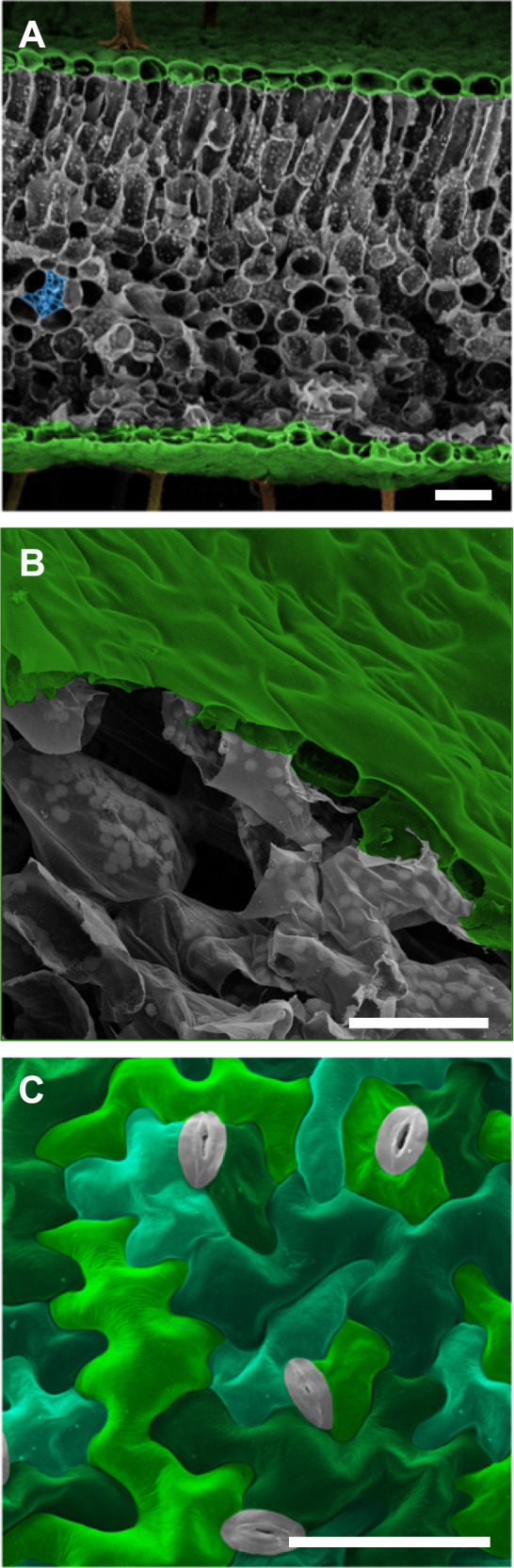
In vascular plants, the epidermal surfaces of leaves and flower petals often display cells with wavy geometries forming intricate jigsaw puzzle patterns. The prevalence and diversity of these complex epidermal patterns, originating from simple polyhedral progenitor cells, suggest adaptive significance. However, despite multiple efforts to explain the evolutionary drivers behind these geometrical features, compelling validation remains elusive. Employing a multidisciplinary approach that integrates microscopic and macroscopic fracture experiments with computational fracture mechanics, we demonstrate that wavy epidermal cells toughen the plants' protective skin. Through a multi-scale framework, we demonstrate that this energy-efficient patterning mechanism is universally applicable for toughening biological and synthetic materials. Our findings reveal a tunable structural-mechanical strategy employed in the microscale design of plants to protect them from deleterious surface fissures while facilitating and strategically directing beneficial ones. These findings hold implications for targeted plant breeding aimed at enhancing resilience in fluctuating environmental conditions. From an engineering perspective, our work highlights the sophisticated design principles the plant kingdom offers to inspire metamaterials.


Conflict of interest statement

The authors declare no competing interests.

Figures

Fig. 1
Fig. 1. Scanning electron micrographs of plant leaf structures.
A Cross-section of Geranium sp. leaf showing epidermal layers (colorized green) sandwiching layers of mesophyll cells (gray) and vascular tissue (blue). B Cutaway of Arabidopsis thaliana leaf demonstrating the epidermis (green) and underlying mesophyll cells (gray). Note the tight adherence between pavement cells compared with the large air spaces between the mesophyll cells. C Orthogonal view of pavement cells at the surface of an Arabidopsis thaliana cotyledon. Pavement cells colorized in green/teal, stomata in gray. Micrographs exemplify cell organization and patterning that are widely found throughout the plant kingdom. Scale bars = 50 µm.
Fig. 2
Fig. 2. Fracture behavior of patterned PMMA sheets.
Specimens featuring brick-shaped cell patterns with crack initiated along (A) and perpendicular (B) to the cells’ long axes. Cracks take approximately straight paths - following cell interfaces if on the crack path (compare green and magenta portions). C, D Cracks in the wavy cell patterned samples take rugged paths following both engraved borders (green) and crossing cells (magenta). Shallow undulations cause the crack to follow interfaces (green) leading to ‘cell-cell separation’ while deeper undulations force cracks to alternate frequently between ‘interface’ and ‘cell’ (magenta). The asterisk marks an arrested crack with a bifurcation which follows the interface briefly before entering the cell. E Force profiles during crack propagation through engraved PMMA sheets. The control (no engraving) experiences a smooth brittle fracture, similar to the onion-patterned samples cracking along cell alignments (brick-long). All other samples continue to bear load after the onset of crack propagation featuring several local maxima. Wavy and Wavy-90 refer to a pavement cell pattern and its 90-degree rotation. F Boxplots of Work-of-Fracture in PMMA sheets (normalized to control specimens) based on engraving pattern with n = 5, 4, 4, 3 and 4 independent samples used for control, longitudinal and transverse brick-shaped, wavy, and its 90-degree rotation patterns, respectively. The central rectangle signifies the Interquartile Range (IQR), with the upper edge denoting the Third Quartile (Q3) and the lower edge representing the First Quartile (Q1). Horizontal green line marks the median, cyan dots indicate the mean, black dots are individual data points. Whiskers extend to 1.5 times the IQR above Q3 and below Q1. Scale bars = 1 cm.
Fig. 3
Fig. 3. Numerical simulation of crack propagation in patterned material using phase-field fracture model.
A Force-displacement curves obtained for control and patterned specimens for Eint=Ecell and Gcint=Gccell/2. The brick-shaped cell patterns oriented parallel to the initiated crack exhibited dramatic brittle behavior as crack propagation followed interfaces (see Methods). The transversely oriented brick-shaped pattern and the wavy cell patterns showed similar force-displacement curves to the control profile. B Force-displacement curves for Eint=Ecell/2 and Gcint=Gccell/2. Brick-shaped cell patterns with longitudinal orientation were brittle as in (A), but the other specimens showed an increased toughness compared to the control. Closeup views of crack paths for longitudinally and transversely aligned brick-shaped cells (C, D) as well as wavy cell patterns (E) and control specimen (F, no engraving). Compare green and magenta segments of crack path marking border and cell wall fracture portions, respectively. Black arrows indicate the direction of initial crack. Purple arrows indicate instances of crack re-initiation when the propagating fracture enters stiffer cells from softer interfaces. See also Supplementary Fig. 4.
Fig. 4
Fig. 4. Tear testing of onion epidermis.
A Geometry and loading of tear tests. Top: Two-leg trouser tear test, Bottom: Notched specimen tear. F denotes the direction of force applied with respect to the xy plane. The orientation of the original blade-cut notch is along x. The two tests differ in the placement of the initial notch and in the orientation of load application (perpendicular to the xy plane for two-leg trouser tear test). B Tearing of epidermis with a notch in the center. Top: tear along the cells’ long axes features a smooth path. Bottom: Tear transverse to cells’ long axes demonstrates that while the path has the global tendency to proceed according to the applied force field, minute, local deviations are induced through microscopic reorientations imposed by cell-cell interface features, leading to rough tear edges and blunted tear tips (see also Supplementary Movies 1,2). Micrographs are representative of over twenty observations. C, D Force displacement profiles of specimens stretched in (A) and (B), respectively. P1 marks the onset of tear growth. P2 marks the peak force after which the tear turned slightly toward the cell direction leading to a drop in force, although the tear did not freely propagate in this direction. Wi and Wp refer to Work for initiation and propagation of tear, respectively. The tear propagation zone for longitudinally-notched specimens was negligible (brittle behavior). E Normalized Work-to-Tear for transversely- and longitudinally-notched specimens with n = 12 and 16 independent samples used in analysis for longitudinal and transverse samples, respectively. The central rectangle signifies the Interquartile Range (IQR), with the upper edge denoting the Third Quartile (Q3) and the lower edge representing the First Quartile (Q1). Horizontal green line marks the median, cyan dots indicate the mean, black dots are individual data points. Whiskers extend to 1.5 times the IQR above Q3 and below Q1. Data points below Q1-1.5*IQR or above Q3 + 1.5*IQR were classified as outliers (not shown). Tearing of two-leg trouser specimens parallel (F) and transverse (G) to cell orientation. While along the cells the tear path remained relatively straight, it showed a marked difficulty crossing from one cell to the next. Type 0 tear path corresponds to the location of the initial notch. Type I marks a tear path parallel to cell lines. Type II is a 90-degree reorientation of the tear path and type III makes a subsequent reorientation of the tear path towards an oblique angle. Different tissue samples showed all or some of these tear path types in varying order. Tears driven perpendicular to cell interfaces were frequently associated with reorientation and rugged edges.
Fig. 5
Fig. 5. Crack propagation at cell-cell interfaces.
A, B A crack traversing a cell (magenta) and arriving at a cell-cell interface is deviated to propagate along the interface (green). In (A) this occurs through a failure in the periclinal wall at the edge towards the anticlinal wall. In (B), the crack traverses an anticlinal wall and failure occurs in the middle lamella, either through adhesive failure (between the middle lamella and anticlinal wall) or cohesive failure (in the middle lamella material). C Crack traverses the cell-cell interface along a path that remains globally straight but locally shows minor reorientations due to changing material properties when traversing the anticlinal wall, middle lamella, and second anticlinal wall.
Fig. 6
Fig. 6. Crack propagation in the plant epidermis.
3D reconstruction of confocal micrographs of cracks in (A) radicle and (B) cotyledon epidermal tissues of an Arabidopsis embryo extracted by removing the seed coat. The embryo was squeezed between the glass slide and the coverslip. Cell-cell detachment can be observed. Cell wall staining was performed using propidium iodide, which at early growth stages can also label nuclei. Fracture at the middle lamella (as in Fig. 5B) rather than at the inside edge (as in Fig. 5A), was confirmed by the observation of the presence of anticlinal walls on both sides of the crack and by the fact that cells had retained their nuclei indicating that the protoplasts were intact. C Scanning electron micrograph of fractured tomato leaves demonstrated a meandering crack path. D Close-up of the box in (C). The arrow points to a shallower lobe with an interface fracture (as in Fig. 5A). Arrow with asterisk demonstrates an arrested crack bifurcation traversing the cell (as in Fig. 5C). Arrowhead points to a crack traversing a cell. E Close-up of a local crack in Arabidopsis leaf induced during application of tensile stress. Crack in the cell wall seems to propagate by splitting the middle lamella (as in Fig. 5B) and the arrow points to a partial view of an anticlinal wall. FH Outer periclinal walls of abaxial onion epidermis split open upon peeling. Brief exposure to boiling water (F, G) or bleach (H) prior to peeling dramatically weakened cell-cell adhesion. As a result, tearing the excised epidermal tissue perpendicular to the cell axes facilitated failure at the middle lamella (as in Fig. 5B). Micrographs in Fig. 6 are representative of approximately 350 observations of fractures in plant epidermal tissues. Scale bars = 20 µm (AD), inset in (E) = 5 µm, 200 µm (F), 30 µm (G), 50 µm (H).

References

    1. Mathur J. Cell shape development in plants. Trends Plant Sci. 2004;9:583–590. doi: 10.1016/j.tplants.2004.10.006. - DOI - PubMed
    1. Bidhendi AJ, Altartouri B, Gosselin FP, Geitmann A. Mechanical stress initiates and sustains the morphogenesis of wavy leaf epidermal cells. Cell Rep. 2019;28:1237–1250.e6. doi: 10.1016/j.celrep.2019.07.006. - DOI - PubMed
    1. Altartouri B, et al. Pectin chemistry and cellulose crystallinity govern pavement cell morphogenesis in a multi-step mechanism. Plant Physiol. 2019;181:127–141. doi: 10.1104/pp.19.00303. - DOI - PMC - PubMed
    1. Coen E, Cosgrove DJ. The mechanics of plant morphogenesis. Science. 2023;379:eade8055. doi: 10.1126/science.ade8055. - DOI - PubMed
    1. Vőfély RV, Gallagher J, Pisano GD, Bartlett M, Braybrook SA. Of puzzles and pavements: a quantitative exploration of leaf epidermal cell shape. N Phytol. 2019;221:540–552. doi: 10.1111/nph.15461. - DOI - PMC - PubMed

Publication types

LinkOut - more resources