. 2023 Dec 14;6(1):1266.
doi: 10.1038/s42003-023-05643-y.

Enzyme-controlled, nutritive hydrogel for mesenchymal stromal cell survival and paracrine functions

Affiliations

Enzyme-controlled, nutritive hydrogel for mesenchymal stromal cell survival and paracrine functions

Cyprien Denoeud et al. Commun Biol. .

Abstract

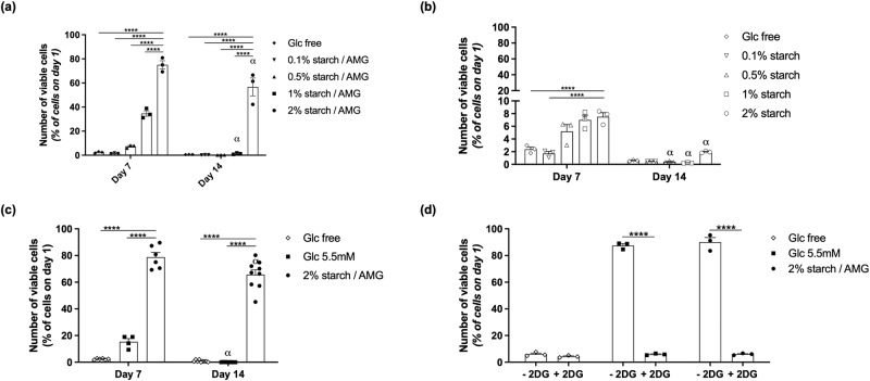
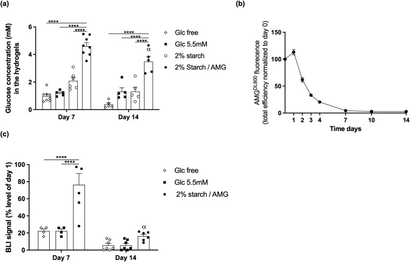
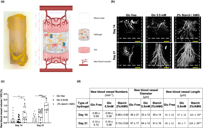
Culture-adapted human mesenchymal stromal cells (hMSCs) are appealing candidates for regenerative medicine applications. However, these cells implanted in lesions as single cells or tissue constructs encounter an ischemic microenvironment responsible for their massive death post-transplantation, a major roadblock to successful clinical therapies. We hereby propose a paradigm shift for enhancing hMSC survival by designing, developing, and testing an enzyme-controlled, nutritive hydrogel with an inbuilt glucose delivery system for the first time. This hydrogel, composed of fibrin, starch (a polymer of glucose), and amyloglucosidase (AMG, an enzyme that hydrolyze glucose from starch), provides physiological glucose levels to fuel hMSCs via glycolysis. hMSCs loaded in these hydrogels and exposed to near anoxia (0.1% pO2) in vitro exhibited improved cell viability and angioinductive functions for up to 14 days. Most importantly, these nutritive hydrogels promoted hMSC viability and paracrine functions when implanted ectopically. Our findings suggest that local glucose delivery via the proposed nutritive hydrogel can be an efficient approach to improve hMSC-based therapeutic efficacy.


Conflict of interest statement

The authors declare the following competing interests: P.J., P.Esther, L.-G.V., and P.H. disclose that the novel enzyme-controlled, nutritive hydrogels used in the current project, were patented globally (EP14306700; 2014). The other authors declare no competing interests.

Figures

Fig. 1
Fig. 1. The starch/AMG hydrogels produce glucose.
a Schematic (not to scale) of the starch/AMG hydrogels. hMSCs were embedded in fibrin hydrogels containing starch enzymatically hydrolyzed by AMG in glucose, fueling hMSCs. (Created with BioRender.com) b. Production of glucose in hydrogels with increasing concentrations of starch loaded with or without AMG (n = 5–7). c Production of glucose in 2% starch hydrogels loaded with increasing concentrations of AMG (n = 4–8). d Representative environmental scanning electron micrographs (eSEM) micrographs of the external architecture of either glucose-free, 5.5 mM glucose, or starch (with or without AMG) hydrogels. Scale bars: 200 µm (for eSEM images in the top row), 50 µm (for eSEM images in bottom row). AMG amyloglucosidase, Glc glucose.
Fig. 2
Fig. 2. The starch/AMG hydrogel environment extends survival of hMSCs by delivering glucose in near anoxia in vitro.
a, b Viability of hMSCs seeded in either glucose-free hydrogels or hydrogels with increasing starch concentrations either with a or without b AMG, after exposure to near-anoxia (0.1% pO2). (n = 3). c Viability of hMSCs seeded in either glucose-free, 5.5 mM glucose, or starch 2%/AMG hydrogels after exposure to near-anoxia for 7 and 14 days. (n = 4–9) d. Viability of hMSCs seeded in either glucose-free, 5.5 mM glucose, or starch 2%/AMG hydrogels after exposure to 2-Deoxy-D-Glucose (a competitive inhibitor for the production of glucose-6-phosphate) in near-anoxia for 3 days. (n = 3).
Fig. 3
Fig. 3. The starch/AMG hydrogels are more effective than glucose hydrogels in enhancing the viability and chemotactic functions of hMSCs in near anoxia in vitro.
a Quantification of hMSC migration in Boyden chambers in response to conditioned media from hMSCs seeded in either glucose-free hydrogels, 5.5 mM glucose hydrogels, or starch 2%/AMG hydrogels and exposed to near-anoxia for 7 and 14 days. (n = 4–6) b. Concentrations of the chemotactic mediators in the conditioned media from hMSCs seeded in either glucose-free, 5.5 mM glucose, or starch 2%/AMG hydrogels and exposed to near-anoxia for 7 and 14 days. (n = 3). c Quantification of HUVEC migration in Boyden chambers in response to conditioned media from hMSCs seeded in either glucose-free, 5.5 mM glucose, or starch 2%/AMG hydrogels, after exposure to near-anoxia conditions for 7 and 14 days. (n = 3) d. Concentrations of the proangiogenic mediators in the conditioned media from hMSCs seeded in either glucose-free, 5.5 mM glucose, or starch 2%/AMG hydrogels and exposed to near-anoxia for 7 and 14 days. (n = 3). IL interleukin, MCP monocyte chemoattractant protein, MMP matrix metalloproteinase, MIF macrophage inhibitory factor, MIP macrophage inflammatory protein, ENA epithelial neutrophil activating protein, IP interferon gamma-induced protein, VEGF vascular endothelial growth factor, ANGPT angiopoietin, ANG angiogenin, FGF-BASIC basic fibroblast growth factor.
Fig. 4
Fig. 4. The starch/AMG hydrogels are more effective than glucose hydrogels in increasing the in vivo survival of hMSCs after subcutaneous implantation.
a Glucose concentration into glucose-free, 5.5 mM glucose, or 2% starch cell free hydrogels with or without AMG after subcutaneous implantation in nude mice for 7 and 14 days (n = 5–8). b Kinetics of AMG fluorescence efficiency after subcutaneous implantation in nude mice up to 14 days (n = 6). c Quantification of the survival of Luc-ZSGreen-hMSCs loaded into glucose-free, 5.5 mM glucose, or starch 2%/AMG hydrogels and implanted in an ectopic mouse model through bioluminescent signal monitoring (n = 4–7).
Fig. 5
Fig. 5. The starch/AMG hydrogels are more effective than glucose hydrogels in increasing the in vivo proangiogenic functions of hMSCs after subcutaneous implantation.
a A gross view of the hydrogel and schematic (not to scale) of the experimental model designed to assess the proangiogenic potential of hMSC-containing hydrogels. (Created with BioRender.com). b, c Micro-CT vasculature analysis in the vicinity of either glucose-free, 5.5 mM glucose, or starch 2%/AMG hydrogels after 14 and 21 days of ectopic implantation in mice. b Representative 3D reconstructions of the new blood vessels into the volume of interest (VOI) of either glucose-free, 5.5 mM glucose, or starch 2%/AMG hydrogels after subcutaneous implantation in nude mice, scale bars: 500 µm. c Quantification of new blood vessel volume into the VOIs of either glucose-free, 5.5 mM glucose, or starch 2%/AMG hydrogels after subcutaneous implantation in nude mice (n = 8). d Table summarizing the number, diameter, and length of newly formed blood vessels into the VOIs of either glucose-free, 5.5 mM glucose, or starch 2%/AMG hydrogels after subcutaneous implantation in nude mice (n = 8).

References

    1. Matthay MA, et al. Treatment with allogeneic mesenchymal stromal cells for moderate to severe acute respiratory distress syndrome (START study): a randomised phase 2a safety trial. Lancet Respir. Med. 2019;7:154–162. doi: 10.1016/S2213-2600(18)30418-1. - DOI - PMC - PubMed
    1. de Luca M, et al. Advances in stem cell research and therapeutic development. Nat. Cell Biol. 2019;21:801–811. doi: 10.1038/s41556-019-0344-z. - DOI - PubMed
    1. Hoogduijn MJ, Lombardo E. Mesenchymal stromal cells Anno 2019: dawn of the therapeutic era? Concise review. Stem Cells Transl. Med. 2019;8:1126–1134. doi: 10.1002/sctm.19-0073. - DOI - PMC - PubMed
    1. Manassero M, et al. Comparison of survival and osteogenic ability of human mesenchymal stem cells in orthotopic and ectopic sites in mice. Tissue Eng. Part A. 2016;22:534–544. doi: 10.1089/ten.tea.2015.0346. - DOI - PubMed
    1. McGinley LM, et al. Mesenchymal stem cell survival in the infarcted heart is enhanced by lentivirus vector-mediated heat shock protein 27 expression. Hum. Gene Ther. 2013;24:840–851. doi: 10.1089/hum.2011.009. - DOI - PMC - PubMed

Publication types

LinkOut - more resources