

Knockdown of MRPL35 promotes cell apoptosis and inhibits cell proliferation in non-small-cell lung cancer
- PMID: 38093266
- PMCID: PMC10720070
- DOI: 10.1186/s12890-023-02677-0
Knockdown of MRPL35 promotes cell apoptosis and inhibits cell proliferation in non-small-cell lung cancer
Abstract
Background: Non-small cell lung cancer (NSCLC) is a major pathological type of lung cancer. However, its pathogenesis remains largely unclear. MRPL35 is a regulatory subunit of the mitoribosome, which can regulate the assembly of cytochrome c oxidases and plays an important role in the occurrence of NSCLC.
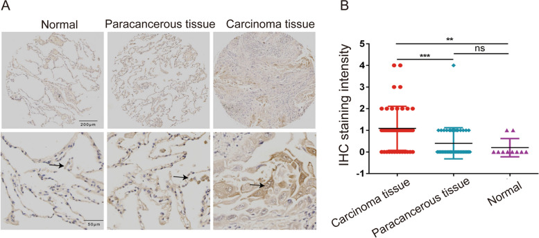
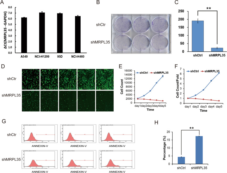
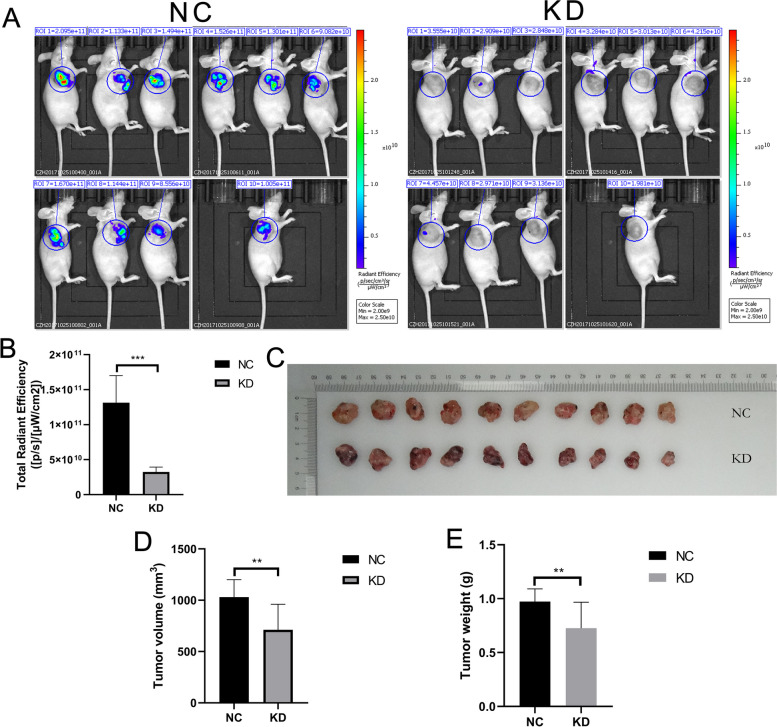
Methods: The expression of MRPL35 in NSCLC was detected by tissue microarray and immunohistochemistry. H1299 cells were infected with lentivirus to knockdown MRPL35, and the cells were subjected to crystal violet staining to assess the results of colony formation assays. A549 cells were infected by lentiviral particles-expressing shMRPL35 or shControl, and then subcutaneously injected into nude mice. Tumorigenesis in mice was detected by in vivo imaging. The potential pathway of MRPL35 in NSCLC was assessed by Western blotting.
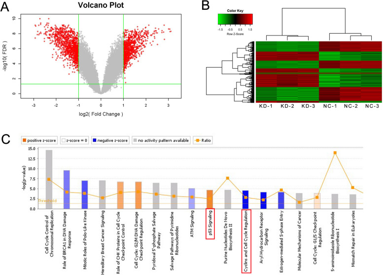
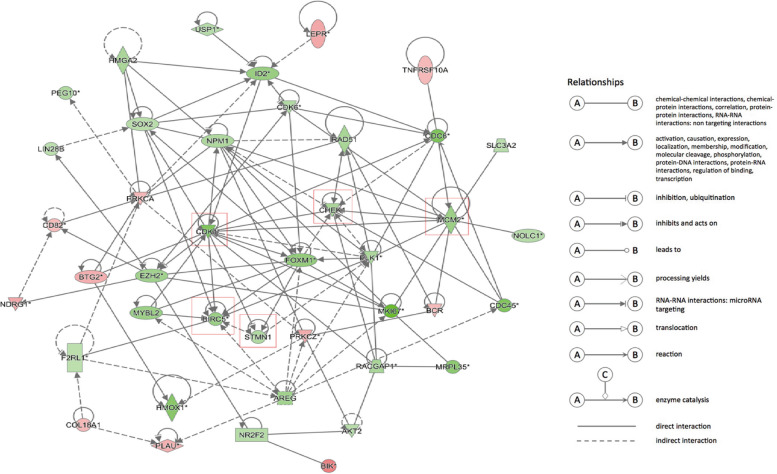
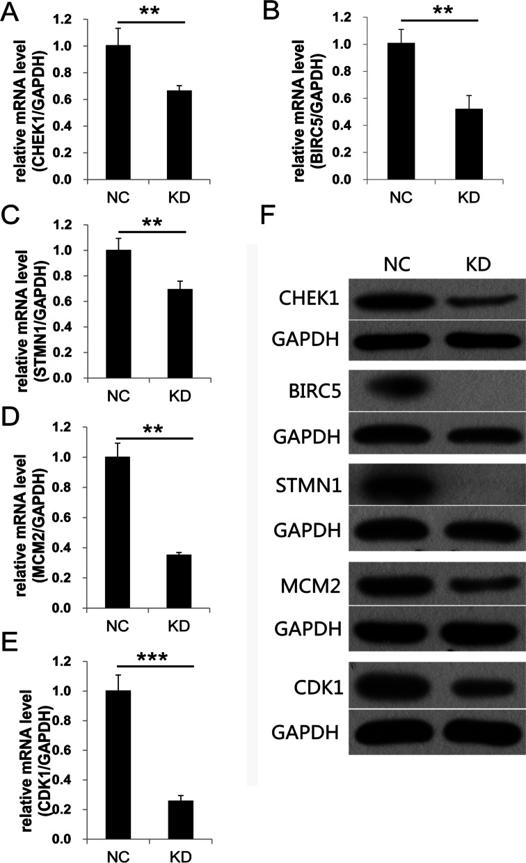
Results: MRPL35 was over-expressed in NSCLC tissue compared to para-cancerous and normal tissues. Knockdown of MRPL35 suppressed cell proliferation and decreased NSCLC progression both in vitro and in vivo. The possible molecular mechanisms were also clarified, which indicated that MRPL35 could be involved in cell apoptosis and proliferation by modulating the expression levels of CDK1, BIRC5, CHEK1, STMN1 and MCM2. Knockdown of MRPL35 activated p53 signaling pathway and inhibited cell cycle regulation.
Conclusions: The oncogenic role of MRPL35 in NSCLC was potentially mediated through the cell cycle regulatory genes such as BIRC5, STMN1, CDK1, CHEK1 and MCM2, as well as activation of P53 signaling pathway.
Keywords: Apoptosis; MRPL35; NSCLC; Proliferation; p53.
© 2023. The Author(s).
Conflict of interest statement
The authors declare no competing interests.
Figures
References
MeSH terms
Substances
Grants and funding
- 82072585/National Natural Science Foundation of China
- 202003a07020024/Anhui Provincial Major Science and Technology Project
- 2020b07030008/Anhui Science and Technology Development Fund Projects guided by China Government in 2021
- gxbjZD2020069/The Project of Anhui Educational Committee for Distinguished Scholars
- 51201108/512 Talent Cultivation Project of Bengbu Medical College
LinkOut - more resources
Full Text Sources
Medical
Molecular Biology Databases
Research Materials
Miscellaneous
