. 2023 Dec 14;22(1):206.
doi: 10.1186/s12943-023-01896-7.

Dynamic altruistic cooperation within breast tumors

Affiliations

Dynamic altruistic cooperation within breast tumors

Muhammad Sufyan Bin Masroni et al. Mol Cancer. .

Abstract

Background: Social behaviors such as altruism, where one self-sacrifices for collective benefits, critically influence an organism's survival and responses to the environment. Such behaviors are widely exemplified in nature but have been underexplored in cancer cells which are conventionally seen as selfish competitive players. This multidisciplinary study explores altruism and its mechanism in breast cancer cells and its contribution to chemoresistance.

Methods: MicroRNA profiling was performed on circulating tumor cells collected from the blood of treated breast cancer patients. Cancer cell lines ectopically expressing candidate miRNA were used in co-culture experiments and treated with docetaxel. Ecological parameters like relative survival and relative fitness were assessed using flow cytometry. Functional studies and characterization performed in vitro and in vivo include proliferation, iTRAQ-mass spectrometry, RNA sequencing, inhibition by small molecules and antibodies, siRNA knockdown, CRISPR/dCas9 inhibition and fluorescence imaging of promoter reporter-expressing cells. Mathematical modeling based on evolutionary game theory was performed to simulate spatial organization of cancer cells.

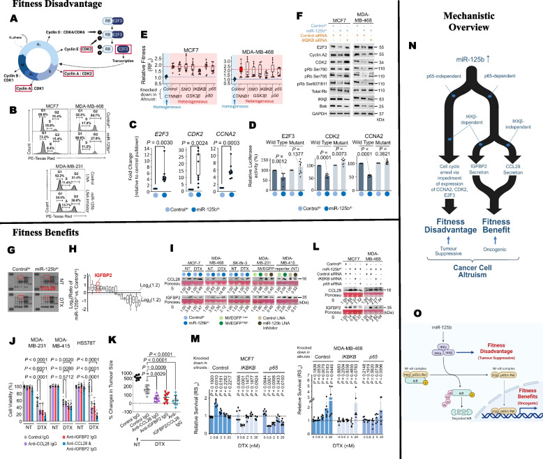
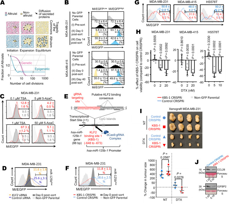
Results: Opposing cancer processes underlie altruism: an oncogenic process involving secretion of IGFBP2 and CCL28 by the altruists to induce survival benefits in neighboring cells under taxane exposure, and a self-sacrificial tumor suppressive process impeding proliferation of altruists via cell cycle arrest. Both processes are regulated concurrently in the altruists by miR-125b, via differential NF-κB signaling specifically through IKKβ. Altruistic cells persist in the tumor despite their self-sacrifice, as they can regenerate epigenetically from non-altruists via a KLF2/PCAF-mediated mechanism. The altruists maintain a sparse spatial organization by inhibiting surrounding cells from adopting the altruistic fate via a lateral inhibition mechanism involving a GAB1-PI3K-AKT-miR-125b signaling circuit.

Conclusions: Our data reveal molecular mechanisms underlying manifestation, persistence and spatial spread of cancer cell altruism. A minor population behave altruistically at a cost to itself producing a collective benefit for the tumor, suggesting tumors to be dynamic social systems governed by the same rules of cooperation in social organisms. Understanding cancer cell altruism may lead to more holistic models of tumor evolution and drug response, as well as therapeutic paradigms that account for social interactions. Cancer cells constitute tractable experimental models for fields beyond oncology, like evolutionary ecology and game theory.


Conflict of interest statement

Lihan Zhou and Mo-Huang Li are employed as Chief Executive Officers of MiRXES Pte Ltd and CellSievo Pte Ltd respectively.

Figures

Fig. 1
Fig. 1
Breast cancer cells exhibit altruistic cooperation. In situ hybridization for miR-125b or U6 in FFPE breast cancer tissues from patients with (n=26) or without (n=27) neoadjuvant taxane treatment. B Staining for miR-125b in MDA-MB-231miR-125bprom-EGFP cells. Left: stained cells with or without docetaxel exposure. Red triangles: cells with high miR-125b. Right: quantification of miR-125b expression. C Left: Higher magnification of (A). Right: Quantification of percentage of cells with miR-125b vs. U6 expression for 24 miR-125b+ patient samples from (A). D Schema showing how “Relative Survival (RSα)” and “Relative Fitness (RFα)” were measured (See “Methods” and Supplementary Note 1). E-G Bar charts depicting ratio of heterogeneous vs. homogeneous total cell count in early post-treatment for indicated breast cancer cell lines. H-J Bar charts depicting RSα ratio for indicated breast cancer cell lines. K-M Box plots of RFα for “heterogeneous” group of indicated breast cancer cell lines (except cells exposed to 5 and 20nM docetaxel, which did not survive past 3 weeks). N Percentage change in tumor size for MCF-7 xenograft of “homogeneous” or “heterogeneous” study, with or without docetaxel treatment. O Bar chart depicting RSα ratio, xenograft mice from (N), with or without docetaxel exposure. P Box plot of RFα for “heterogeneous” xenograft tumor, with or without docetaxel treatment. Dot: single section read; dots of same colour: sections imaged from same animal. Q Representative images of xenograft tumor biopsy from “homogeneous” and “heterogeneous” group. Black arrows: cells stained positive Mi/EGFP. R Matrix for classifying social behaviour based on RS and RF (See Supplementary Note 1). S Viability of MDA-MB-231miR-125bprom-EGFP cells of cultures of Mi/EGFPHigh and Mi/EGFPLow pure and mixed culture. Red dashed lines show the expected cell viabilities of mixed culture under hypothesis H0 (Upper, without docetaxel) or H1 (Lower, with 5 nM docetaxel). Without docetaxel, cell viability for mixed culture is not significantly different from H0 (two-tailed, T= -0.7241, degree of freedom = 2, P = 0.5442). With docetaxel, cell viability was significantly greater than H1 (two-tailed, T= 4.9884, degree of freedom = 2, P = 0.0379), indicating cooperative effect of minority Mi/EGFPHigh cells in promoting population-wide survival. Experiment repeated three times, representative set shown (B). Data are mean ± s.d. from three independent biological sets of three technical replicates (E-M). n = six independent animals (N), from which three were analyzed for (O), and remaining three for (P,Q). Statistical analysis was performed using Chi-square test (A), two-tailed Mann Whitney U test (B), two-tailed one sample t-test against 1.0 (E-M, P), two-tailed unpaired t-test (N) and one-way ANOVA with post-hoc Tukey HSD (S). NT: no treatment; DTX: docetaxel treatment. Exact P values are shown
Fig. 2
Fig. 2
miR-125b dichotomizes via NF-κB signaling into altruistic fitness benefits and disadvantage. A Schema showing cell cycle process and regulators. Those indicated with red boxes are involved in G1-S transition and which were selected for further investigation as inhibitory targets of miR-125b. B Cell cycle analysis of miR-125bm or Controlm-transfected MCF7 or MDA-MB-468 cells, or control LNA or miR-125b LNA inhibitor-transfected MDA-MB-231 cells. C Box plots of fold change in enrichment of indicated gene transcripts pulled-down using biotinylated miR-125b. D Relative luciferase activities of HEK293T cells following transfection of wild-type or mutant reporter construct for indicated genes and mimics. E Homogeneous and heterogeneous mixtures of MCF7 (Left) or MDA-MB-468 (Right) were transfected with siRNA against indicated genes, exposed to sublethal 0.8 nM docetaxel, and the RFα measured. Blue or red dotted line/box plot indicate respective levels of RFα for homogenous mixture or heterogenous mixture with control KD. IKBKB gene codes for IKKβ. F Immunoblotting to detect for indicated proteins extracted from indicated cell lines transfected with combination of indicated mimics and siRNAs for IKBKB. G, H Conditioned media was harvested from cell culture of miR-125bm or Controlm-transfected MCF-7 cells and analyzed using cytokine array kit (G) and iTRAQ (H). I Immunoblotting to detect CCL28 and IGFBP2 in conditioned media from mimic or LNA inhibitor-transfected, or EGFP reporter-sorted cell fractions of indicated cell lines. Ponceau S was used to visualize protein load. Quantification of band intensities (relative to Controlm/ Control LNA / Mi/EGFPLow) is shown. See Fig. S6F for results of plasmid-transfected MCF7 cells. J Viability of indicated cell lines with indicated exposure to docetaxel and/or neutralizing antibodies to IGFBP2 and/or CCL28. K Percentage change in size of MDA-MB-231 xenografted tumors in NSG mice with indicated treatment of docetaxel and/or neutralizing antibodies to IGFBP2 and/or CCL28. L Immunoblotting to detect for CCL28 and IGFBP2 in conditioned media from indicated cell lines transfected with combination of indicated mimics and siRNA. Ponceau S for protein load normalization. Quantification of band intensities (relative to first band of each set) is shown. M RSα of homogeneous and heterogeneous mixture of MCF7 (Left) or MDA-MB-468 (Right) cells transfected with indicated siRNA and exposed to indicated docetaxel concentration. N Schema showing proposed mechanism of how miR-125b dichotomizes into fitness benefit and disadvantage of cancer cell altruism. O Schematic showing the different components of the NF-κB signaling pathway and how the oncogenic (fitness benefits) and tumor suppressive (fitness disadvantage) events may be mediated by different parts of the same signaling pathway. Experiment repeated two times, representative result shown (B,F,L). Data are mean ± s.d. from three independent biological sets of triplicates (C,D,E,M). Experiment performed once (G), and results validated in (I). Representative blots from three independent replicates (I). Data are mean ± s.d. from three independent biological duplicates (H) or quadruplicates (J). n = 8-9 independent animals (K). Statistical analysis was performed using two-tailed one sample t-test against 1 (C,M) or 100 (D) or one-way ANOVA with post-hoc Tukey HSD (J,K). NT: no treatment; DTX: docetaxel treatment. Schematic created using Biorender (A,O). Exact P values are shown.
Fig. 3
Fig. 3
Altruistic cancer cells regenerate via epigenetic mechanism. A Mathematical model explains the persistence of altruistic subclone in breast cancer cell population (Upper), and a spatial model simulates changes in the percentage of altruistic cells over time for hypothesized genetic- and epigenetics-mediated altruism (Lower). Dotted lines link events in upper panel to corresponding points in lower panel. See Supplementary Note 2 & 3. B Indicated cancer cell lines were sorted according to Mi/EGFP levels and the fluorescence monitored over indicated time. C Mi/EGFP fluorescence of non-sorted MDA-MB-231miR-125bprom-EGFP cells, treated as indicated, as determined by FACS. D Mi/EGFPLow cells were transfected with indicated siRNAs and the Mi/EGFP fluorescence determined after four days. E Schema showing putative consensus sites for KLF2 binding along the hsa-miR-125b-1 promoter and gRNA targeting sequence of CRISPRi. F Mi/EGFPLow cells were transduced with lentivirus for expression of CRISPRi targeting KBS-1 or non-specific sequence, and Mi/EGFP fluorescence determined after four days. G,H Changes in Mi/EGFP fluorescence as treated in (F) were monitored in indicated cell lines (G). Percentage effect of KBS-1 CRISPRi expression on cell viability relative to control CRISPRi was also measured. Cancer cells were exposed to indicated concentration of docetaxel (H). I MDA-MB-231miR-125bprom-EGFP cells, as treated in (F), were grown as xenografts in NSG mice. Upper: excised tumors at end point of monitoring period. Lower: Percentage change in tumor size with and without docetaxel treatment. J Immunoblotting to detect for IGFBP2 and CCL28 in conditioned media from MDA-MB-231miR-125bprom-EGFP cells as treated in (F) and used to establish xenograft model for (I). Ponceau S was used to visualize protein load. Quantification of band intensities (relative to Control CRISPRi) is shown. Experiments repeated two times (B, C, D, F, G, H, J). Representative data are shown for (J). Mean percentage ± s.d. cells for technical triplicates of representative set are shown in same colours as corresponding histograms (C, D, F, G) or in black only (B). Data are mean ± s.d. from two independent biological sets of triplicates (H). n = 4 independent animals per group (I). Statistical analysis was performed using two-tailed one sample t-test against 0 (H) and two-tailed unpaired t-test (I lower). NT: no treatment; DTX: docetaxel treatment. Exact P values are shown
Fig. 4
Fig. 4
Lateral inhibition maintains a sparse spatial organization of altruists. A Immunoblotting to detect indicated proteins extracted from Mi/EGFPHigh and Mi/EGFPLow fractions from indicated cancer cell lines. B,C Mi/EGFPLow and Mi/EGFPHigh cells from indicated cancer cell lines were exposed to conditioned media harvested from separate batches of Mi/EGFPLow and Mi/EGFPHigh cells. After four days, the formers’ fluorescence levels were analyzed by FACS (B) and extracted protein of exposed Mi/EGFPLow cell studied by immunoblotting (C). D, E Mi/EGFPLow cells from indicated cell lines were treated with indicated recombinant proteins in combination with neutralizing antibodies and the fluorescence level analyzed by FACS after four days. F-H Mi/EGFPLow cell fractions from indicated cancer cell lines, transfected with control or GAB1 siRNA, were exposed to recombinant IGFBP2 or CCL28. After four days, their fluorescence levels were analyzed by FACS (F, IGFBP2; G, CCL28) and their extracted protein by immunoblotting (H). CM-R: Mi/EGFPLow recipient cells exposed to recombinant protein. I Box plots of fold change in enrichment of GAB1 mRNA pulled-down using biotinylated miR-125b mimics. J Relative luciferase activities of HEK293T cells following transfection of wild-type or mutant reporter construct for GAB1 and indicated mimics. WT: wild type. K Immunoblotting to detect expression of indicated proteins extracted from miR-125bm or Controlm-transfected MCF7 or MDA-MB-468 cells with or without docetaxel treatment (Upper), or miR-125b LNA-inhibitor- or control-LNA-transfected MDA-MB-231 or MDA-MB-415 cells (Lower). L,M Schema of lateral inhibition model mediated by diffusible IGFBP2 and CCL28 secreted by miR-125bHigh altruists. Upon exposure to IGFBP2 and CCL28, heightened PI3K-AKT signaling is induced in the recipient cells, resulting in reduction in miR-125b expression and adoption of the non-altruistic social fate (L). Hypothesized level of the diffusible proteins, extent of PI3K activation and probability of altruist arising as the distance from altruist increases is depicted, in association with the altruist’s ability to influence social fates beyond the immediate neighboring cells (M). N Simulation of lateral inhibition dynamics showing pattern generation when diffusion coefficient d = 1 (as in the case of Notch-Delta signaling) and d>1 (mediated by diffusible proteins such as IGFBP2 and CCL28) (Left column). Spatial patterns of Mi/EGFPHigh and Mi/EGFPLow cells in indicated cell lines, with or without IGFBP2 & CCL28 antibodies treatment, are shown (Center and Right columns). Experiments repeated two times, representative data are shown for (A-H, K, N). Mean percentage ± s.d. cells for technical triplicates of representative set are shown in same colour as corresponding histograms (B,D-G). Data are mean ± s.d. from three independent biological sets of triplicates (I, J). Statistical analysis was performed using two-tailed one sample t-test against 1 (I) or 100 (J). NT: no treatment; DTX: docetaxel treatment. Exact P values are shown
Fig. 5
Fig. 5
Breast tumor as a dynamic social system manifesting altruistic cooperation. A In altruistic cooperation, a small subpopulation of altruistic cells (blue) confers communal protection against taxane exposure by secreting trophic factors (IGFBP2 and CCL28) that activate PI3K/AKT signaling and thus leading to heightened chemotolerance in neighboring cells (yellow). During post-treatment expansion phase, the altruistic subpopulation, saddled with a fitness disadvantage due to miR-125b-mediated cell cycle impediment, risks becoming extinct due to competition from the faster growing neighboring non-altruists. B Conferment of survival benefits to others (an oncogenic event) and incurring of fitness cost to self (a tumor suppressive event), both of which are defining attributes of altruism, are found to be commonly mediated by heightened miR-125b expression, via differential NF-κB signaling, in the altruistic cancer cells. C The altruistic subpopulation persists, due to phenotypic conversion from the neighboring non-altruists via a KLF2/PCAF-mediated epigenetic mechanism acting on the promoter of the hsa-miR-125b-1 gene. D The tumor cell population actively self-organizes via a lateral inhibition mechanism mediated by IGFBP2/CCL28-induced GAB1-PI3K-AKT-miR-125b signaling circuit. This limits the altruistic subpopulation to a minority presence and a sparse spatial arrangement. A closer look at the lateral inhibition model (below) shows inhibition of GAB1 by high miR-125b expression in the altruist, which prevents self-activation of PI3K/AKT by the altruists-secreted IGFBP2 and CCL28, thus averting self-benefiting and instability of the altruistic phenotype. E The lateral inhibition mechanism, coupled with epigenetic regenerability of the altruists, permits stable co-existence of functionally distinct subpopulations: an altruistic miR-125bHigh minority confers costly communal protection during chemotherapeutic crisis while the miR-125bLow majority undergoes aggressive proliferation post-crisis to re-colonize the tumor. Cooperation between these different phenotypes suggests the existence of division of labor, a hallmark of complex biological societies, within the breast tumor. F One possible explanation of the origin of altruistic tumor society is evolution from a homogeneous population of generalist cancer cells. The composition of the resulting altruistic society of cancer cells can theoretically be perturbed with varying ecological consequences. Without epigenetic regeneration of altruists, non-altruists or “cheats” would dominate and deplete existing resources, leading to a situation called the “tragedy of the commons” [53]. Conversely, without lateral inhibition, altruists would dominate the population, hence inflicting a fitness burden on the tumor. Breast tumor may thus constitute a potential model to study how tumor-specific ecological factors can affect evolution and manifestation of altruistic cooperation. G Examples of altruistic social systems and how the social dynamics is regulated. Above: In honeybee (Apis Mellifera), the queen bee secretes primer pheromone such as CHCs and other glandular compounds that suppress worker ovarian development, thus maintaining the workers as reproductive altruists [64]. In Dictyostelium amoeba, the pre-spores secrete differentiation-inducing factor-1 (DIF-1) to prevent the altruistic pre-stalk cells from developing into spores, thus maintaining a 80:20 spore-to-stalk cell ratio in the fruiting body that is formed eventually [65]. Such secretion-mediated regulation of cell fate is similarly observed in the altruistic breast cancer cells. Below: Epigenetic regulation is known to underlie behavioral plasticity in Apis Mellifera [57], and we likewise observed how epigenetic mechanism regulates social fate plasticity in breast cancer cells

References

    1. Greaves M, Maley CC. Clonal evolution in cancer. Nature. 2012;481:306–313. doi: 10.1038/nature10762. - DOI - PMC - PubMed
    1. Heppner GH. Tumor cell societies. J Natl Cancer Inst. 1989;81:648–649. doi: 10.1093/jnci/81.9.648. - DOI - PubMed
    1. Tabassum DP, Polyak K. Tumorigenesis: it takes a village. Nat Rev Cancer. 2015;15:473–483. doi: 10.1038/nrc3971. - DOI - PubMed
    1. Archetti M, Pienta KJ. Cooperation among cancer cells: applying game theory to cancer. Nat Rev Cancer. 2019;19:110–117. doi: 10.1038/s41568-018-0083-7. - DOI - PMC - PubMed
    1. Miller BE, Miller FR, Wilburn D, Heppner GH. Dominance of a tumor subpopulation line in mixed heterogeneous mouse mammary tumors. Cancer Res. 1988;48:5747–5753. - PubMed

Publication types