

Case Reports
doi: 10.1016/j.jaccas.2023.102078.
eCollection 2023 Nov 15.
Hemorrhage Pleural Effusion During Percutaneous Coronary Intervention Due to Pericardial Agenesis
Affiliations
- PMID: 38094180
- PMCID: PMC10715972
- DOI: 10.1016/j.jaccas.2023.102078
Item in Clipboard
Case Reports
Hemorrhage Pleural Effusion During Percutaneous Coronary Intervention Due to Pericardial Agenesis
JACC Case Rep.
.
Abstract
Pericardial agenesis is a rare congenital defect that is generally asymptomatic. We describe a case of pericardial agenesis that was incidentally discovered through the development of hemorrhagic pleural effusion as a complication during percutaneous coronary intervention. (Level of Difficulty: Advanced.).
Keywords: coronary perforation; percutaneous coronary intervention; pericardial agenesis; pericardial effusion; pleural effusion.
© 2023 The Authors.
Conflict of interest statement
The authors have reported that they have no relationships relevant to the contents of this paper to disclose.
Figures

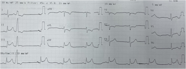
Initial Electrocardiogram Initial electrocardiogram shows ST-segment elevation in all inferior leads.

Coronary Angiogram of the Right Coronary Artery Angiography shows acute total occlusion of the right coronary artery.

Coronary Angiogram of the Left Anterior Descending Coronary Artery Angiography shows significant stenosis at the proximal left anterior descending coronary artery.

Postintervention Result No significant abnormalities are noted postintervention.

Distal Right Coronary Artery Perforation Angiography reveals extravasation of contrast at the distal right coronary artery.

Embolization Using Coils Two 4 × 3.7-mm coils are used for embolization.

Pleural Effusion Imaging shows presence of pleural and pericardial effusion. The left lung is severely compressed (red arrow). Note that the pleural space (yellow arrow) contains fluid with the same radiopacity as the contrast at the perforation site (green arrow).

After Thoracentesis Flow of contrast medium from the pericardial to the pleural space is noted. Note that the perforation site (green arrow) has the same radiopacity as the remaining fluid in the pleural space (yellow arrows), compared with the ipsilateral pleural space (red arrow). The contrast medium is also lining the left pleural space (yellow arrows).

Multislice Computed Tomography of the Pericardium The pericardial defect is identified (arrow).

Possible Mechanism
References
-
- Escaned J., Ahmad R., Shiu M. Pleural effusion following coronary perforation during balloon angioplasty: an unusual presentation of the postpericardiotomy syndrome. Eur Heart J. 1992;13(5):716–717. - PubMed
-
- Nasser W. Congenital diseases of the pericardium. Cardiovasc Clin. 1976;7(3):271–286. - PubMed
-
- Klein A.L., Abbara S., Agler D.A., et al. American Society of Echocardiography clinical recommendations for multimodality cardiovascular imaging of patients with pericardial disease: endorsed by the Society for Cardiovascular Magnetic Resonance and Society of Cardiovascular Computed Tomography. J Am Soc Echocardiogr. 2013;26(9):965–1012.e15. doi: 10.1016/j.echo.2013.06.023. - DOI - PubMed
Publication types
LinkOut - more resources
Full Text Sources
