. 2023 Sep 13;8(10):1334-1353.
doi: 10.1016/j.jacbts.2023.06.005. eCollection 2023 Oct.

PCSK9, A Promising Novel Target for Age-Related Cardiovascular Dysfunction

Affiliations

PCSK9, A Promising Novel Target for Age-Related Cardiovascular Dysfunction

Csaba Matyas et al. JACC Basic Transl Sci. .

Abstract

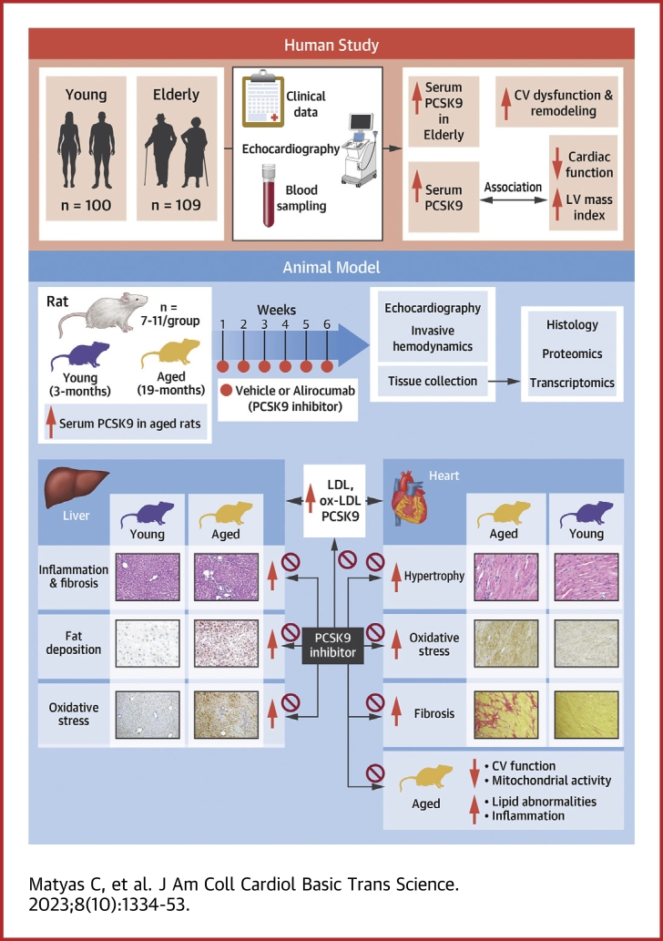
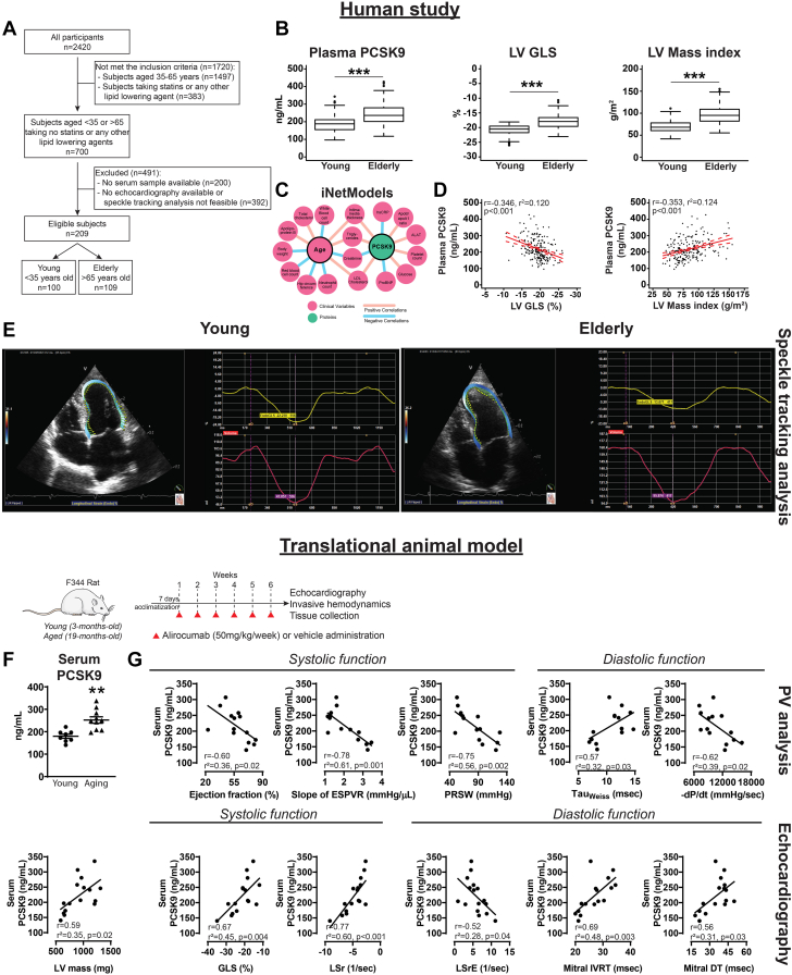
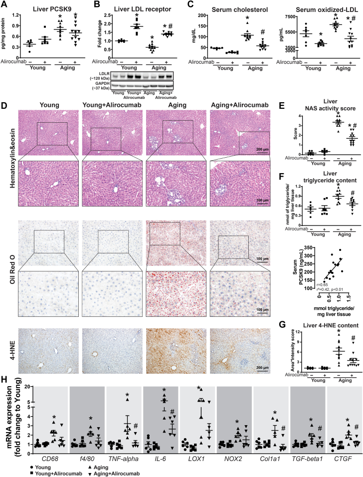
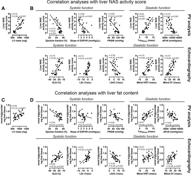
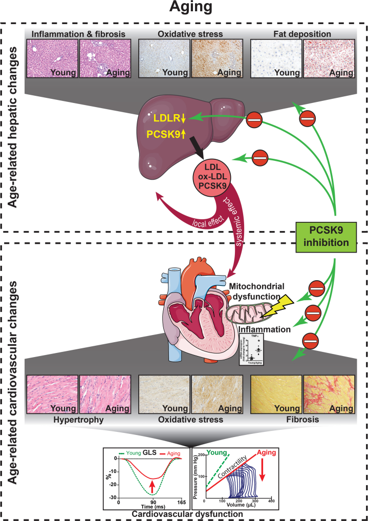
Cardiovascular diseases (CVDs) are the leading cause of death among elderly people. Proprotein convertase subtilisin/kexin type 9 (PCSK9) is an important regulator of cholesterol metabolism. Herein, we investigated the role of PCSK9 in age-related CVD. Both in humans and rats, blood PCSK9 level correlated positively with increasing age and the development of cardiovascular dysfunction. Age-related fatty degeneration of liver tissue positively correlated with serum PCSK9 levels in the rat model, while development of age-related nonalcoholic fatty liver disease correlated with cardiovascular functional impairment. Network analysis identified PCSK9 as an important factor in age-associated lipid alterations and it correlated positively with intima-media thickness, a clinical parameter of CVD risk. PCSK9 inhibition with alirocumab effectively reduced the CVD progression in aging rats, suggesting that PCSK9 plays an important role in cardiovascular aging.

Keywords: NASH; PCSK9; cardiovascular aging; heart failure; nonalcoholic steatohepatitis; transcriptomics.


Conflict of interest statement

The study was supported by the intramural program of National Institute on Alcohol Abuse and Alcoholism/National Institutes of Health Grant 1ZIAAA000375-17 (to Dr Pachar) and partly by the National Institute on Alcohol Abuse and Alcoholism/National Institutes of Health Grant R01 AG072895 (to WXD). The research was supported by project NKFIH-1277-2/2020 by the Thematic Excellence Programme (2020-4.1.1.-TKP2020) of the Ministry for Innovation and Technology in Hungary, within the framework of the Bioimaging Thematic Programme of Semmelweis University. Project no. RRF-2.3.1-21-2022-00003 has been implemented with the support provided by the European Union. Drs Fabian and Kovacs have received personal fees from Argus Cognitive, Inc, outside the submitted work. Dr Tokodi was formerly an employee of Argus Cognitive, Inc. Dr Merkely has received grants from Boston Scientific and Medtronic and personal fees from Biotronik, Abbott, AstraZeneca, Novartis, and Boehringer Ingelheim, outside the submitted work. All other authors have reported that they have no relationships relevant to the contents of this paper to disclose.

Figures

None
Graphical abstract
Figure 1
Figure 1
Increased PCSK9 Levels Are Associated With Age-Related Cardiovascular Dysfunction Human study (top). (A) Study design. (B) Box plots for plasma proprotein convertase subtilisin/kexin type 9 (PCSK9) levels, left ventricular (LV) global longitudinal strain (GLS), and LV mass index values. Statistics: for PCSK9 and GLS, Mann-Whitney U test; for LV mass index, Student’s unpaired t test. (C) Network from the iNetModels database. (D) Correlation of PCSK9 levels with LV GLS and LV mass index. (E) Representative speckle-tracking strain analyses from the study groups. Translational animal model (bottom, with study design). (F) Serum PCSK9 levels (Student’s unpaired t test). Scattered dot plots show mean ± SEM. Correlation of PCSK9 levels with functional parameters derived from (G) pressure-volume (PV) analysis and echocardiography. ∗P < .05, ∗∗P < .01, ∗∗∗P < .001. −dP/dt = minimal slope of maximal rate of left ventricular pressure rise; DT = deceleration time; ESPVR = end-systolic pressure-volume relationship; IVRT = isovolumic relaxation time; LSr = longitudinal strain rate; LSrE = longitudinal strain rate E-wave; PRSW = preload recruitable stroke work; TauWeiss = left ventricular diastolic time constant.
Figure 2
Figure 2
Increased PCSK9 Levels Are Associated With Age-Related Liver Changes (A) Liver proprotein convertase subtilisin/kexin type 9 (PCSK9) levels. (B) Liver low-density lipoprotein receptor (LDLR) levels and representative Western blot. (C) Serum cholesterol and oxidized low-density lipoprotein (LDL) levels. (D) Liver histology, liver Oil red O staining, and 4-hydroxynonenal (4-HNE) immunohistochemistry. (E) Nonalcoholic fatty liver disease activity score (NAS). (F) Liver triglyceride content and its correlation with serum PCSK9 levels. (G) Liver 4-HNE scoring. (H) Gene expression of CD68, f4/80, tumor necrosis factor (TNF)-alpha, interleukin (IL)-6, oxidized LDL receptor 1 (LOX1), NADPH oxidase 2 (NOX2), collagen 1a1 (Col1a1), transforming growth factor (TGF)-beta1, and connective tissue growth factor (CTGF). ∗P < 0.05 vs young; #P < 0.05 vs aging. One-way analysis of variance with Tukey’s post hoc test. Scattered dot plots, show mean ± SEM. mRNA = messenger RNA.
Figure 3
Figure 3
NAFLD Development Correlates With the Impairment of Cardiovascular Dysfunction Correlation analyses of liver NAS with (A) LV mass and with (B) functional parameters derived from PV analysis and echocardiography. Correlation analyses of liver triglyceride content with (C) LV mass and (D) functional parameters derived from PV analysis and echocardiography. Abbreviations as in Figure 1.
Figure 4
Figure 4
PCSK9 Inhibition Improves Age-Related Cardiovascular Dysfunction (A) Representative echocardiography recordings and echocardiography results. (B) Strain analysis. (C) PV analysis with representative recordings. ∗P < 0.05 vs young; #P < 0.05 vs aging. One-way analysis of variance with Tukey’s post hoc test. Scattered dot plots show mean ± SEM. AW = anterior wall; CSr = circumferential strain rate; CSrE = circumferential strain rate E-wave; d = diastole; dP/dt = maximal rate of left ventricular pressure rise; EDV = end-diastolic volume; E/A = E-wave to A-wave ratio; GCS = global circumferential strain; ID = internal diameter; PW = posterior wall; s = systole; other abbreviations as in Figure 1.
Figure 5
Figure 5
PCSK9 Inhibition Attenuates Cardiac Hypertrophy (A) Hematoxylin and eosin histology and (B) cardiomyocyte diameter. (C) Heatmap of hypertrophy- and contractility-related genes from transcriptomic analysis. (D) Gene expression levels of Myh-7 and Myh-6 (alpha and beta myosin heavy chain [MHC], respectively). (E) Serum B-type natriuretic peptide (BNP) levels. ∗P < 0.05 vs young; #P < 0.05 vs aging. One-way analysis of variance with Tukey’s post hoc test. Scattered dot plots show mean ± SEM. FPKM = fragments per kilobase of transcript per Million mapped reads; mRNA = messenger RNA; PCSK9 = proprotein convertase subtilisin/kexin type 9.
Figure 6
Figure 6
PCSK9 Inhibition Attenuates Cardiac Fibrosis (A) Sirius red and Masson trichrome histology and scoring. (B) Heatmap showing fibrosis-related genes from transcriptomic analysis. (C) Col1a1, fibronectin, and CTGF gene expression levels. ∗P < 0.05 vs young; #P < 0.05 vs aging. One-way analysis of variance with Tukey’s post hoc test. Scattered dot plots show mean ± SEM. Abbreviations as in Figures 1, 2, and 5.
Figure 7
Figure 7
PCSK9 Inhibition Improves Age-Related Oxidative/Nitrative Stress (A) Malondialdehyde and nitrotyrosine immunohistochemistry and scoring. (B) Heatmap showing gene expression levels from transcriptomic analysis related to oxidative stress and apoptosis. (C) PARP1 and caspase3 activity levels. (D) NOX1, NOX2, NOX3 and catalase gene expression levels. (E) TNF-alpha and IL-6 receptor and (F) LOX1 and vascular cell adhesion molecule (VCAM) gene expression levels. ∗P < 0.05 vs young; #P < 0.05 vs aging. One-way analysis of variance with Tukey’s post hoc test. Scattered dot plots show mean ± SEM. Abbreviations as in Figures 1, 2, and 5.
Figure 8
Figure 8
PCSK9 Inhibition Improves Mitochondrial Dysfunction (A) Selected Gene Ontology Cellular Component (GO CC) terms related to mitochondria and (B) selected Kyoto Encyclopedia of Genes and Genomes (KEGG) pathways related to heart and expression heatmap of the corresponding genes. (C) Mitochondrial complex I, II, IV activities. (D) Oxidative phosphorylation (KEGG map00190): gene expression changes in aging (A) vs young (Y). Blue = down-regulation, red = up-regulation. ∗P < 0.05 vs Y; #P < 0.05 vs A. One-way analysis of variance with Tukey’s post hoc test. Scattered dot plots show mean ± SEM. A+Ali = aging+alirocumab; PCSK9 = proprotein convertase subtilisin/kexin type 9.
Figure 9
Figure 9
Proposed Mechanism of Beneficial Effects of PCSK9 Inhibition on Aging-Related Cardiovascular Dysfunction Aging is associated with characteristic changes of the hepatic tissue, such as inflammation, fibrosis, oxidative stress, and fat deposition. These pathological events induce increased PCSK9 production and consequently decreased number of LDLRs. LDL, oxidized low-density lipoprotein (ox-LDL), and PCSK9 are secreted from the liver and promote local and systemic effects, possibly contributing to the development of age-related cardiovascular changes including mitochondrial injury and dysfunction, myocardial hypertrophy, oxidative stress, inflammation (TNF-α) and fibrotic remodeling, which eventually culminate in cardiovascular dysfunction. PCSK9 inhibition, as a novel interventional strategy, might improve age-related cardiac and hepatic pathologies by acting on systemic and local targets. Student’s unpaired t test. Scattered dot plots show mean ± SEM, ∗P < 0.05. Abbreviations as in Figures 1 and 2.

References

    1. Ungvari Z., Tarantini S., Sorond F., Merkely B., Csiszar A. Mechanisms of vascular aging, a geroscience perspective: JACC Focus Seminar. J Am Coll Cardiol. 2020;75:931–941. - PMC - PubMed
    1. Kim I.H., Kisseleva T., Brenner D.A. Aging and liver disease. Curr Opin Gastroenterol. 2015;31:184–191. - PMC - PubMed
    1. Paneni F., Diaz Canestro C., Libby P., Luscher T.F., Camici G.G. The aging cardiovascular system: understanding it at the cellular and clinical levels. J Am Coll Cardiol. 2017;69:1952–1967. - PubMed
    1. Younossi Z.M., Koenig A.B., Abdelatif D., Fazel Y., Henry L., Wymer M. Global epidemiology of nonalcoholic fatty liver disease-Meta-analytic assessment of prevalence, incidence, and outcomes. Hepatology. 2016;64:73–84. - PubMed
    1. Przybyszewski E.M., Targher G., Roden M., Corey K.E. Nonalcoholic fatty liver disease and cardiovascular disease. Clin Liver Dis (Hoboken) 2021;17:19–22. - PMC - PubMed