. 2023 Oct 23;12(11):e2049-e2058.
doi: 10.1016/j.eats.2023.07.025. eCollection 2023 Nov.

A Modified Technique for All-Inside Anterior Cruciate Ligament Reconstruction (ACLR): True Femoral Socket

Affiliations

A Modified Technique for All-Inside Anterior Cruciate Ligament Reconstruction (ACLR): True Femoral Socket

Wenbo Yang et al. Arthrosc Tech. .

Abstract

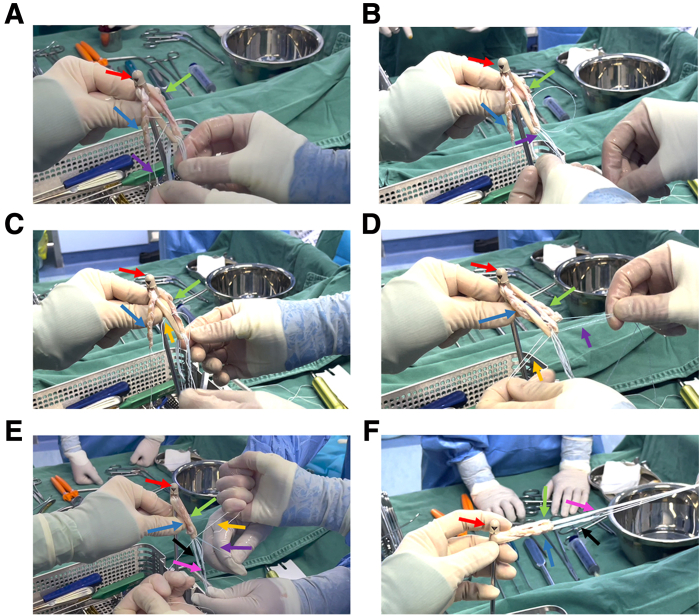
Anterior cruciate ligament reconstruction (ACLR) performed via arthroscopy is the primary treatment for anterior cruciate ligament injury. In traditional ACLR, the surgeon must create bone tunnels in both the femur and tibia, which increases the risk of bleeding and pain. The advent of all-inside technology has introduced the concept of bone sockets. However, the femoral socket created by the traditional all-inside technique is not a true femoral socket since the tunnel ends are still connected to achieve suspensory fixation. We are dedicated to achieving a true femoral socket in the all-inside ACLR technique. The AperFix Implant fixation system offers the potential for a genuine femoral socket by securely holding the ligaments in place through compression fixation. In this report, we present an all-inside ACLR using the AperFix Implant fixation system, which allows for a single exit of the femur side tunnel. This technique effectively reduces "windshield wiper" effect, "bungee cord" effect, as well as surgical time and minimizes the risk of bleeding, pain, and local microfractures.


Figures

Fig 1
Fig 1
An animated scheme of our improved technology.
Fig 2
Fig 2
Anterior cruciate ligament rupture was observed under direct arthroscopy. This field of vision is observed through the anterolateral approach. The red arrow indicates the location of the torn anterior cruciate ligament.
Fig 3
Fig 3
Acquisition and weaving of single semitendinosus tendon graft. (A) Harvest the semitendinosus tendon. The surgical incision was positioned medially to the tibial tubercle and measured 3 cm in length. (B and C) The 2 free ends of the tendon were sutured and braided separately. The green arrow indicates the tendon graft. The purple arrows indicate the suture used to braid the tendon.
Fig 4
Fig 4
The preparation of AperFix implant + tendon graft + adjustable-loop cortical suspension devices complex. (A) Suture at one end of the graft is threaded through the AperFix implant. The red arrow indicates the AperFix implant. Purple arrows indicate the suture. The green arrow indicates the tendon graft. (B) Graft is threaded through the AperFix system. (C) The suture go through the cortical suspension device. The blue arrow indicates the adjustable loop of the cortical suspension device. The pink arrow indicates the button. (D) The tendon graft passes through the AperFix system a second time. (E and F) General view and detailed presentation of the formed complex. The yellow arrow represents the suture for pulling the button. The orange arrows represent the suture used to tighten the loop.
Fig 5
Fig 5
The loop-shape tendon graft complex fixed by knotting. (A) One of the sutures at one of the free ends of the semitendinosus graft is threaded through the loop of the tendon graft. The red arrow indicates the AperFix implant. Purple arrows indicate the suture of one of the free ends. The green arrow indicates one of the free ends of the semitendinosus graft. The blue arrow indicates the other free end of the semitendinosus graft. (B) Suture is then immediately threaded through adjustable-loop cortical suspension devices. (C) One of the sutures at the other free end of the graft passes through the loop of the tendon graft and immediately passes through the adjustable loop cortical suspension devices. Orange arrows indicate the suture of the other free end of the semitendinosus graft. (D) The structure of the complex before it is ready to be knotted and fixed. (E and F) The two sutures at each free end of the graft are knotted and fixed in pairs, and the graft is prepared to form a loop structure required for the all-inside technique. The black arrow indicates the adjustable loop, and the pink arrow indicates the button.
Fig 6
Fig 6
Establishment of bone sockets. The anterolateral approach is used as the observation approach, and the anteromedial approach is used as the operational approach. (A) Determination of position by the offset positioner. (B) Through the offset positioner, the 2-mm guide needle is drilled to the lateral femur cortex. This step does not require penetrating the lateral cortex of the femur. (C) Using a 9-mm femur drill bit, the surgeon prepares a 28-mm deep bone socket at the site where the guide needle was drilled. (D) The socket was fully exposed after being cleaned with a radiofrequency. The blue arrows indicate the offset positioner. The yellow arrow indicates the guide needle. The green arrow indicates the 9-mm femur drill bit. The red arrow indicates the femoral socket. (E and F) Establishment of lateral tibial bone socket. The tibial socket was achieved using the 8-mm retrograde drill (pink arrow). A traction suture (orange arrow) was inserted through the tibial socket to the intra-articular to anteromedial approach.
Fig 7
Fig 7
Fixation of femoral end with autogenous tendon graft complex. The anterolateral approach (green arrow) is used as the observation approach, and the anteromedial approach is used as the operational approach. (A-C) The complex is delivered into the joint cavity through the anteromedial approach, and the AperFix implant (blue arrow) is positioned into the femoral socket. AperFix implant should be fully inserted into the femoral socket and reach the bottom as far as possible. The tendon length located in the femoral socket should be maintained at 15 mm. The red arrow indicates the tendon graft. (D and E) Expand and squeeze the fixed wing of the implant through AperFix handle (yellow arrow) to secure and pull out the auxiliary device. (F) After the AperFix system is fixed, the ligament position was observed arthroscopically, and the ligament stability at femur side was tested.
Fig 8
Fig 8
Fixation of tibial end with autogenous tendon graft complex. (A and B) Assistant tensions the braided suture and pulls the graft into the tibial socket under the direct vision of the arthroscopy. The anterolateral approach is used as the observation approach. (C-E) Tighten the adjustable loop, knot the suture, and fix the tibial cortical suspensory fixation button after 20 times of flexion and extension. The purple arrow represents the braided suture at the end of the tendon graft. The green arrows represent the tightened suture of the adjustable-loop. The red arrows represent the traction suture of the button. The yellow arrow indicates the button. (F) Examine the graft location using the anteromedial approach. The pink arrow represents the graft.

References

    1. Khan I., Ullah S., Khattak S.K., Khan Z., Ahmad I., Khan A. Functional outcome of arthroscopic anterior cruciate ligament reconstruction using hamstring autograft with fixation using Endobutton at femoral end and bioabsorbable screw at tibial end. Mymensingh Med J. 2022;31:1142–1147. - PubMed
    1. Zhu J., Marshall B., Tang X., Linde M.A., Fu F.H., Smolinski P. ACL graft with extra-cortical fixation rotates around the femoral tunnel aperture during knee flexion. Knee Surg Sports Traumatol Arthrosc. 2022;30:116–123. - PubMed
    1. Lin R., Zhong Q., Wu X., Cui L., Huang R., Deng Q., et al. Randomized controlled trial of all-inside and standard single-bundle anterior cruciate ligament reconstruction with functional, MRI-based graft maturity and patient-reported outcome measures. BMC Musculoskel Disord. 2022;23:289. - PMC - PubMed
    1. Lubowitz J.H. All-inside anterior cruciate ligament graft link: Graft preparation technique. Arthrosc Tech. 2012;1:e165–168. - PMC - PubMed
    1. Lin T.Y., Chung C.C., Chen W.C., Su C.W., Fang H.W., Lu Y.C. Complications following all-inside anterior cruciate ligament reconstruction. Int Orthop. 2022;46:2569–2576. - PubMed

LinkOut - more resources