. 2023 Dec 12;15(23):13593-13607.
doi: 10.18632/aging.205297. Epub 2023 Dec 12.

Uncovering a unique pathogenic mechanism of SARS-CoV-2 omicron variant: selective induction of cellular senescence

Affiliations

Uncovering a unique pathogenic mechanism of SARS-CoV-2 omicron variant: selective induction of cellular senescence

Franziska Hornung et al. Aging (Albany NY). .

Abstract

Background: SARS-CoV-2 variants are constantly emerging with a variety of changes in the conformation of the spike protein, resulting in alterations of virus entry mechanisms. Solely omicron variants use the endosomal clathrin-mediated entry. Here, we investigate the influence of defined altered spike formations to study their impact on premature cellular senescence.

Methods: In our study, in vitro infections of SARS-CoV-2 variants delta (B.1.617.2) and omicron (B.1.1.529) were analyzed by using human primary small alveolar epithelial cells and human ex vivo lung slices. We confirmed cellular senescence in human lungs of COVID-19 patients. Hence, global gene expression patterns of infected human primary alveolar epithelial cells were identified via mRNA sequencing.

Results: Solely omicron variants of SARS-CoV-2 influenced the expression of cell cycle genes, highlighted by an increased p21 expression in human primary lung cells and human ex vivo lungs. Additionally, an upregulated senescence-associated secretory phenotype (SASP) was detected. Transcriptomic data indicate an increased gene expression of p16, and p38 in omicron-infected lung cells.

Conclusions: Significant changes due to different SARS-CoV-2 infections in human primary alveolar epithelial cells with an overall impact on premature aging could be identified. A substantially different cellular response with an upregulation of cell cycle, inflammation- and integrin-associated pathways in omicron infected cells indicates premature cellular senescence.

Keywords: SARS-CoV-2; cellular senescence; lung airway cells; variant of concern.


Conflict of interest statement

CONFLICTS OF INTEREST: The authors declare that the research was conducted in the absence of any commercial or financial relationships that could be construed as a potential conflict of interest.

Figures

Figure 1
Figure 1
(A) Immunofluorescence staining of lungs from deceased COVID-19 patients. Lung tissue was stained with p21 (green), actin (red), and nuclei (blue) staining. Scalebar indicates 50 μm. (B) Gene expression of senescence marker CDKN1A and CDKN2A in three distinct lung biopsies of three deceased patients per SARS-CoV-2 variant. Expression levels are displayed normalized to healthy lung tissue from healthy lung transplantation tissue. P calculated by ANOVA with Kruskal-Wallis multiple comparisons tests (B), **p < 0.05.
Figure 2
Figure 2
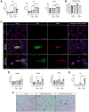
(A) Increased levels of IL-6, IL-8, and MCP-1 of infected supernatants of SAECs (n = 3) determined 8 h and 24 h p.i. compared to non-infected mock. (B) SAECs were infected with SARS-CoV-2 delta or omicron variant with an MOI of 5 for 24 h. Infected cells show clear signals for intracellular spike protein (green) and dsRNA (red). (C) SARS-CoV-2 RNA detected in cell lysates of infected SAECs after 8 h and 24 h p.i.. (D) Gene expression of senescence markers CDKN1A, CDKN2A, and LMNB1, normalized to mock cells. CDKN1A is significantly upregulated in cells infected with omicron variant compared to the delta variant. (E) Levels of the senescence-associated secretory phenotype (SASP) protein IL-1β are significantly increasing in omicron-infected cells from 8 h to 24 h p.i.. (F) β-Galactosidase staining of SAECs after 24 h p.i. with SARS-CoV-2. Scalebar indicates 100 μm. P calculated by one-way ANOVA with Tukey’s multiple comparisons tests (A, E) and Welch’s t-test (D), *p < 0.01, **p < 0.05, ***p < 0.001, ****p < 0.0001.
Figure 3
Figure 3
(A) Infection with SARS-CoV-2 variants alters the gene expression patterns of SAEC cells. Co-expression Venn Diagram showing regulated genes of mock, omicron- and delta variant infected cells identified via mRNA-sequencing 8 h p.i.. (B) Pathways with significant enrichment scores (-log (p-value)) revealed by QIAGEN Ingenuity Pathway Analysis 8 h p.i. indicates significant increase of gene regulation in omicron-infected cells. (C, D) Representative heatmaps of genes regulated due to omicron variant or delta variant, after 8 h (C) or 24 h (D) p.i. shown in relation to pathway or gene families. Here, selective genes for cyclins, senescence-associated secretory phenotype (SASP), Damage-associated molecular patterns (DAMPS), integrins, interleukins, and cell cycle are shown.
Figure 4
Figure 4
(A) Schematic overview of the experimental setup for the infection of human PCLSs with SARS-CoV-2 delta and omicron variant. Created with https://www.biorender.com/. (B) SARS-CoV-2 RNA detected in supernatants of infected human PCLSs after 2 d p.i.. (C) Immunofluorescent staining of mock and infected PCLS. Surfactant protein a (SP-A, green) positive cells are visible in the alveoli of mock and infected slices 4 d p.i.. Colocalization of spike protein (red) and SP-A positive cells in the alveolus of infected slices. Scalebar indicates 100 μm. (D) Gene expression of senescence markers CDKN1A, CDKN2A, and LMNB1, normalized to mock PCLSs. CDKN1A is significantly upregulated in cells infected with omicron variant compared to delta. LMNB1 expression is significantly downregulated in omicron slices compared to delta and mock (E) Levels of the senescence-associated secretory phenotype (SASP) proteins IL-1β, IL-6, IL-8, TNF-α, IL-10 are significantly upregulated in omicron- and delta-infected PCLSs at 2 d p.i. MCP-1 levels are on average highest in omicron-infected slices. P calculated by one-way ANOVA with Multiple comparisons (D, E), *p < 0.01, **p < 0.05, ***p < 0.001.
Figure 5
Figure 5
Graphical abstract of known differences of SARS-CoV-2 delta and omicron variant entry and findings of our study obtained from mRNA sequence data of 24 h post infection. Schematic overview of entry mechanisms of SARS-CoV-2 delta and omicron variants [5]. Delta variant (right panel) uses cell surface entry, by ACE2 and the protease TMPRSS2. Own data indicate a downregulation of the integrin activation without affecting p38 and p16 expression resulting in normal cell cycle. Omicron variant (right panel) prefers to use the clathrin-mediated endocytosis (CME) and cathepsin L as protease. Our results suggest the expression and activation of integrins (ITGB1, ITGB4, ITGA1, ITGA2, ITGA3, ITGA6) resulting an increase in p38 and p16. That increase in central kinases affects several cyclins, which in turn downregulates the retinoblastoma, increases the E2F transcription factors and results in cell cycle arrest and cellular senescence. Additionally, these changes lead to an increase in senescence-associated secretory phenotype (SASP). Thus, our findings indicate an influence of the altered cell entry mechanism of the omicron variant on the cell cycle. Created with https://www.BioRender.com.

References

    1. Harvey WT, Carabelli AM, Jackson B, Gupta RK, Thomson EC, Harrison EM, Ludden C, Reeve R, Rambaut A, Peacock SJ, Robertson DL, and COVID-19 Genomics UK (COG-UK) Consortium. SARS-CoV-2 variants, spike mutations and immune escape. Nat Rev Microbiol. 2021; 19:409–24. 10.1038/s41579-021-00573-0 - DOI - PMC - PubMed
    1. Du X, Tang H, Gao L, Wu Z, Meng F, Yan R, Qiao S, An J, Wang C, Qin FX. Omicron adopts a different strategy from Delta and other variants to adapt to host. Signal Transduct Target Ther. 2022; 7:45. 10.1038/s41392-022-00903-5 - DOI - PMC - PubMed
    1. Kumar S, Thambiraja TS, Karuppanan K, Subramaniam G. Omicron and Delta variant of SARS-CoV-2: A comparative computational study of spike protein. J Med Virol. 2022; 94:1641–9. 10.1002/jmv.27526 - DOI - PubMed
    1. Meng B, Abdullahi A, Ferreira IAT, Goonawardane N, Saito A, Kimura I, Yamasoba D, Gerber PP, Fatihi S, Rathore S, Zepeda SK, Papa G, Kemp SA, et al., and CITIID-NIHR BioResource COVID-19 Collaboration, and Genotype to Phenotype Japan (G2P-Japan) Consortium, and Ecuador-COVID19 Consortium. Altered TMPRSS2 usage by SARS-CoV-2 Omicron impacts infectivity and fusogenicity. Nature. 2022; 603:706–14. 10.1038/s41586-022-04474-x - DOI - PMC - PubMed
    1. Jackson CB, Farzan M, Chen B, Choe H. Mechanisms of SARS-CoV-2 entry into cells. Nat Rev Mol Cell Biol. 2022; 23:3–20. 10.1038/s41580-021-00418-x - DOI - PMC - PubMed

Publication types

Supplementary concepts